
Mycobacteria Fungal Respiratory Tract Pathogens Prof Dr Asem

Mycobacteria & Fungal Respiratory Tract Pathogens Prof. Dr. Asem Shehabi Faculty of Medicine University of Jordan
Global Prevalence of Tuberculosis n n n The World Health Organization (WHO) estimates that approximately one-third of the global population is infected asymptomatic with M. tuberculosis (TB). Around 10 million new cases of TB are being reported each year, 2 -3 million deaths occur each year worldwide. . 95% in developing countries After emerging (HIV)/AIDS, TB is the second most common cause of death in AIDS patients due to an infectious disease.

Mycobacterium Tuberculosis n. Tubercle Bacilli. . Acid-Fast Bacilli. . Widely distributed in Human, Animals, Birds, Environment. . TB bacilli grow slowly, Resistant to Dryness, low Acidity. . survive years in nature. . But Susceptible to UV-light, Heat. M. tuberculosis. . Causes 95% of human TB cases. . mostly pulmonary. . Respiratory infection. . Few cells. . Lung positive person may infect hundred of susceptible person. . All ages. . mostly children. . with malnutrition. Optimal conditions for transmission include: overcrowding, Large cities, poor conditions & Low standard public hygiene. . n n
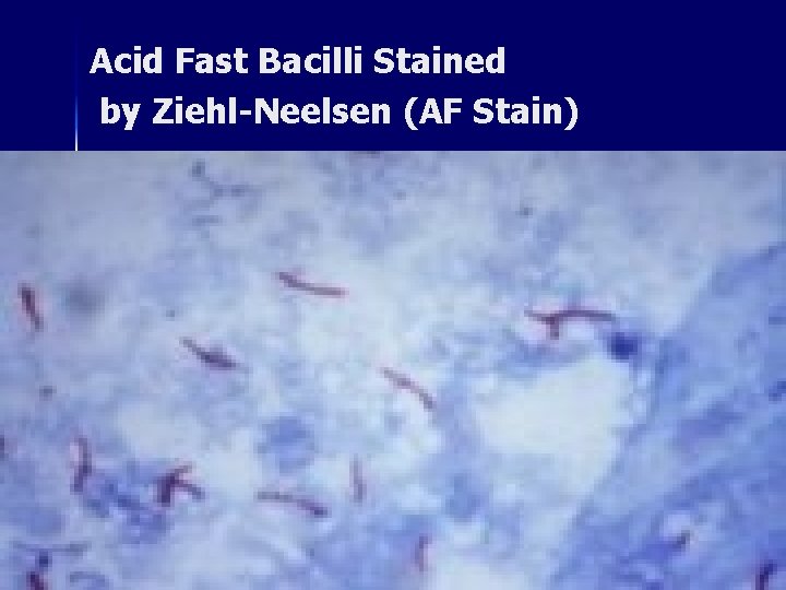
Acid Fast Bacilli Stained by Ziehl-Neelsen (AF Stain)
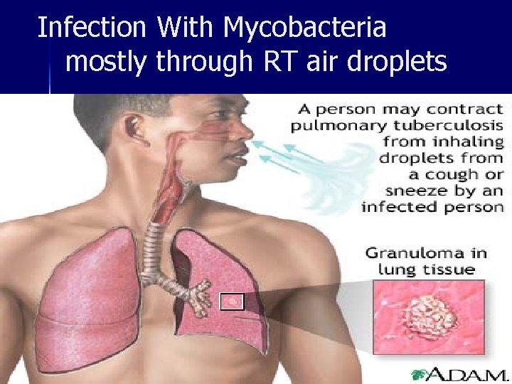
Infection With Mycobacteria mostly through RT air droplets
Pulmonary Infection Primary Tuberculosis: 90% Pulmonary TB , Children, Asymptomatic, TB Bacilli infect Alveolar macrophages. . Develop small lung lesions. . Fibrosis, Calcification, Hypersensitivity. . Infected person becomes Positive for Tuberculin Skin test. n Few cases Primary TB infection may spread from Lesion by direct extension to lymphatic system, bronchi, blood, Kidneys, Meningitis (children). . Gastrointestinal. . causing localized lesions but rarely Military tuberculosis. n
Post-Pulmonary Infection-2 n n n Post primary tuberculosis: Reactivation old lesions/infection. . Common in young adults & elderly persons. . Developing Large Lung lesions. . Cavities , Less lymphatic involvement , intensive Granuloma & Caseation. . May spread rapidly to other body part, CNS, Gastrointestinal/Urinary Tract. Clinical Features Pulmonary TB: Productive Cough. . bloody sputum, Low continuous fever, Night-sweating , Loss weight & Appetite. . General weakness, Breath shortness. Lesions/ Cavities can be detected easily by Chest xray, Sputum culture , Positive Tuberculin test
Tuberculin Test n n Both Symptomatic/ asymptomatic infected persons with any type of Mycobacteria. . develop positive Tuberculin skin test. . Reaction to TB glycerol extract ( Mycolic acids + lipoproteins ) Mantoux -Tuberculin skin test. . More specific TB antigens produced from boiling culture of M. tuberculosis. The test consists of intradermal injection of 5 tuberculin -units (0. 1 ml) of PPD in the forearm. . The test is read after 48 -72 hours. Positive tuberculin: Indurations, Edema & Erythematic skin > 1 cm, Interpretation of test. Vaccination with BCG. . Protection 30 -70%. . result in positive Tuberculin test.
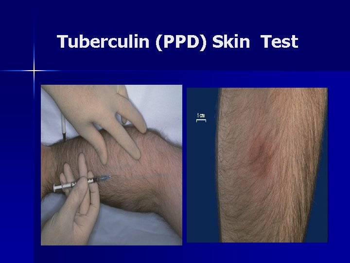
Tuberculin (PPD) Skin Test
Other Human Pathogenic Mycobacteria species n n n M. bovis: common in domestic animal. . rare human. . Infection. . source: milk, dairy products, meat. . begins mostly intestinal infection. . may spread to other parts. . Slow grower Atypical mycobacteria: Widely distributed in nature. . water, soil, birds, animals , mostly slow grower (1 -3 weeks) M. kansasii: Soil, Photochromogenic, Produce yellow/orange color during incubation in light. . Mostly Lung tuberculosis. . immuno-suppressed persons, AIDS. M. marinum : Water , Fish , localized Skin, ulcers-soft tissues, Swimming pool, aquarium granuloma. . Lymph nodes. M. avium complex : Animals, water, Skin Lesions, rarely Pulmonary disease. M. ulcernas: Soil in Tropical countries, Skin lesions, necrosis, More Resistant to anti-tuberculosis drugs. .
Diagnosis & Treatment-1 n n Rapidly growing Mycobacteria species: Rarely cause skin ulceration, mostly non-pathogens. . M. smegmatis. . Found in on extragenital tract. . May contaminate urine culture. Diagnosis & treatment: Tuberculosis is confirmed by positive Direct AF Smear/ Culture, PCR X-ray, Positive tuberculin Test. Clinical specimens: Sputum, Urine, CSF, Tissues, Culture Loewenstein-Jensen Agar. . 4 -8 Weeks. . No Blood Serological test. Treatment Multiple Antibiotics: 6 -24 Months. . Rifampicin, Isonaized, Pyrazinamid, Ethambutol, development of Multidrug resistant MB tuberculosis. . At present 1 -5% worldwide. . Completing treatment is essential for cure
Nocardiosis n n Nocardia asteroids/ N. barsiliensis. Aerobic G+ve Pleomorphic Bacilli & Branched short Filaments. . Slightly Acid Fast. . Common as Environmental Saprophytes. Human Exogenous Infection. . Mostly Pulmonary localized abscesses. . Necrosis. . small cavities. . spread to Brain, Kidneys. . Common in Immunosuppressed , Lung malignancy, patients with renal dialysis. Chronic suppuration. . Abscess. . Granulomas, Draining sinuses containing granules. . Muscles, Bones, Feet, Hands and other body parts. Diagnosis & Treatment: Sputum/biopsies culture on blood 1 -4 weeks at room temperature , Cotrimoxazole, Rifampicin, Amikacin. . 4 -6 Weeks.
Respiratory Fungal Agents n n Fungal respiratory diseases can be divided into: Fungi/Molds. . Their spores/filaments widely distributed in Environment. . Cause mostly infection in immunocompromised individuals. . receiving immunosuppressive therapy. . undergoing bone marrow transplantation or solid-organ transplant. . HIV infection. Clinical presentations : Fungal respiratory infections are non-specific. . often overlap with other infectious and noninfectious processes. . mostly without fever The causative agents can be opportunistic endogenous Yeast or exogenous filmentous Fungi /Molds
Yeast Form: Candidiasis n n n Part of oral /intestinal/vaginal flora. . causes characteristic mucosa patches of a creamy-white to grey pseudomembrane composed of Blastospores and Pseudohyphae. Candidasis often develop after long antibiotics treatment. Oral candidasis may spread. to Esophagus, Bronchi, Lungs, Gastro-intestinal tract, or become systemic. . Candidiaemia. . may results in endocarditis. . meningitis. Systemic Candidiasis is common in patients with cellmediated immune deficiency, receiving aggressive cancer, immunosuppression, transplantation therapy. C. albicans, C. glabrata, C. tropicalis, C. krusei,
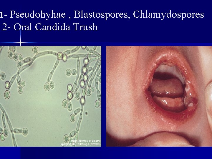
1 - Pseudohyhae , Blastospores, Chlamydospores 2 - Oral Candida Trush
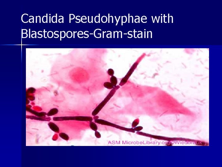
Candida Pseudohyphae with Blastospores-Gram-stain
Predisposing Factors for the Development of Candidiasis n n n Impaired epithelial barrier: Burns, Wounds / abrasions, Hydration/maceration, Indwelling catheters, Foreign bodies (Dentures, etc), Increased gastric p. H, Cytotoxic/ Antibiotics agents. . Radiation Systemic disorders: Diabetes mellitus, Pregnancy/oral contraceptives, Malnutrition, Malabsorption, Iron deficiency. Malignancy / Haematologic disorders: Neutropnea / Leukemia, Lymphoma, advanced cancer, AIDS all immunodeficiency conditions. Sytemic treatment: fluconazole , amphotericin B, Caspofungin. . effect on Ergosterol Cell membrane Local Ointment: Nystatin, micronazole, clotrimazole
Yeast: Cryptococcosis n n Encapsulated C. neoformans. . cause a chronic, subacute -acute pulmonary. . systemic or meningitic disease. . Meningoencephalitis. . Often isolated from Pigeon & Birds excreta. Primary pulmonary infections: Mostly pneumonia-like illness, with symptoms such as cough, fever, chest pain. . dissemination may include central nervous system. . Lesions in brain, skin, bones and other visceral organs. . Common significant opportunistic pathogens in immunodifficient humans , AIDS patients. Diagnosis: Culture + Antigens detection in Serum , CSF & Biopsy.
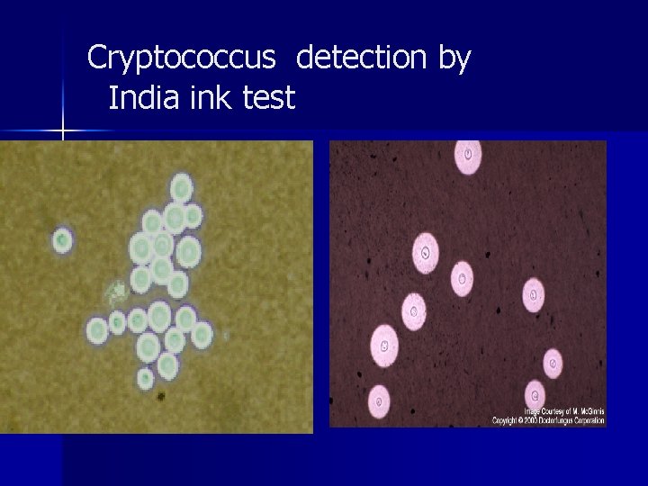
Cryptococcus detection by India ink test
Molds: Aspergillosis-1 n Most common human Aspergillus species A. fumigatus, A. flavus, A. niger. n Aspergillus spores and filaments are widely distributed in nature. . Survive harsh environmental conditions. . Dryness for months. . Contaminate rapidly dead animal, plants , fruits, Nuts, Grains, soil, Milk powder, spread via small spores that are extremely light and float easily in the air and with dust particles. n Allergic Bronchopulmonary: Repeat exposing of Sinuses & lung tissues to conidia & fungal filaments often associated with development Allergic reaction. . Eosinophilia & Asthma.
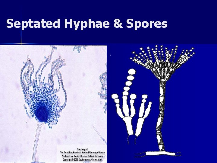
Septated Hyphae & Spores
Aspergillosis-2 3) Pulmonary Aspergilloma ( Fungus Ball). . Invasive Aspergillosis. . Pre-existing lung cavity (Tuberculosis). . inflammatory, granulomatous, necrotizing disease of lungs. . May spread to other organs. . causing mostly Thrombosis. . Rhino-cerebral lesions rarely systemic and fatal disease. n Treatment: Fluconazole, Itraconazole, Caspofungin Amphotericin B. . n Less common Respiratory Fungi: Mucor, Rhizopus Fusarium spp. . (Mucormycosis). . Causing Commonly Rhino-cerebral lesions.
Dimorphic Fungus: Histoplasmosis-1 n n Histoplasma capsulatum. . Dimorphic fungus with yeast cells & Microconidia in vivo at body temperature. . Hyphae & marcoconidia in vitro culture. . Common in soil enriched with excreta of birds. Endemic in southern U. S. A, Australia. . Less common in other countries. The primary site of infection is usually Lung. . inhalation dust with microconidia. . Phagocytosed by macrophages, obligate intracellular parasites. . Causing slight inflammatory reaction. . Most cases of histoplasmosis are asymptomatic /subclinical, benign as Flu-like syndrome. Few may develop chronic progressive lung disease. . Granuloma & fibrosis, chronic cutaneous or systemic disease involve any internal organ. . Fatal systemic disease. All infected persons become positive by histoplasmin skin test.
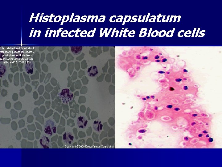
Histoplasma capsulatum in infected White Blood cells
Coccidioidomycosis & Blastomycosis-2 n Coccidioides immitis & Blastomyces dermatitidis. . soil inhabiting Dimorphic Fungus. . Endemic in south-western U. S. A. , northern Mexico and various parts South America. n n Respiratory infection. . inhalation of microconidia, often resolves rapidly leaving the patient with a strong specific immunity to re-infection. Some individuals the disease may progress to a chronic pulmonary condition or a systemic disease involving the meninges, bones, joints, subcutaneous & cutaneous tissues. . Antigen Skin test positive. . Not significant in diagnosis.
Laboratory Diagnosis n n n Direct microscopy and culture should be performed on all specimens (sputum, bronchial washings, CSF, pleural fluid tissue biopsies from various visceral organs ). wet mounts in 10% KOH with india ink. . Ovoidbudding yeast cells (b) Gram-stain smear. . Cultures on Sabouraud dextrose agar should be maintained for one month at 25 C. . fungal growths & Wet Mount. . Identification. . produces hyphae-like conidio-phores & Spores. . Color of fungal growth Serological tests are of limited value. . not significant Detection of Histoplasm antigen in blood & urine is significant
Pneumocystis pneumonia Small yeast-like cells. . No filaments or Capsules n P. jiroveci (human type). . Old name P. carinii n Pneumocystis infection occurs by inhalation contaminated dust. . It is commonly found in the lungs of healthy individuals without symptoms n Asymptomatic infection started mostly in children & increased in Adults. . Worldwide. n Clinical Disease occurs only associated with both decrease of cellular immunity & humoral immunity or suppressed immunity. n
Pneumocystis-2 n n n Infectious trophic form of the organism attaches to the lung alveoli. . Encyst & multiple in host tissues. Clinical disease. . Fever, non-productive cough similar atypical Pneumonia. . Organism is usually found in the interstitial fluid & tissue of the lungs. immunocompromised patients. . AIDS Organism may disseminate to other internal body organs & Associated with high mortality. Sputum /lung biopsy specimens are usually used for PCP detection in Pathology examination. . Silver & Giemsa. Stain. . Immunofluorescent Antigen (IFA). . No culture Treatment: Cotrimoxazole alone or with intravenous Pentamidine in sever cases.
- Slides: 28