Mycobacteria Chlamydia Mycoplasma Rickettsia Fungus Mycobacteria Acidfast bacilli

Mycobacteria Chlamydia, Mycoplasma, Rickettsia Fungus 진단검사의학과 김명희

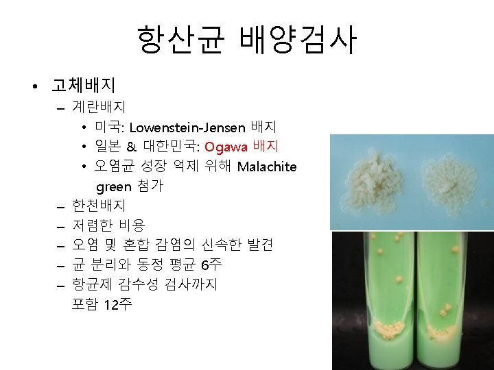
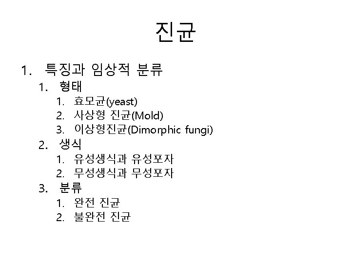
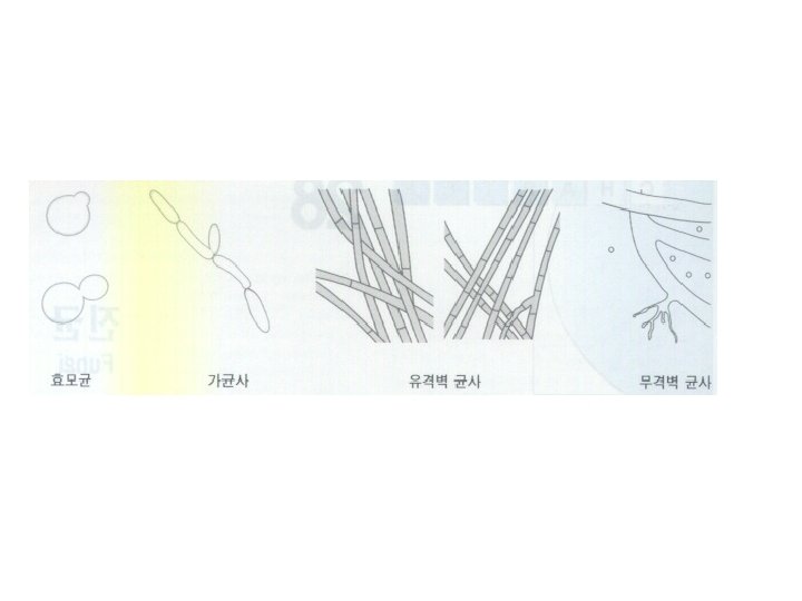
Mycobacteria

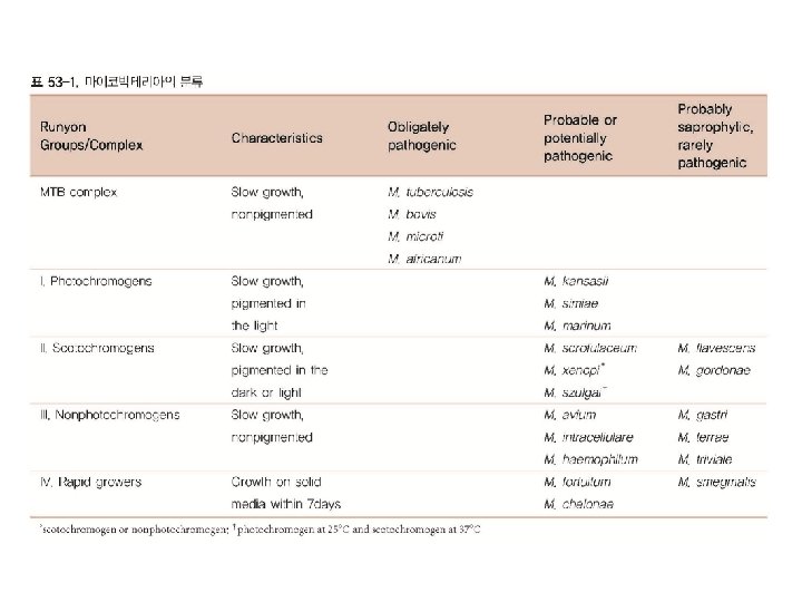
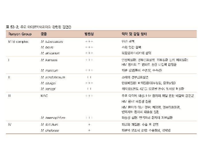
• Acid-fast bacilli • 200여 균종 – MTB complex – Non-tuberculous mycobacteria, NTM
 carbolfuchsin methods : Ziehl-Neelsen (most reliable) & Kinyoun methods 2) fluorochrome")
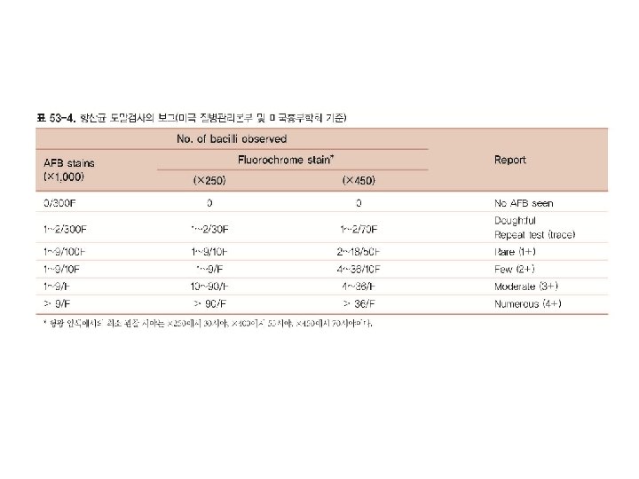
항산균 도말검사 1) carbolfuchsin methods : Ziehl-Neelsen (most reliable) & Kinyoun methods 2) fluorochrome procedure : auramine-O (green) or auramine-rhodamine dyes (yellow/orange)

• 분자생물학적 방법 – PCR, real-time PCR, nested PCR • 잠복감염 진단 – Mantoux skin test • Purified protein derivative (PPD) – IFN-γ release assy (IGRA) • Quanti. FERON Gold IT, Elispot

Chlamydia, Mycoplasma, Rickettsia
 – RNA, DNA 모두")
Chlamydia • Obligate intracellular parasite – 바이러스보다는 세균에 가까움 (그람음성) – RNA, DNA 모두 가짐 – 항균제에 감수성 • 기본체 (elementary body) • 망상체 (reticulate body) • 봉입체 (inclusion)

C. trachomatis • 면역형: 15가지이상 존재 – – Inclusion conjunctivitis: D-K Trachoma: A, B, Ba, C Lymphogranuloma venereum, LGV: L 1, L 2, L 3 Nongonococcal urethritis & prostitis • 비임균성 요도염의 50~60% • D-K – Gynecological infection • D-K • 자궁 경관염, 자궁내막염, 나팔관염 – 신생아 폐렴: 산도로부터 감염 • D-K
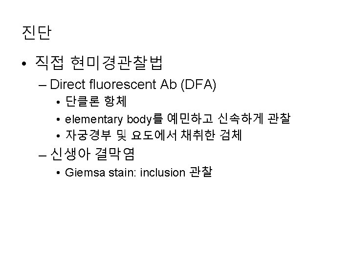
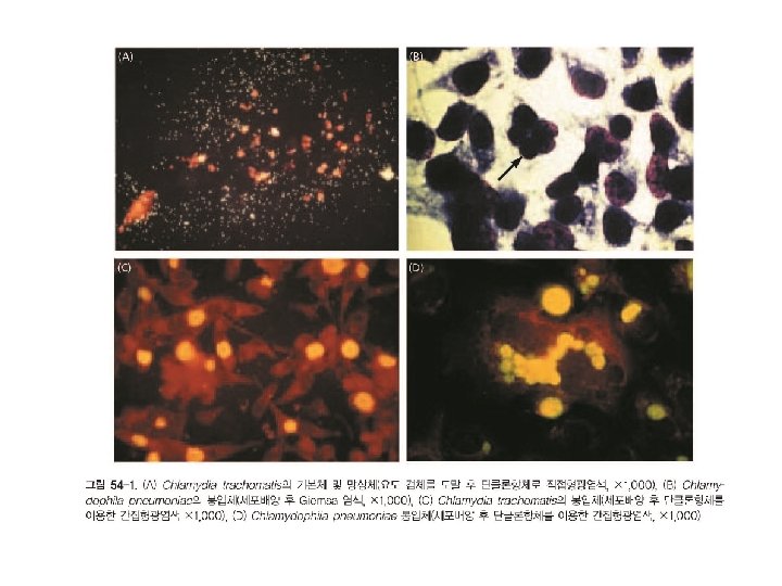

배양 • C. trachomatis – Mc. Coy cell, Hela cell – inclusion 관찰 • C. pneumoniae – Hela cell, Hep 2 cell

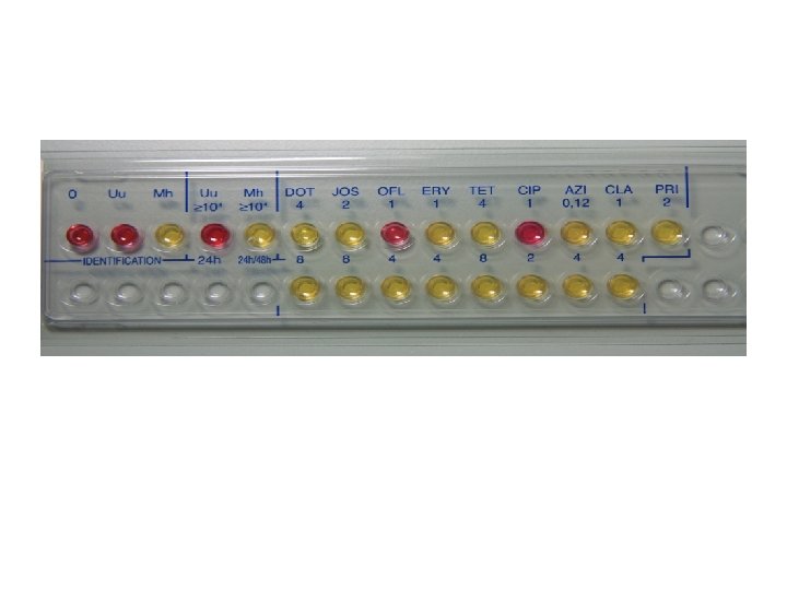
Mycoplasma • 인체에서 10여가지 균종 검출 • 병원균 – M. pneumoniae • Primary atypical pneumonia, tracheobronchitis, . . – M. hominis • 신우신염, 난관염, 요도염, 구인두염 – U. urealyticum • 비임균성 요도염

진단 • 배양 • 분자유전검사 – Sequence specific oligonucleotide probe hybridization – Sequence specific primer • 혈청학적 검사 – Anti-I Ig. M cold agglutinin (선별검사) – Anti-mycoplasmal Ig. M, Ig. G 특이항체 (확진)
 R. rickettsia Rocky산 홍반열 서반구")
Rickettsia, Coxiella, Rochalimaea 병인 인체감염증 분포지역 전파방법 Spotted fevers(홍반열군) R. rickettsia Rocky산 홍반열 서반구 Tick bite R. cororii Boutonneuse fever 지중해, 아프리카, 인도, 서 남아시아 Tick bite R. sibrica Siberian tick typhus 시베리아, 몽고, 중앙아시아 Tick bite R. australis Queensland tick typhus 호주 Tick bite R. akari Rickettsial pox 미국, 러시아, 한국 Mite bite (좀진드기)
 전세계 Infected louse feces R.")
병인 인체감염증 분포지역 전파방법 R. prowazekii Epidemic typhus(발진티푸 스) 전세계 Infected louse feces R. prowazekii Brill-Zinsser병 북아메리카, 유럽 Reactivation of epidemic typhus R. typhi Murine typhus( 발진열) 전세계 Infected flea feces R. tsutsugamushi Scrub typhus 아시아, 인디아, 호주, 태평양제도 Chigger bite(좀진 드기) Coxiella burnettii Q fever 전세계 Infectious aerosol, Tick bite Rochalimaea qiuntana Trench fever 유럽, 아프리카, 북아메리카 Scratching louse feces Typhus fever

Fungus
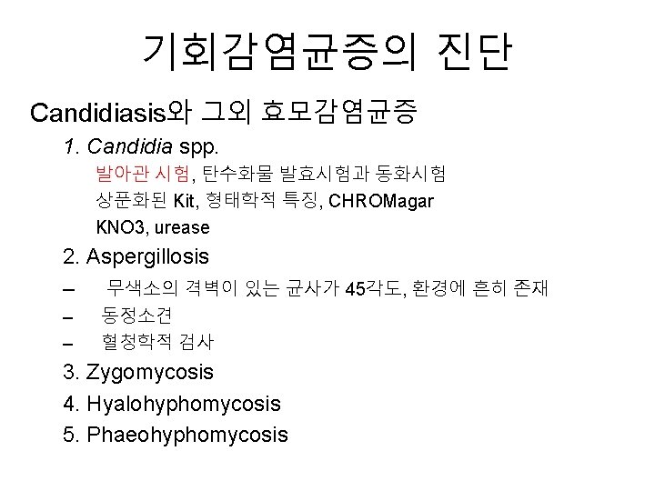

2. 직접검사 – 10 -20% KOH : 피부, 조갑, 모발 – India ink : 뇌척수액, Cryptococcus neoformans (민감도 50% 이하) – Calcofluor white : Pneumocystis jirovecii – Fluorescent monoclonal Ab 3. 조직, 병리학적 검사 – Methenamine silver, PAS 4. 혈청학적 검사 – 면역확산법, 보체고정법, latex 응집법, ELISA – Crytococcosis, Aspergillosis, Candidiasis, Sporotrichosis, Blastomycosis, Coccidioidosis, Histoplasmosis

India ink 염색

Calcofluor white • rapid detection of yeasts, fungi & parasitic organisms • non-specific fluorochrom – binds to cellucose and chitin in cell walls A. fumigatus Yeast
 비선택배지 : 항균제와 cycloheximide")
5. 배양검사 1. 배지 • • • Sabouraud dextrose agar(SDA) 비선택배지 : 항균제와 cycloheximide 2. 배양조건 30 o. C에서 최소 30일간 3. 사상형 진균의 동정법 Lactophenol cotton blue 4. 효모균 진균의 동정법 Germ tube test : C. albicans API 20 C Corn meal tween 80 agar Nitrate(KNO 3) 동화반응, urease검사, CHROMagar Candida

Lactophenol cotton blue 염색 • Phenol: kills any live organisms; • Lactic acid : It preserves fungal structures, and • Cotton blue : It stains the chitin in the fungal cell walls. Aspergillus spp. Micosporum gypseum Yeast, C. neoformans

Germ tube test • Differentiate Candida albicans from other yeast • synthesis of protein and ribonucleic acid • Germ tube: one of the virulence factors of C. albicans • C. tropicalis: pseudohyphae

진균의 분자 진단 • 핵산 추출 – 세포벽의 파괴: 물리적 방법, 효소처리, 화학적 방법 • 주요 표적 부위 – – – – Mitochondrial r. DNA 5 S r. DNA Nontranscribed spacer Intergenic spacer r. DNA 28 S r. DNA 18 S r. DNA Internal transcribed spacer • PCR, multiplex PCR, RQ PCR
- Slides: 42