Mustansiriyah University College of science Biology Dept Zoology

Mustansiriyah University College of science Biology Dept. Zoology th 4 class Organs Histology LAB. (5) NAME : 1

Histology of the Digestive System • The digestive system consists of the • 1. gastrointestinal (GI) tract (or gut), which includes the oral cavity (mouth), esophagus, stomach, small intestine, and large intestine, and the • 2. accessory organs, which include the salivary glands, pancreas, liver, and gallbladder.

The digestive system • The digestive system consists of all organs of the GI tract and associated organs that participate in the process of digestion—the breakdown of ingested food into its nutrient molecules, the absorption of those molecules into the blood and lymphatic capillaries, and the elimination of waste products.

GASTROINTESTINAL TRACT The GI tract is a continuous hollow muscular tube structurally modified into discrete regions and organs that carry out specific functions, as follows: • Oral cavity (mouth). Contains the tongue, teeth, and minor salivary glands. The ducts of the major salivary glands open in the oral cavity, where the process of digestion is initiated. • Pharynx and esophagus. Transport food from the mouth to the stomach. • Stomach. Digests food and secretes hormones. • Small intestine (duodenum, jejunum, ileum). Completes the digestive process and absorbs nutrient molecules. • Large intestine (cecum, colon, rectum, anus). Absorbs water and electrolytes and compacts and eliminates feces.

ACCESSORY ORGANS The following accessory organs are located outside of the GI tract proper and deliver their secretions, which aid digestion, to the gut via long ducts: • Salivary glands (parotid, submandibular, sublingual) Produce saliva. • Pancreas. Produces digestive enzymes, which act in the small intestine, and hormones, which are important for glucose and lipid metabolism. • Liver. Removes and secretes substances into the blood and produces bile. • Gallbladder. Concentrates and stores bile.

ORGANIZATION OF THE GASTROINTESTINAL TRACT • The wall of the GI tract distal to the pharynx consists of four concentric layers: the mucosa, submucosa, muscularis externa, and serosa or adventitia. • The layers are similar throughout the length of the GI tract, but display regional modifications needed to perform specific functions

Histology of the Mucosa Consists of the epithelium that lines the lumen of the GI tract, the underlying lamina propria, and a thin layer of smooth muscle, called the muscularis mucosae. Organ Mouth Epithelium Non-keratinized Stratified Squamous Pharynx Non-keratinized Stratified Squamous Esophagus Non-keratinized Stratified Squamous Stomach Simple Columnar Small Intestine Simple Columnar Large Intestine Simple Columnar Anus Non-keratinized Stratified Squamous

• Submucosa. • A layer of dense, fibroelastic connective tissue with numerous blood vessels and lymphatics and Meissner’s submucosal nerve plexus. • Muscularis externa. • A thick layer of smooth muscle organized into an inner circular sublayer and an outer longitudinal sublayer. • Serosa or adventitia. • A thin layer of loose connective tissue.

Oesophagus • Mucosa: Stratified squamous non - keratinized epithelium • Submucosa: contains Meissner’s plexus and oesophageal glands • Muscularis externa: Upper one-third: skeletal fibres Middle one-third: mixed fibres Lower one-third: smooth fibres • Adventitia: loose areolar connective tissue

Oesophagus

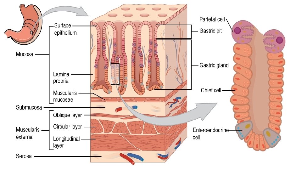
Stomach • Mucosa: simple columnar epithelium and presence of gastric pits. • Stomach is divided into three histological regions on the basis of nature of glands: ØCardiac region ØFundic region (fundus & body) ØPyloric region

DIFFERENCE BETWEEN CARDIA, FUNDUS & BODY, AND PYLORUS CARDIA FUNDUS & BODY PYLORUS Contain cardiac gland Contain gastric gland Contain pyloric gland Gastric pit less deeper than pyloric gland Gastric pit more deeper than gastric or cardiac gland Parietal cells absent Parietal cells more or very few Parietal cells few 13
- Slides: 13