Musculoskeletal System Dr Nalan Alan Selcuk Bone scan

Musculoskeletal System Dr. Nalan Alan Selcuk

Bone scan n Intraarticular radionuclid synovectomy n Palliative treatment n

n Bone scintigraphy is one of the most frequently performed of all radionuclide procedures. n Radionuclide bone imaging is quick, relatively inexpensive, widely available, and exquisitely sensitive and is invaluable in the diagnostic evaluation of numerous pathologic conditions.

Bone Scan n The procedure is performed with technetium-99 m– labeled diphosphonates; – Tc-99 m-MDP – Tc-99 m-HMDP – Tc-99 m-PP n These compounds accumulate rapidly in bone, and by 2– 6 hours after injection, about 50% of the injected dose is in the skeletal system. n The uptake mechanisms of diphosphonates have not been completely elucidated.

n n Presumably they are adsorbed to the mineral phase of bone, with relatively little binding to the organic phase. The degree of radiotracer uptake depends primarily on two factors: – blood flow and – the rate of new bone formation
 and posterior (right) whole-body bone scintigrams obtained in an adult demonstrate")
Figure: Anterior (left) and posterior (right) whole-body bone scintigrams obtained in an adult demonstrate normal anatomy.
 and posterior (right) whole-body bone scintigrams obtained in a child demonstrate normal")
Anterior (left) and posterior (right) whole-body bone scintigrams obtained in a child demonstrate normal anatomy. Note the increased activity in the physes of the long bones and in the hematopoietically active facial bones.

Bone scan n n Metastatic Disease Trauma Infection Miscellaneous Conditions – – – Paget disease Degenerative disorder Reflex Sympathetic Dystrophy Avascular Necrosis Hypertrophic Osteoarthropathy

n Dynamics images can be useful in detecting infections.

Technique n n n Imaging is typically performed 2– 6 hours after intravenous administration of 740– 925 MBq (20– 25 m. Ci) of Tc-99 m–labeled diphosphonates. The delay between injection and imaging allows clearance of the radiotracer from the soft tissues, resulting in a higher target-to-background ratio and improved visualization of bone. Skeletal detail can be further enhanced by encouraging patients to drink copious amounts of fluid after radiotracer injection.

Acquisition n n A gamma camera equipped with a lowenergy, high-resolution collimator will yield the highest-resolution images. Additional anterior and posterior whole -body images are often obtained as needed.
 ; – Breast – Prostate – Lung –")
n Bone metastasis caused by (80%) ; – Breast – Prostate – Lung – Thyroid – Renal tm

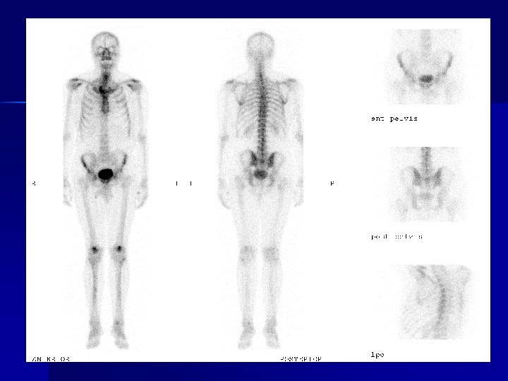
n n 57 yaşında Erkek hasta Prostat ca Multiple kemik met.

n Metastazların yaygın olduğu bazı durumlarda “super scan” ismi verilen, aksiyel iskeletin diffüz olarak artmış radyoaktivite tutulumu gösterdiği, distal ekstremite bölgelerinin ve böbreklerin hiç izlenmediği bir patern izlenebilir

n n n Super scan Aksiel iskelette diffüz aktivite artışı Böbrekler ve mesane izlenmiyor

Travma: Kemik sintigrafisi onkolojiden sonra ikinci sırada, travmaların değerlendirilmesinde kullanılır n Genellikle kemik travmasından sonraki ilk 24 saat içinde travmatik bölgede artmış radyoaktivite tutulumu izlenir n

n Spinal kırıklarda, kuboid, kalkaneus ve sesamoid kemik kırıklarında, çocuklarda yaş ağaç kırıklarında x-ray normal iken de sintigrafi pozitif olabilir.

Stress fracturu

Osteomiyelit: n n Kemik sintigrafisi üç fazlı olarak uygulandığında kemik infeksiyonlarını tanımada çok duyarlı bir yöntemdir Akut osteomiyelitte radyolojik olarak yumuşak dokuya ait değişikliklerin ortaya çıkması için minimum 3 gün, diagnostik periostal yeni kemik yapımının görünür olması için 14 gün geçmesi gerekir

n İnfeksiyonunun başlamasından 24 -72 saat sonra sintigrafik olarak pozitif bulgu elde edilebileceğini göstermiştir

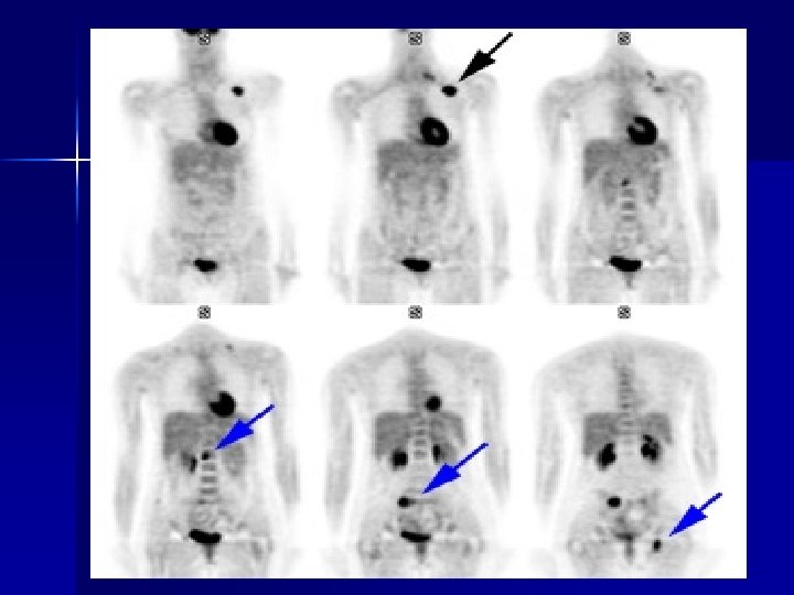
Pozitron Emisyon Tomografi: n n n Kas-iskelet sistemi hastalıklarında son yıllarda daha yaygın olarak kullanılmaya başlanılan başka bir tanı yöntemide pozitron emisyon tomografi (PET)’dir En sık kullanılan radyafarmasötik glikolizi gösteren Flor-18 işaretli deoksiglkoz (FDG)’dir FDG ile PET çalışmaları yaygın olarak onkoloji alanında uygulanmaktadır
 Anterior bone scintigram shows discrete focal activity in the left")
Figure 3 a. (a) Anterior bone scintigram shows discrete focal activity in the left maxilla (arrowhead) due to a dental process and heart-shaped activity in the anterior neck (arrow) representing the thyroid cartilage, both of which are normal variants. (b) Posterior bone scintigram shows focal activity in the right side of the neck (arrow) caused by a cervical osteophyte.
 and posterior (right) whole-body")
Figure 4. Extensive osseous metastases from lung carcinoma. Anterior (left) and posterior (right) whole-body bone scintigrams show multiple, randomly distributed foci of abnormal radiotracer uptake. The foci vary in size and intensity.
 and posterior (right) whole-body scintigrams obtained in a patient who")
Figure 5. Anterior (left) and posterior (right) whole-body scintigrams obtained in a patient who fell demonstrate multiple foci of increased radiotracer uptake. The linearly distributed rib foci and H-shaped sacral activity indicate trauma as the cause of these foci. The increased activity in the right proximal humerus is due to a fracture.
 and posterior (right) whole-body scintigrams")
Figure 6. Renal osteodystrophy and secondary hyperparathyroidism. Anterior (left) and posterior (right) whole-body scintigrams demonstrate uniformly increased activity throughout the skeleton that is especially intense in the calvaria. These images show the superscan pattern associated with metabolic bone disease.

Figure 7. Paget disease. Whole-body scintigram demonstrates increased radiotracer accumulation in the proximal right femur and in the deformed and enlarged tibias. Figure 8. Lung carcinoma with hypertrophic osteoarthropathy. Anterior (left) and posterior (right) whole-body scintigrams demonstrate pericortical stripes of activity (tramline sign) in the lower extremities. Hypertrophic osteoarthropathy is also present in the bones of the forearms.

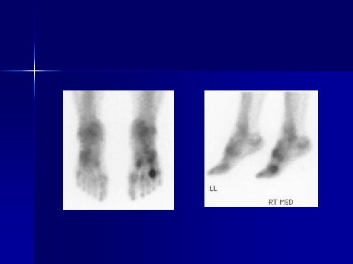
Figure 9. Reflex sympathetic dystrophy. Scintigram shows diffusely increased uptake in the distal right upper extremity.
- Slides: 30