MUSCULOSKELETAL BLOCK Pathology Noninfectious arthritis Objectives Know the

MUSCULOSKELETAL BLOCK Pathology Non-infectious arthritis
, Know the")
Objectives Know the pathogenesis and clinicopathological features of osteoarthritis (degenerative joint disease), Know the pathogenesis and clinicopathological features of rheumatoid arthritis Know the pathogenesis and clinicopathological features of gout and calcium pyrophosphate arthropathy [pseudogout]
 has four main causes 1. Degeneration, e.")
Inflammatory disease of joints (arthritis and synovitis) has four main causes 1. Degeneration, e. g. osteoarthritis. 2. Autoimmity, e. g. rheumatoid arthritis, SLE, rheumatic fever 3. Crystal deposition, e. g. gout and other crystalline arthropathies. 4. Infection, e. g. septic arthritis, tuberculous arthritis.

Osteoarthritis Definition and Incidence Osteoarthritis is the most common type of joint disease and is characterized by the progressive erosion of articular cartilage in weightbearing joints. The incidence increases with age. Osteoarthritis can be primary or secondary

Osteoarthritis Types Primary osteoarthritis Secondary osteoarthritis

Osteoarthritis Types Primary osteoarthritis: appears insidiously with age and without apparent initiating cause usually affecting only a few jonts.

Osteoarthritis Types Secondary osteoarthritis: some predisposing condition, such as previous traumatic injury, developmental deformity, or underlying systemic disease such as diabetes, ochronosis, hemochromatosis, or marked obesity Secondary often less osteoarthritis affect young involves one or several predisposed joints than 5% of cases

Osteoarthritis Pathogenesis In general, osteoarthritis affects joints that are constantly exposed to wear and tear. Articular cartilage bears the brunt of the degenerative changes in osteoarthritis. Normal articular cartilage performs two functions: (1) Along with the synovial fluid, it provides virtually friction-free movement within the joint (2) in weight-bearing joints, it spreads the load across the joint surface

Pathogenesis Early osteoarthritis is marked by degenerating cartilage In hyaline cartilage affected by osteoarthritis, the water content is increased and the proteoglycan content is decreased. The elasticity and compliance of the cartilage is, therefore, reduced. The very first change seen in osteoarthritis is proliferation of chondroblasts, and it has been proposed that these cells produce enzymes that induce these biochemical changes in the hyaline cartilage.

Osteoarthritis Common sites usually one joint or same joint bilaterally Gender has some influence: Women : knees and hands Men : hips

Osteoarthritis The pathological changes involve: cartilage bone synovium joint capsule with secondary effects on muscle ( atrophy)
, becomes eroded")
Osteoarthritis Pathogenesis The early change: destruction of articular cartilage, which splits (fibrillation), becomes eroded There is inflammation and thickening of the joint capsule and synovium Small fractures can dislodge pieces of cartilage and subchondral bone into the joint, forming loose bodies(joint mice).

Small cysts develop in the bone osteophytes constant friction of bone surfaces, leading to a polished ivory bony articular surface (eburnation)

Osteoarthritis. : Histologic demonstration of the characteristic fibrillation of the articular cartilage.

Cracking and fibrillation of cartilage

Severe Osteoarthritis Eburnated articular surface exposing subchondral bone Subchondral cyst Residual articular cartilage

Pathological changes in osteoarthritis normal synovial joint early change in osteoarthritis 'Heberden's nodes (osteophytes on the interphalangeal joints of the fingers)

Osteoarthritis Clinical features The most frequently affected joints are the hips, the knees, the cervical and lumbar vertebrae, the proximal and distal interphalangeal (PIP and DIP) joint of the hands, the first metacarpophalangeal joint and the first metatarsophalangeal joint. Osteophytes at the DIP joints produce nodular swellings called Hebderden’s nodes. With increasing deformity of the joint the typical symptoms develop, which are pain (which is worse with use), morning stiffness and limitation in joint movement. With involvement of the cervical and lumbar spine, osteophytes may impinge on the nerve roots causing symptoms such as pain and pins and needles in the arms or legs.

Osteophyte

Course & Prognosis Osteoarthrosis is a slowly progressive, chronic joint disability Eventually, elderly sufferers may become confined to wheelchairs Recent advancements in the technique of joint replacement with prostheses have improved the outlook of these patients


Rheumatoid arthritis Definition etiology pathological features clinical features radiological features

Rheumatoid arthritis Definition Rheumatoid arthritis is a chronic inflammatory multisystem disorders but the joints are always involved. The condition can affect all age groups. When children are affected, the condition is designated Still’s disease. Females are affected more often than males. Produces nonsuppurative proliferative synovitis, may progress to destruction of articular cartilage and joint ankylosis

Extra-Articular Manifestations

Rheumatoid arthritis Aetiology The pathogenesis is not well understood, but it is thought that an initiating agent, possibly an organism, triggers immunological dysfunction resulting in persistent chronic inflammation in generatically susceptible individuals. In the joints, the ongoing inflammation causes destruction of the articular cartilage. Circulating autoantibodies (rheumatoid factors). The exact role of these autoantibodies is uncertain.
 Antibodies against cyclic citrullinated peptides (CCP protein antibodies) is")
HLA-DR 4, DR 1 (65%) Antibodies against cyclic citrullinated peptides (CCP protein antibodies) is the most specific for a diagnosis of rheumatoid arthritis

Rheumatoid arthritis Laboratory Findings: Rheumatoid factor: 80% have Ig. M autoantibodies to Fc portion of Ig. G not sensitive or specific Anti-CCP (cyclic citrullinated peptides)protein antibodies most specific for a diagnosis of rheumatoid arthritis ESR and C-reactive proteien

Rheumatoid arthritis Pathologic Features 1. synovial cell hyperplasia and proliferation 2. dense perivascular inflammatory cell infiltrates (frequently forming lymphoid follicles) in the synovium composed of CD 4+ T cells, plasma cells, and macrophages 3. increased vascularity due to angiogenesis 4. neutrophils and aggregates of organizing fibrin on the synovial surface 5. increased osteoclast activity in the underlying bone erosion.

Rheumatoid arthritis Pathologic Features Pannus formed by proliferating synovial-lining cells admixed with inflammatory cells, granulation tissue, and fibrous connective tissue Eventually the pannus fills the joint space, and subsequent fibrosis and calcification may cause permanent ankylosis.
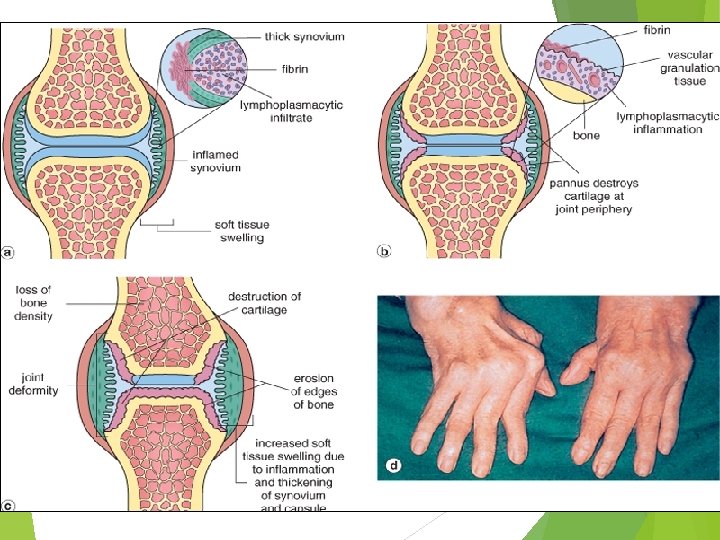

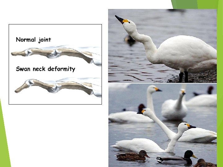
dense perivascular inflammatory cell infiltrates increased vascularity neutrophils and aggregates of organizing fibrin on the synovial surface synovial cell hyperplasia and proliferation Pannus formation increased osteoclast activity in the underlying bone erosion swan neck finger

Rheumatoid arthritis Pathologic Features

Rheumatoid arthritis Microscopic Hyperplastic synovial cells Dense chronic inflammation consisting of lymphocytes and plasma cells

Rheumatoid arthritis Clinical Feaures joints are warm, swollen, painful with morning stiffness symmetric arthritis characteristic derformities develop. These include: Radial deviation at the wrists. Ulnar deviation at the fingers. Flexion and hyperextension deformities of the fingers (swan neck and boutonniere deformities).

Rheumatoid arthritis X-ray: Loss of articular cartilage leading to narrowing of the joint space. Joint effusions. Localized osteoporosis. Erosions.

Subcutaneous rheumatoid nodule Palisading Granulomas

Rheumatoid arthritis Prognosis Reduces life expectancy by 3 -7 years Death due to amyloidosis, vasculitis, GI bleeds from NSAIDs, infections from steroids.

Comparison of the morphologic features of RA and osteoarthritis

Comparison of Osteoarthrosis & Rheumatoid Arthritis


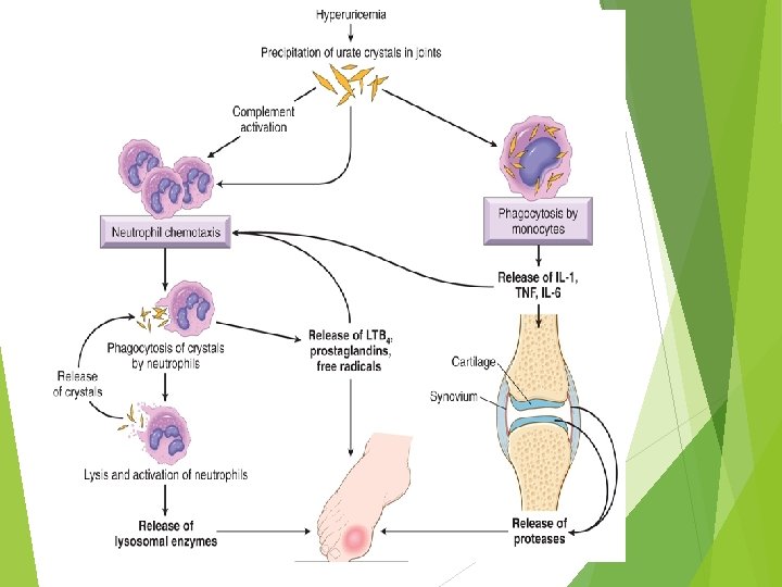
Gout is an inflammatory disease. The most commonly affected site is: first metatarsophalangeal joint. It is swollen, red, and very painful. Sodium urate crystals have precipitated into the joint, producing an acute inflammatory response.

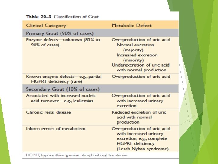
Clinical feauters Gout is more common in men than in women; it does not usually cause symptoms before the age of 30. Risk factors for the disease include obesity, excess alcohol intake, consumption of purine-rich foods, diabetes, the metabolic syndrome, and renal failure. Polymorphisms in genes involved in the transport and homeostasis of urate

Tophi are large aggregates of urate crystals which are visible with the naked eye. They occur in the joints and soft tissues of people with persistent hyperuricaemia. A common site for tophi is the pinna of the ear.

Tophus, gout – Histology Tophi consist of crystals that are surrounded by macrophages, lymphocytes, and often foreign body giant cells. In routinely processed sections, the crystals are removed during processing. Long, slender, needleshaped monosodium urate crystals

Gout Besides joints, what other organ is affected in gout? approximately 20% of patients die of renal failure. Renal lesions are many: precipitation of urates in the medulla forms tophi uric acid stones acute renal failure due to precipitation of urates in the collecting tubes

Pseudogout Calcium pyrophosphate crystals This condition is due to the deposition of calcium pyrophosphate crystals in the synovium (pseudogout) and articular cartilage (chondrocalcinosis). It can occur in three main settings: Sporadic (more common in the elderly). Hereditary. Secondary to other conditions, such as previous joint damage, hyperparathyroidism, hypothyroidism, haemochromatosis and diabetes. The crystals first develop in the articular cartilage (chondrocalcinosis), which is usually asymptomatic. From here, the crystals may shed into the joint cavity resulting in an acute arthritis, which mimics gout and is therefore called pseudogout.

Pseudogout can be differentiated from gout in three ways: The knee is most commonly involved. X-rays show the characteristic line of calcification of the articular cartilage. The crystals look different under polarizing microscopy, they are rhomboid in shape.

- Slides: 51