MUSCULAR SYSTEM Produce movement Maintain posture Stabilize joints

MUSCULAR SYSTEM

Produce movement Maintain posture Stabilize joints Generate heat

1. Skeletal 2. Cardiac 3. Attach to and cover skeleton Dominant tissue in the walls of the heart Smooth Walls of hollow visceral organs

Longest muscle fibers Striated Voluntary muscle Conscious control Responsible for overall body mobility Has the ability to contract rapidly but must rest after short periods of activity

Found only in the heart Bulk of the heart walls Striated Involuntary

Found in stomach, urinary bladder, intestines, respiratory passageways Forces fluids & other substances through the internal body channel NO striations involuntary

Skeletal muscle with obvious striations Smooth Muscle Cardiac Muscle
 – the ability to respond to a stimulus Contractility – the")
Excitability (Irritability) – the ability to respond to a stimulus Contractility – the ability to shorten forcibly when adequately stimulated Extensibility – the ability to be stretched or extended Elasticity – the ability of a muscle fiber to recoil & resume its resting length after being stretched

SKELETAL MUSCLES NOTES TODAY…GET OUT NOTEBOOK.

Contraction= pulling joints together Muscles always work in groups Will always have: Agonists Antagonists synergists
 of the")
The Agonist – direct mover. The Antagonist – is the muscle(s) of the same joint that relaxes when the agonist is contracting The Synergists – are any other muscles that aid the prime mover (bracing a joint or adding more power when required etc).

Q - Which muscle needs to contract to produce this movement? A– The bicep. Q – which muscle acts as the antagonist? A – triceps

Q - Which muscle needs to contract to produce this movement? A– The triceps. Q - Is this the agonist or antagonist? A – now it’s the agonist (prime mover)

Which bone is staying fixed and which bone is moving? Attachment of the muscle to the immoveable bone in a joint is its origin Attachment to the moveable bone is its insertion.

ANATOMY OF SKELETAL MUSCLES myology

Muscle is surrounded by dense CT called epimysium. Muscle made of bundles of muscle fibers called fascicles Fascicles surrounded by perimysium
 surrounded by endomysium. The epi-, peri-, and endomysium are all")
Muscle fibers (cells) surrounded by endomysium. The epi-, peri-, and endomysium are all continuous with one another.

- A muscle fiber is made of myofibrils - A Myofibrils is made of myofilaments

sarcolemma like membrane Sarcoplasm like cytoplasm Sarcolemma membrane pinches in and creates tubes that penetrate through the cell called transverse tubules or T tubules. Sarcoplasm has lots of mitochondria (why? )

Myofibrils are composed of myofilaments 2 kinds of myofilaments Thick Thin

Figure 9. 4 a, b

ACTIN thin filament- made up of 3 different types of protein: actin, tropomyosin, and troponin. ACTIN “bead” Tropomyosin “string” myosin binding site Troponin bound to Actin and Tropomyosin.

Figure 9. 4 d
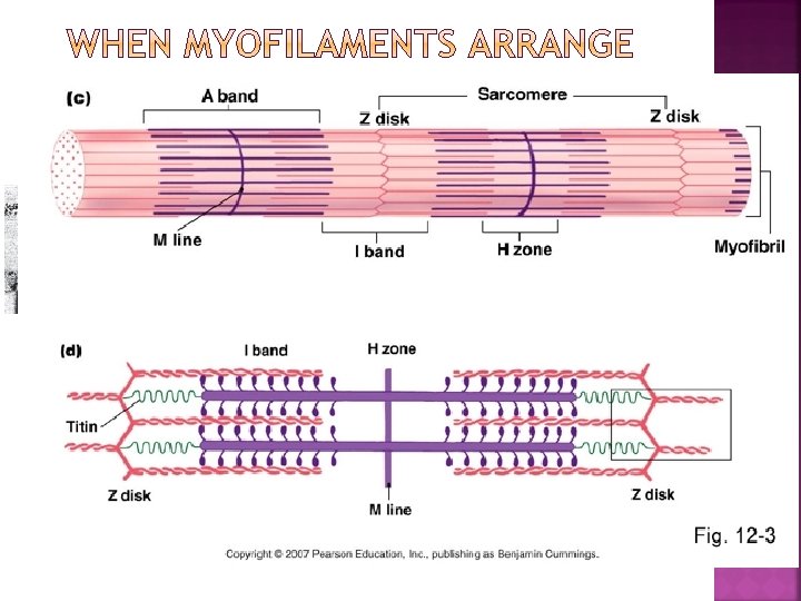

REGION MYOFILAMENTS CONTAINED A Band I Band M line Z disc H zone What constitutes a sarcomere?

A Band: thick and thin. Also contains M line I Band: thin only M Line: runs in the middle of the A band/H zone. Thick filaments. Looks dark due to desmin protein. Attachment point for thick filaments Z-disc –sheet of proteins that attaches thin filaments together H zone: thick filaments Sarcomere: from Z to Z

Sarcoplasmic reticulum and T tubules Made of smooth ER Functions in the regulation/store intracellular calcium levels

Sarcoplasmic reticulum and T tubules are continuous with the sarcolemma They conduct impulses to the deepest regions of the muscle These impulses signal for the release of Ca 2+ from adjacent terminal cisternae

Skeletal muscles must be stimulated by a NERVE IMPULSE to contract. The nerve comes very close to touching the muscle This is called the neuromuscular junction.

Helpful Vocabulary SYNAPTIC CLEFT: MOTOR UNIT: ACH RECEPTOR: NMJXN

EXCITATION 1. 2. 1. A nerve signal arrives at the synaptic end of neuron Ach released 2. ACh will diffuse across the synaptic cleft and bind to the ACh receptors on muscle cell. The binding of ACh causes Na+ channels to open 3. Na+ will rush into the muscle cell, making the. Cell is now depolarized demo

4. 4. If enough of Na+ diffuses…action potential fires and muscle contracts. 5. Action Potential continues down sarcolema (muscle cell membrane) to T tubules 6. T-tube picks up AP signal and tells Sarcoplasmic Reticulum to release Ca+ demo
 needs the following: 2 straws 1 string of yarn")
Each group (of 4) needs the following: 2 straws 1 string of yarn (see demo strip to determine long) 6 different colors of play doh Direction Sheet

Axon ACH Sarcolemma SR T-tubules Myofibril Terminal Cisternae Action Potential Calcium

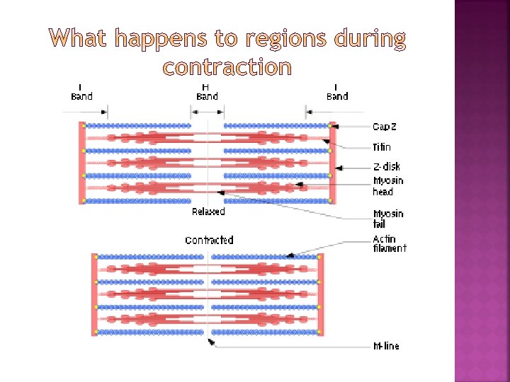
Calcium allows the muscle fibers to contract Once Ca+ is released from terminal cisternae, a series of steps occur. This process is called the sliding filament theory Named because Actin slides past Myosin Sliding Filament Theory

1. CA+ released from Terminal Cisternae of SR binds to troponin 2. Troponin moves the tropomyosin off of myosin binding site on actin filament. 3. ATP breaks down and releases energy. Myosin head is released from “standing” position and binds to actin.

4. Myosin head pulls on actin filament: “power stroke” 5. ATP binds to Myosin head and provides energy needed for Myosin head to release from actin 6. Cycle stops when CA+ is reabsorbed into SR and tropomyosin recovers myosin binding sites on actin Sliding Filament Theory SFT-3 SFT-5

Practice the sliding filament theory by doing one of the following right now: 1. Get into a group of 6 and develop a quick skit that shows the steps of the sliding filament theory OR 2. Develop a frame-by-frame drawing showing the steps of the sliding filament theory. Color pictures
- Slides: 39