Muscular System Overview Types of Muscle Skeletal striated


")
 Contains protein filaments – ACTIN (thin) and MYOSIN (thick)")
 • Actin and Myosin filaments")


 The theory of how muscle contracts is the sliding filament")

 ●Creatine phosphate increases regeneration of")


 Atrophy - muscles become small")


- Slides: 40
Muscular System Overview
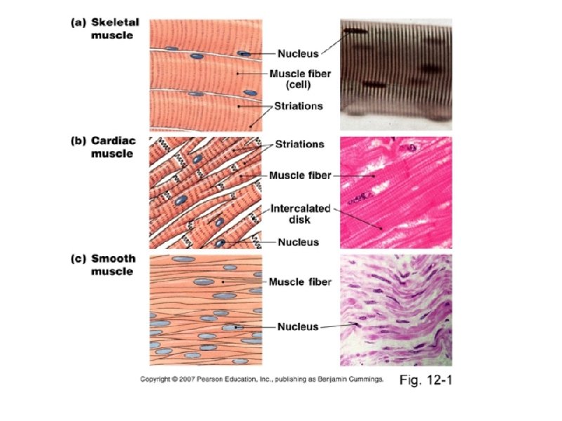
Types of Muscle ●Skeletal – striated & voluntary ●Smooth – non-striated & involuntary ●Cardiac – striated & involuntary
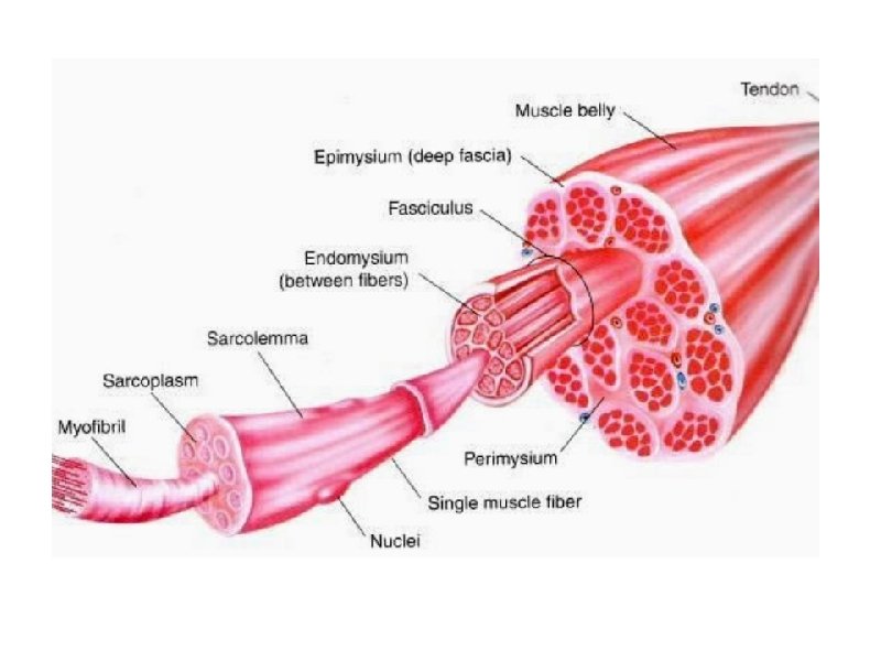
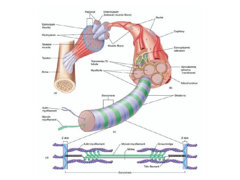
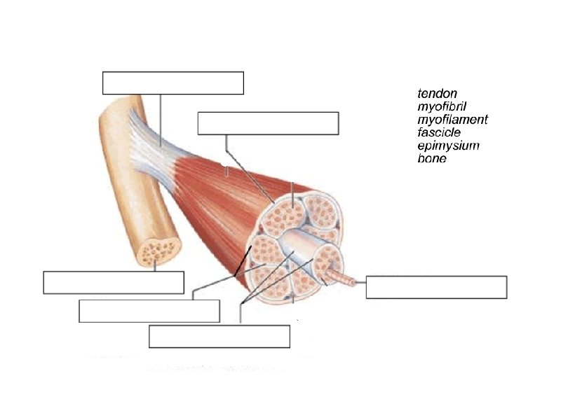
Muscles and Muscle Fiber Structure Muscles are composed of many fibers that are arranged in bundles called FASCICLES Individual muscles are separated by FASCIA, which also forms tendons and ligaments.
Muscle Layers: Epimysium, Perimysium, and Endomysium
ENERGY Fibers contain multiple mitochondria for energy Most fibers have multiple nuclei
SARCOLEMMA Sarcolemma = muscle fiber membrane Sarcoplasm = inner material surrounding fibers (like cytoplasm) Myofibrils = individual muscle fibers --> made of myofilaments
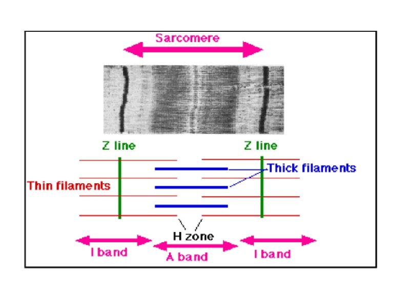
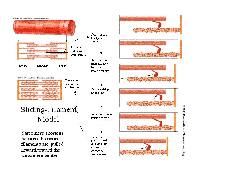
Myofibril (Muscle fiber or cell) Contains protein filaments – ACTIN (thin) and MYOSIN (thick) These filaments overlap to form dark and light bands on the muscle fiber § A band = d. Ark • thick (myosin) § I band = l. Ight • th. In (actin) ●In the middle of each I band are Z lines. A sarcomere is one Z line to the other
Structure of a Sarcomere (functional unit of a muscle) • Actin and Myosin filaments are arranged in an overlapping pattern of light (I bands) and dark (A bands). • In the middle of each I band is a line called a Z line. • The section of a myofibril from one Z line to the next Z line is the Sarcomere. • *The arrangement of these sarcomeres next to each other produces the striations of the skeletal muscle fibers. • Note: Each Myofibril is surrounded by a network of channels called Sarcoplasmic Reticulum. Transverse tubules pass through the fibers.
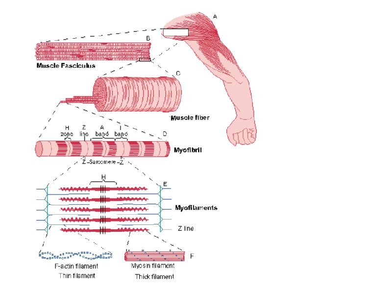
myosin It is important to remember the heirarchy myofibrils fasicles myofilaments actin
It is important to remember the heirarchy fasicles myofibrils myofilaments actin myosin
muscle fiber myofilament epimysium muscle myofibrils sarcomere
myofilament muscle sarcomere epimysium myofibrils muscle fiber
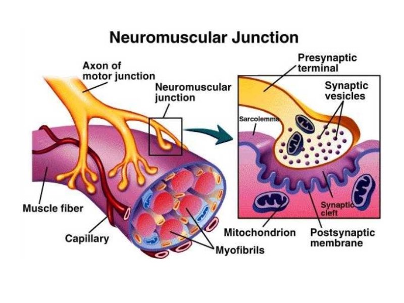
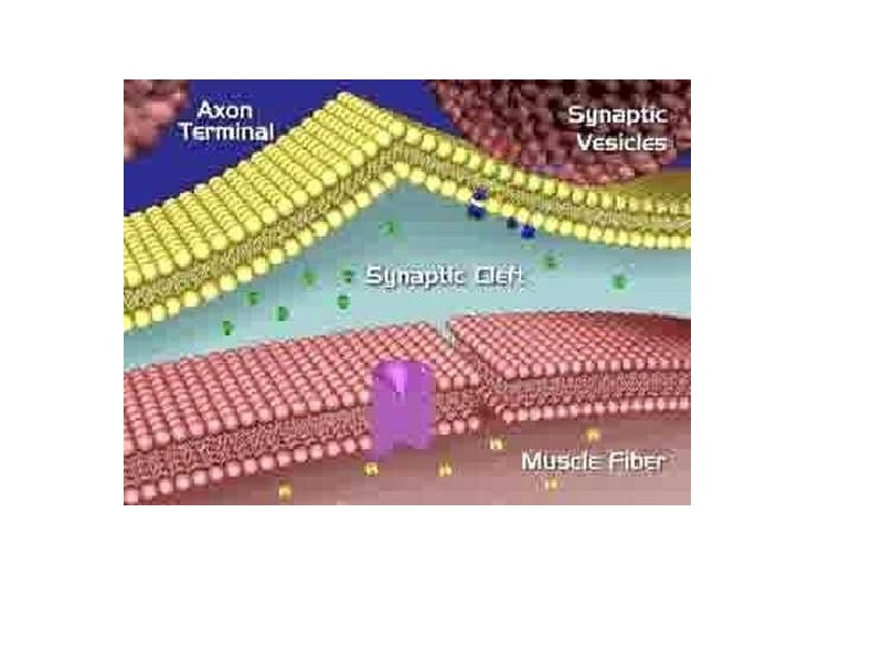
Muscles & the Nervous System 1. NEUROMUSCULAR JUNCTION - where a NERVE FIBER and muscle fiber come together. A. K. A. Myoneural junction. 2. MOTOR NEURON ENDINGS - nerve fiber caries the impulse that stimulates the muscle fibers 3. MOTOR END PLATE - specialized part of muscle fiber membrane (sarcolemma) located at the neuromuscular junction, has many folds 4. SYNAPTIC CLEFT - An actual "gap" or cleft which exits between the motor neuron endings and the motor end plate. 5. SYNAPTIC VESICLES - numerous vesicles in motor neuron endings, where neurotransmitters are stored before being released into the synaptic cleft. 6. NEUROTRANSMITTER - substance that is released from nerve endings into synaptic cleft. Stimulates an impulse. In this case, a "muscle impulse". One of the major neurotransmitters is ACETYLCHOLINE. This brings about muscle contractions. ACETYLCHOLINESTERASE is an enzyme that breaks down acetylcholine
Motor Unit or Neuromuscular Junction 1. Neuron 3. Vesicle 2. Sarcolemma (or motor end plate) 4. Synapse 5. Mitochondria
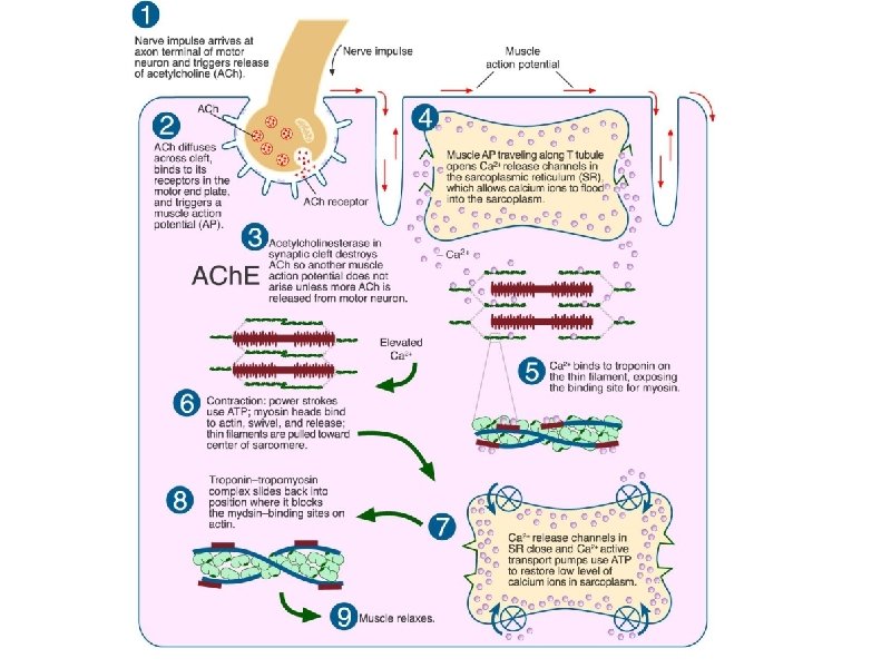
Events in a Muscle Contraction 1. A nerve impulse stimulates the release of a neurotransmitter (acetylcholine) from synaptic vesicles into the synaptic cleft. 2. The impulse spreads across the sarcolemma and into the fiber. This impulse causes an increase in the permeability to calcium ions. The S. R. has a high conc. of Ca++. 3. Calcium ions diffuse into the sarcoplasm. The Ca++ causes the formation of "cross bridges" between the actin and myosin filaments. 4. The filaments slide between each other, and this shortens the myofibrils, which in turn shorten the muscle fibers, which shortens the muscle. 5. A "Calcium Pump" returns the CA++ into the S. R. (requires energy. ATP) 6. The enzyme Acetylcholinesterase stops the action of Acetylcholine
The neurotransmitter that crosses the gap is ACETYLCHOLINE. This is what activates the muscle. Acetylcholine is stored in vesicles
Motor Unit The muscle fiber and the motor neuron
SLIDING FILAMENT THEORY (MODEL) The theory of how muscle contracts is the sliding filament theory. The contraction of a muscle occurs as the thin filament slide past the thick filaments. The sliding filament theory involves five different molecules plus calcium ions. The five molecules are: myosin actin tropomyosin troponin ATP
ANIMATION OF SLIDING FILAMENT http: //www. blackwellpublishing. com/matthews/myosin. html
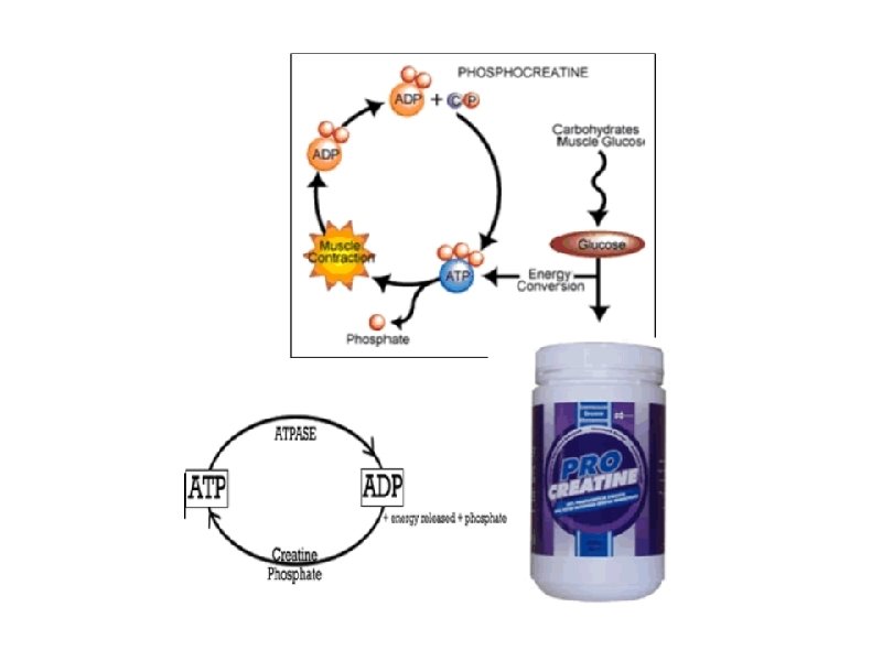
Energy Source ●Provided by ATP from cellular respiration (mitochondria) ●Creatine phosphate increases regeneration of ATP ●Much of the energy forms heat, which keeps our bodies warm
Other Terms ● 1. Threshold Stimulus ● 2. All-or-None Response ● 3. Motor Unit ● 5. Recruitment ● 6. Muscle Tone ● 7. Muscular Hypertrophy ● 8. Muscular Atrophy ● 9. Muscle Fatigue ● 10. Muscle Cramp ● 11. Oxygen Debt
Threshold Stimulus Minimal strength required to cause a contraction Motor neuron releases enough acetylcholine to reach threshold All-or-None Response Fibers do not contract partially, they either do or don't
Motor Unit The muscle fiber + the motor neuron Recruitment more and more fibers contract as the intensity of the stimulus increases Muscle Tone Sustained contraction of individual fibers, even when muscle is at rest
Hypertrophy - muscles enlarge (working out or certain disorders) Atrophy - muscles become small and weak due to disuse
Muscle Fatigue - muscle loses ability to contract after prolonged exercise or strain Muscle Cramp - a sustained involuntary contraction Oxygen Debt oxygen is used to create ATP, during exercise you may not have enough oxygen -> this causes Lactic Acid to accumulate in the muscles - -
Origin and Insertion Origin = the immovable end of the muscle Insertion = the movable end of the muscle **when a muscle contracts the insertion is moved toward the origin The biceps brachii has two origins (or two heads).
What is rigor mortis? A few hours after a person or animal dies, the joints of the body stiffen and become locked in place. This stiffening is called rigor mortis. Depending on temperature and other conditions, rigor mortis lasts approximately 72 hours. The phenomenon is caused by the skeletal muscles partially contracting. The muscles are unable to relax, so the joints become fixed in place.
What is tetanus? Tetanus causes cholinosterase to not break down the acetylcholine in the synapse. This results in a person's muscles contracting and not relaxing. A tetanus shot must be administered shortly after exposure to the bacteria. Once you develop tetanus, there is no cure.