
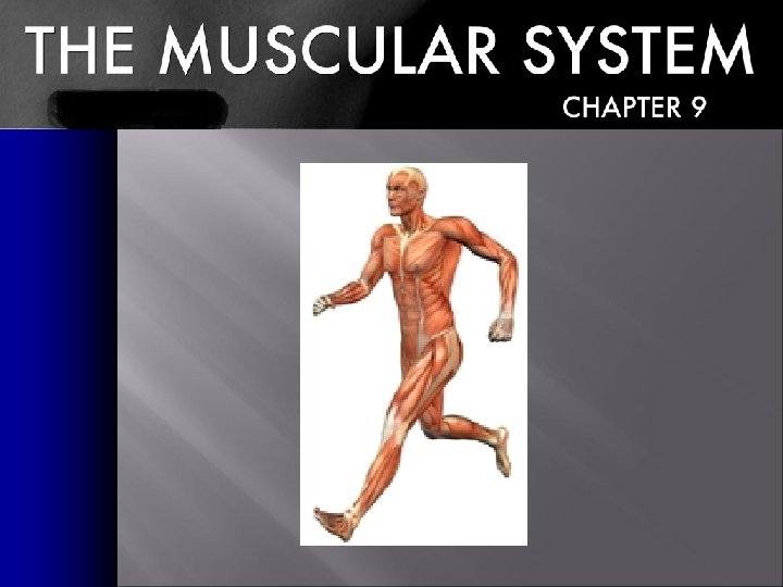
Muscular System Objectives TSWBAT Identify the three types
Muscular System Objectives: TSWBAT : • Identify the three types of muscle tissue and its components. • Identify the six functions of the muscular system. • Label the major muscles in the human body. • Analyze the movements of the joints and what muscles create the movements. • Explain common injuries and medical conditions of the muscular system.
MUSCLES � � � Approximately 40% of your body weight Approximately 650 muscles Muscles only pull (they can’t push) You have over 30 facial muscles Eye muscles move more than 100, 000 times a day
Skeletal Muscle Functions Muscle plays six important roles in the body: 1. Produce skeletal movement 2. Maintains posture and body position 3. Support soft tissues (abdominal wall & pelvic cavity) 4. Guard entrances and exits (digestive and urinary tracts) 5. Maintain body temperature (energy is converted to heat) 6. Store nutrient reserves (proteins are broken down & amino acids are used)
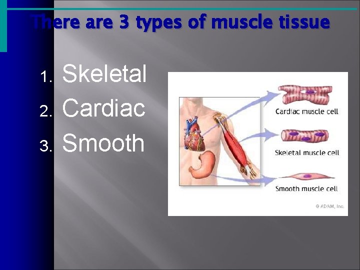
There are 3 types of muscle tissue 1. 2. 3. Skeletal Cardiac Smooth

Skeletal Muscles � � Definition - organs that are composed mainly of skeletal muscle tissue, but they also contain connective tissues, nerves, and blood vessels. Each cell is a single muscle fiber. Muscle fibers form bundles called fascicles. Directly or indirectly attached to the bones of the skeleton
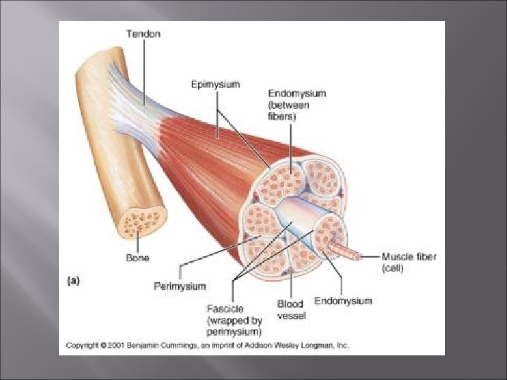
Organization of Skeletal Muscle Tissue Three layers of connective tissue are part of each muscle: 1. Epimysium – dense layer of collagen fibers that surround the entire muscle 2. Perimysium – divides the muscle into a series of compartments each containing a bundle of muscle fibers (fascicle); contains collagen & elastic fibers, blood vessels and nerves that maintain blood flow 3. Endomysium – flexible, elastic connective tissue layer; surrounds the individual skeletal muscle cells and interconnects adjacent muscle fibers
Fascicle Arrangement � � � 1. 2. 3. 4. The muscle fibers in a single fascicle are parallel, but the organization of fascicles in skeletal muscles can vary The arrangement is correlated with muscle power and range of motion (structure determines function) Skeletal muscles are classified as: Parallel muscles (most common) – fascicles are parallel to the long axis of the muscle Convergent muscles – converge at a common attachment site; fibers spread out and pull in different directions Pennate muscles – form a common angle with the tendon Circular muscles – cocentrically arranged around an opening
Organization of Skeletal Muscle Tissue � � At the end of the muscle, the collagen fibers of the epimysium, perimysium, and endomysium come together to form a tendon or aponeurosis Tendons and aponeuroses attach muscles to bone Origin – where the fixed end of the muscle attaches to the bone (cartilage or connective tissue) Insertion – where the movable end of the muscle attaches to another structure
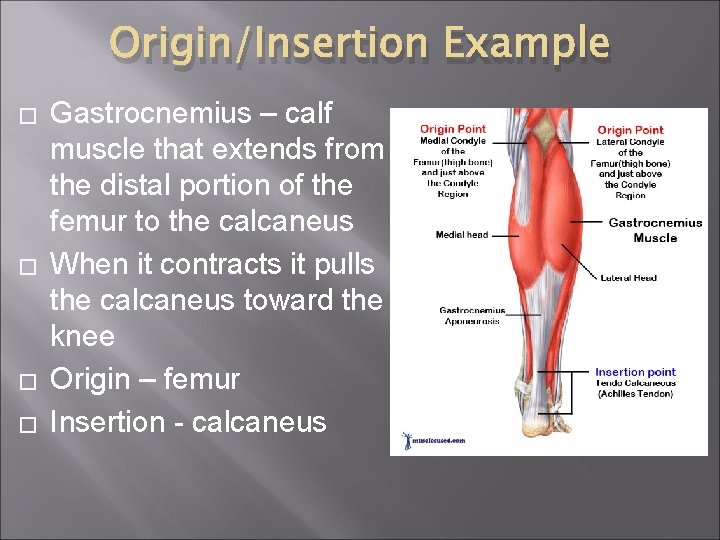
Origin/Insertion Example � � Gastrocnemius – calf muscle that extends from the distal portion of the femur to the calcaneus When it contracts it pulls the calcaneus toward the knee Origin – femur Insertion - calcaneus
Muscle Actions Agonist (prime mover) – a muscle whose contraction is mostly responsible for producing a particular movement Example – biceps brachii � Antagonist – a muscle whose action opposes that of a particular agonist Example – triceps brachii � Agonists and antagonists are functional opposites � Synergists – help a larger agonist work efficiently �
Muscle Tone and Contractions � � � Muscle Tone – resting tension in a skeletal muscle Isotonic muscle contraction – tension rises and the skeletal muscle’s length changes (lifting) Isometric muscle contraction – the muscle as a whole does not change length, and the tension produced never exceeds the load (holding)
Types of Ordinary Body Movements · Flexion – bending at the joint · Extension - straightening at the joint · Hyperextension – over extension of joint · Rotation – rotating on axis · Abduction – moving away from the midline · Adduction – moving toward the body · Circumduction – circular movement Copyright © 2003 Pearson Education, Inc. publishing as Benjamin Cummings Slide 6. 32
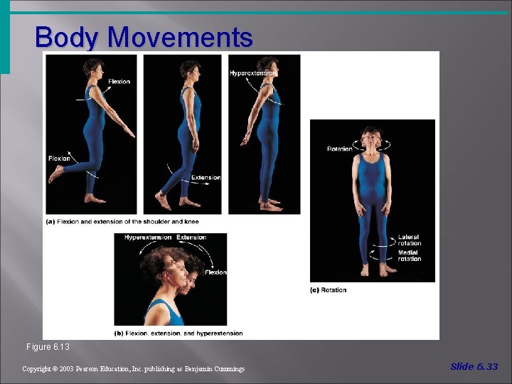
Body Movements Figure 6. 13 Copyright © 2003 Pearson Education, Inc. publishing as Benjamin Cummings Slide 6. 33
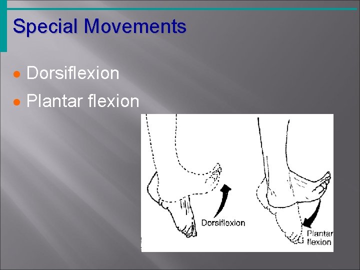
Special Movements · Dorsiflexion · Plantar flexion
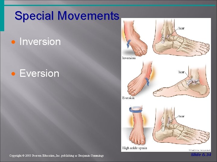
Special Movements · Inversion · Eversion Copyright © 2003 Pearson Education, Inc. publishing as Benjamin Cummings Slide 6. 34
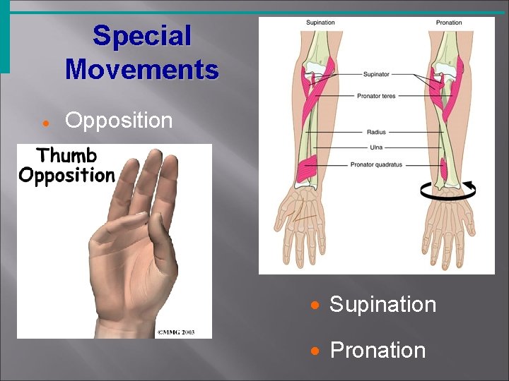
Special Movements · Opposition · Supination · Pronation
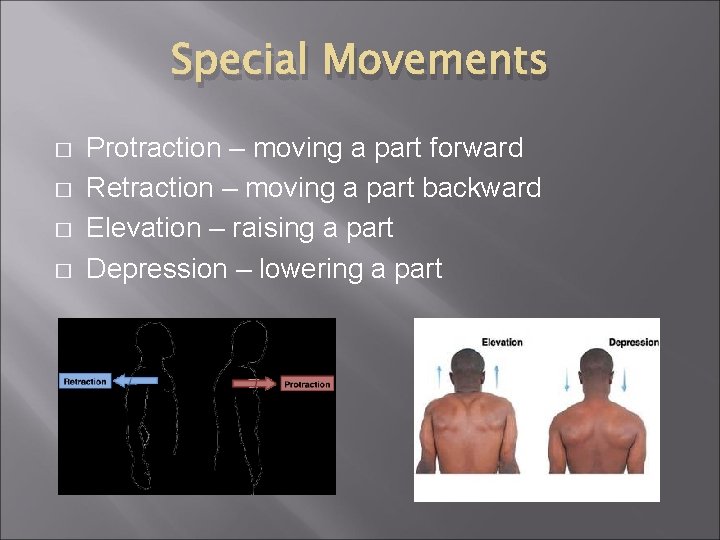
Special Movements � � Protraction – moving a part forward Retraction – moving a part backward Elevation – raising a part Depression – lowering a part
Naming of Skeletal Muscles · Direction of muscle fibers üExample: rectus (straight) · Relative size of the muscle üExample: maximus (largest) Copyright © 2003 Pearson Education, Inc. publishing as Benjamin Cummings Slide 6. 36 a
Naming of Skeletal Muscles · Location of the muscle üExample: many muscles are named for bones (e. g. , temporalis) · Number of origins üExample: triceps (three heads) Copyright © 2003 Pearson Education, Inc. publishing as Benjamin Cummings Slide 6. 36 b
Naming of Skeletal Muscles · Location of the muscles origin and insertion ü Example: sterno (on the sternum) · Shape of the muscle ü Example: deltoid (triangular) · Action of the muscle ü Example: flexor and extensor Copyright © 2003 Pearson Education, Inc. publishing as Benjamin Cummings Slide 6. 37
Axial and Appendicular Muscles � � Axial muscles arise on the axial skeleton (60% of skeletal muscles) ; position the head and spinal column and move the rib cage Appendicular muscles stabilize and move the appendicular skeleton (40% of skeletal muscles)
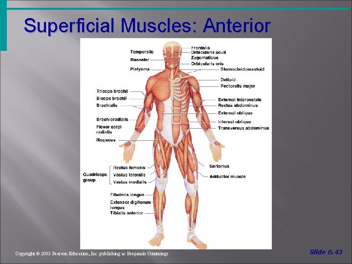
Superficial Muscles: Anterior Figure 6. 20 Copyright © 2003 Pearson Education, Inc. publishing as Benjamin Cummings Slide 6. 43
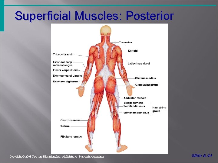
Superficial Muscles: Posterior Figure 6. 21 Copyright © 2003 Pearson Education, Inc. publishing as Benjamin Cummings Slide 6. 44
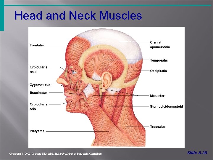
Head and Neck Muscles Figure 6. 14 Copyright © 2003 Pearson Education, Inc. publishing as Benjamin Cummings Slide 6. 38
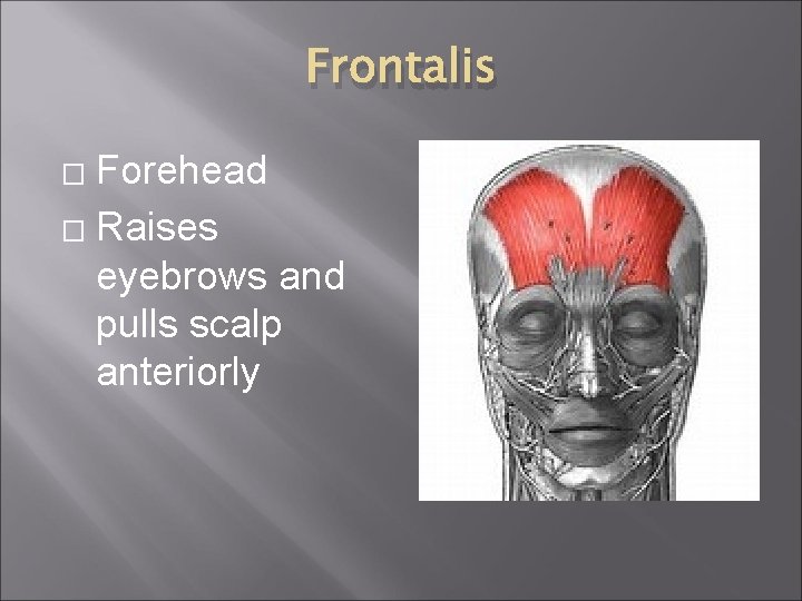
Frontalis Forehead � Raises eyebrows and pulls scalp anteriorly �

Temporalis � � Lateral muscles of the skull Closes mandible
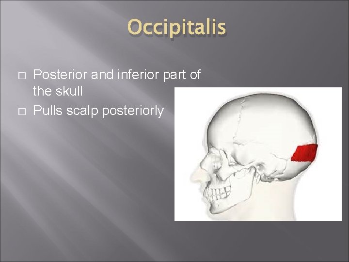
Occipitalis � � Posterior and inferior part of the skull Pulls scalp posteriorly
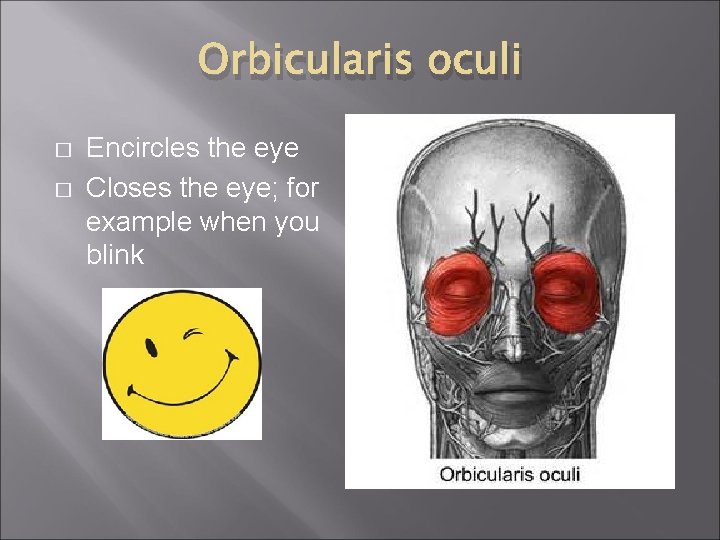
Orbicularis oculi � � Encircles the eye Closes the eye; for example when you blink
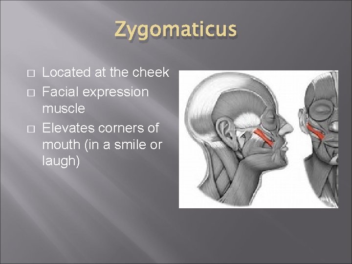
Zygomaticus � � � Located at the cheek Facial expression muscle Elevates corners of mouth (in a smile or laugh)
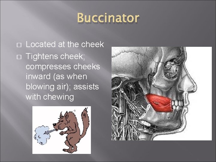
Buccinator � � Located at the cheek Tightens cheek; compresses cheeks inward (as when blowing air); assists with chewing
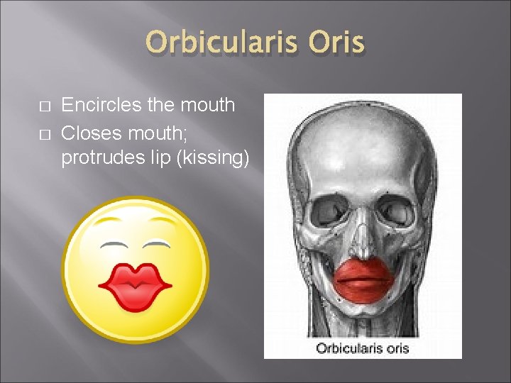
Orbicularis Oris � � Encircles the mouth Closes mouth; protrudes lip (kissing)
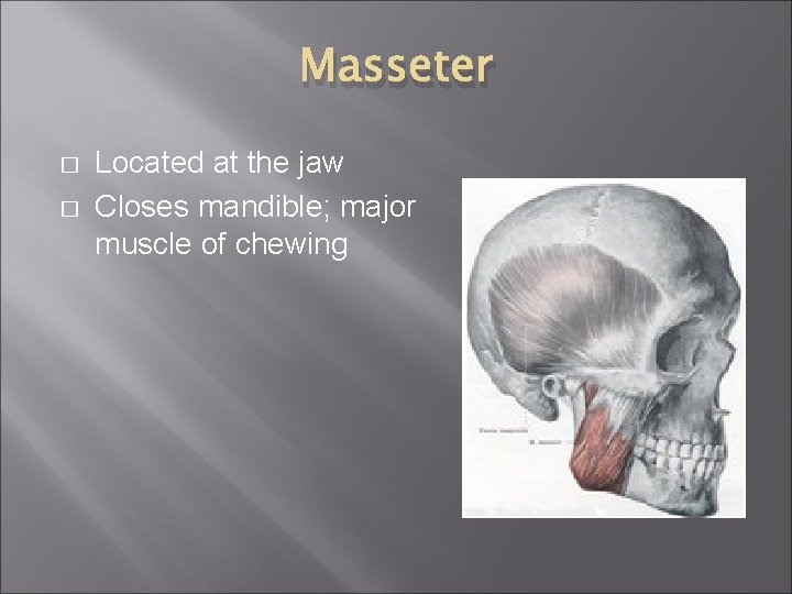
Masseter � � Located at the jaw Closes mandible; major muscle of chewing
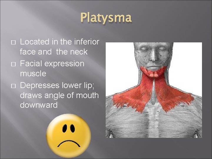
Platysma � � � Located in the inferior face and the neck Facial expression muscle Depresses lower lip; draws angle of mouth downward

Sternocleidomastoid � � � Located in the neck Rotates and extends head Flexes neck

Deltoid � � Located at the shoulder, clavicle area Abducts, extends, and flexes arm
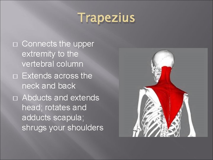
Trapezius � � � Connects the upper extremity to the vertebral column Extends across the neck and back Abducts and extends head; rotates and adducts scapula; shrugs your shoulders

Trunk Muscles Figure 6. 15 Copyright © 2003 Pearson Education, Inc. publishing as Benjamin Cummings Slide 6. 39
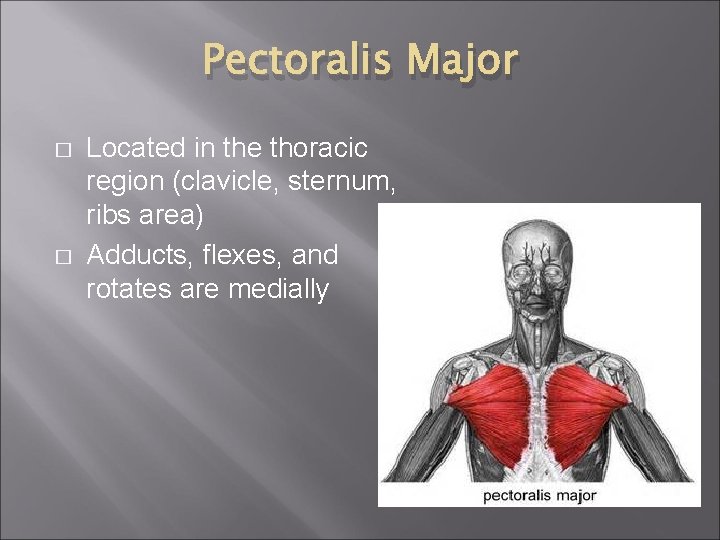
Pectoralis Major � � Located in the thoracic region (clavicle, sternum, ribs area) Adducts, flexes, and rotates are medially
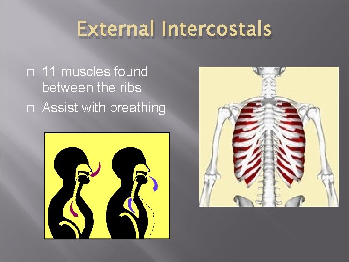
External Intercostals � � 11 muscles found between the ribs Assist with breathing
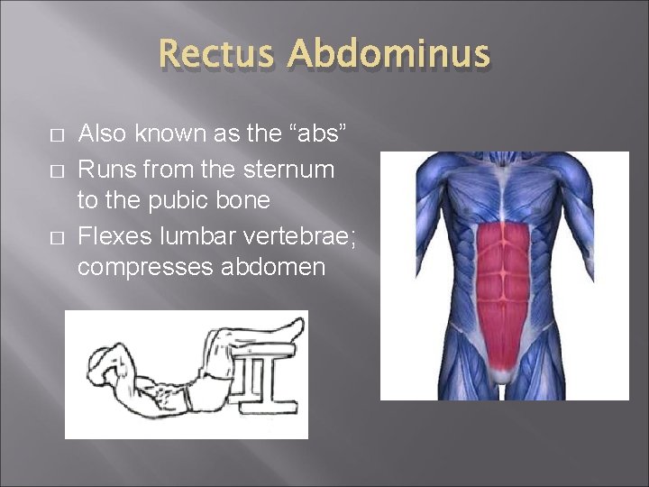
Rectus Abdominus � � � Also known as the “abs” Runs from the sternum to the pubic bone Flexes lumbar vertebrae; compresses abdomen
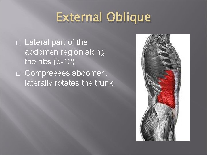
External Oblique � � Lateral part of the abdomen region along the ribs (5 -12) Compresses abdomen, laterally rotates the trunk
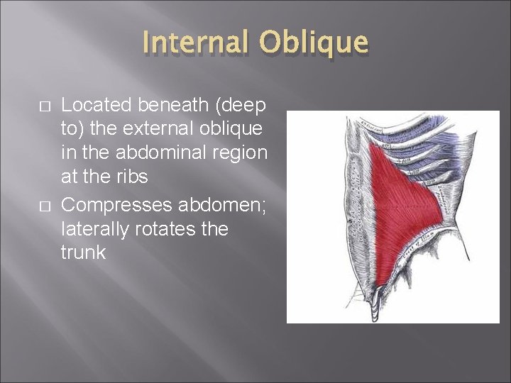
Internal Oblique � � Located beneath (deep to) the external oblique in the abdominal region at the ribs Compresses abdomen; laterally rotates the trunk
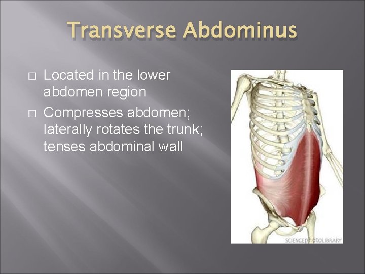
Transverse Abdominus � � Located in the lower abdomen region Compresses abdomen; laterally rotates the trunk; tenses abdominal wall
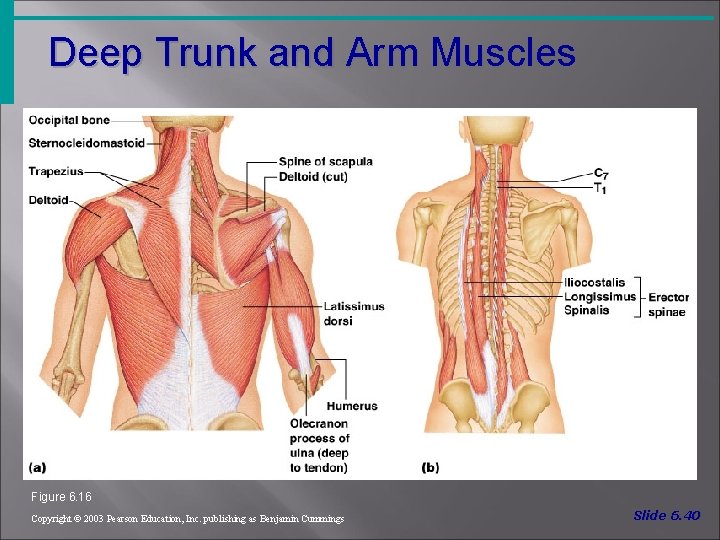
Deep Trunk and Arm Muscles Figure 6. 16 Copyright © 2003 Pearson Education, Inc. publishing as Benjamin Cummings Slide 6. 40
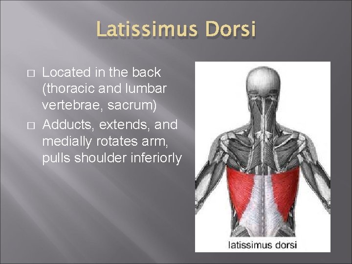
Latissimus Dorsi � � Located in the back (thoracic and lumbar vertebrae, sacrum) Adducts, extends, and medially rotates arm, pulls shoulder inferiorly
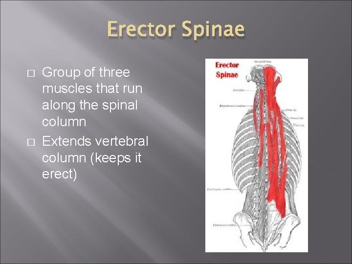
Erector Spinae � � Group of three muscles that run along the spinal column Extends vertebral column (keeps it erect)
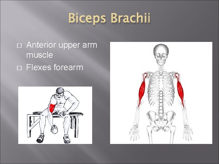
Biceps Brachii � � Anterior upper arm muscle Flexes forearm
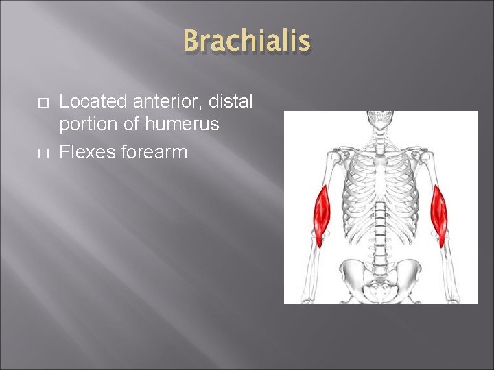
Brachialis � � Located anterior, distal portion of humerus Flexes forearm
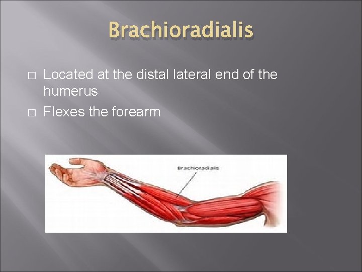
Brachioradialis � � Located at the distal lateral end of the humerus Flexes the forearm
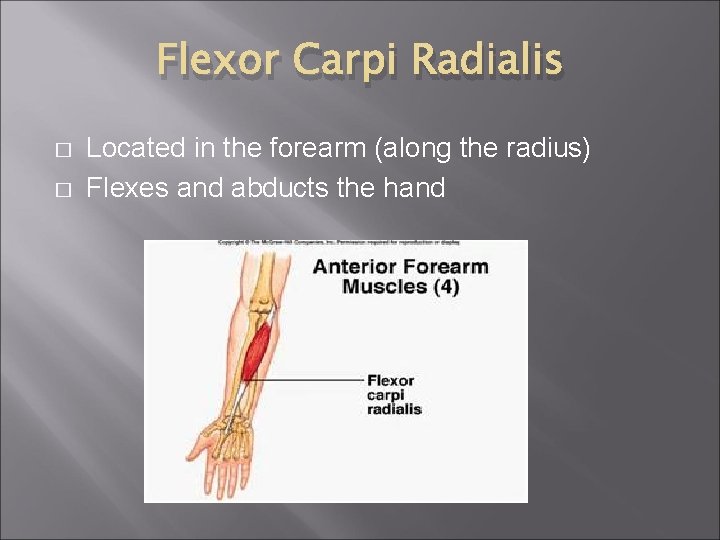
Flexor Carpi Radialis � � Located in the forearm (along the radius) Flexes and abducts the hand

Flexor Carpi Ulnaris � � Located in the forearm (along the ulna) Flexes and adducts hand
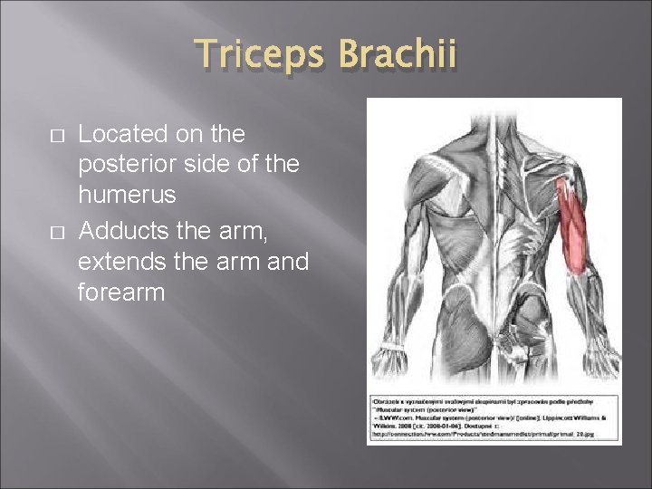
Triceps Brachii � � Located on the posterior side of the humerus Adducts the arm, extends the arm and forearm

Extensor Carpi Radialis Longus � � Located in the forearm Extends wrist and abducts hand
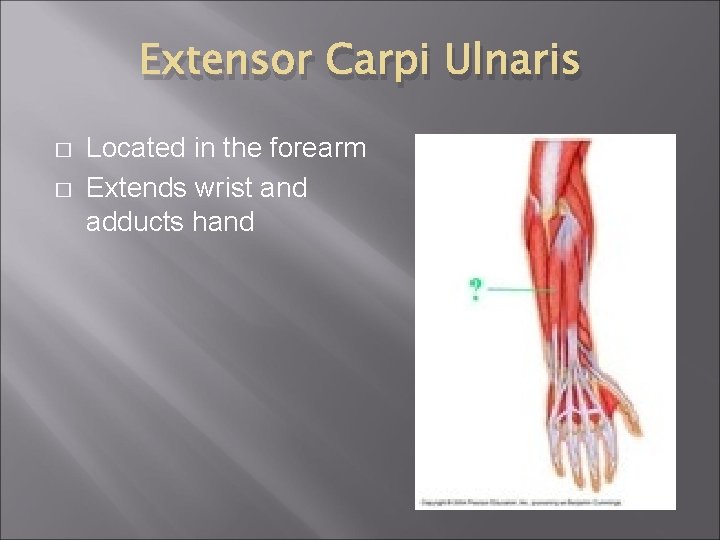
Extensor Carpi Ulnaris � � Located in the forearm Extends wrist and adducts hand
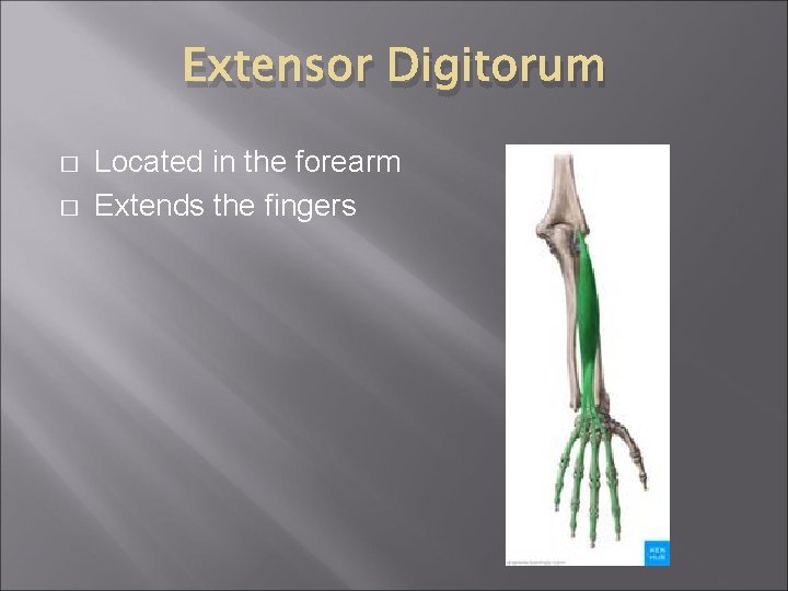
Extensor Digitorum � � Located in the forearm Extends the fingers
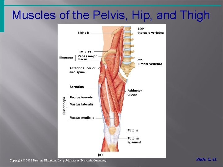
Muscles of the Pelvis, Hip, and Thigh Figure 6. 18 c Copyright © 2003 Pearson Education, Inc. publishing as Benjamin Cummings Slide 6. 41
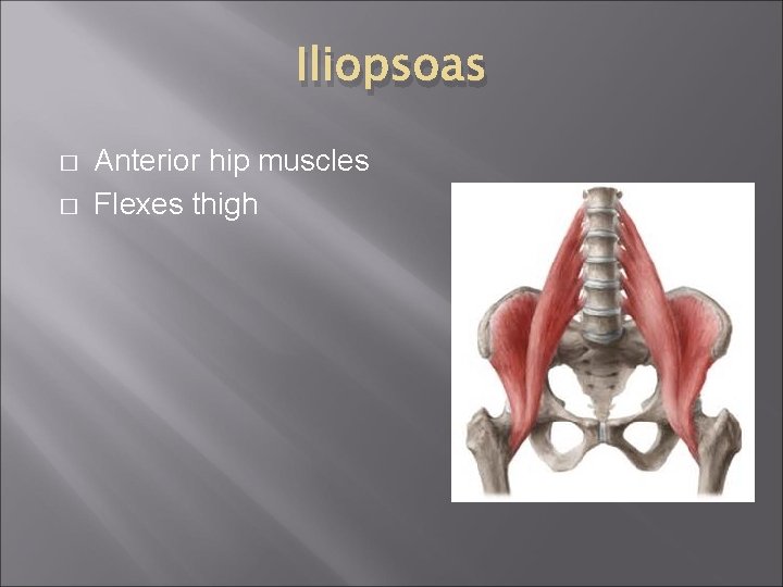
Iliopsoas � � Anterior hip muscles Flexes thigh
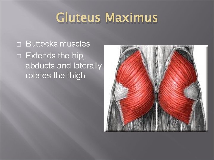
Gluteus Maximus � � Buttocks muscles Extends the hip, abducts and laterally rotates the thigh
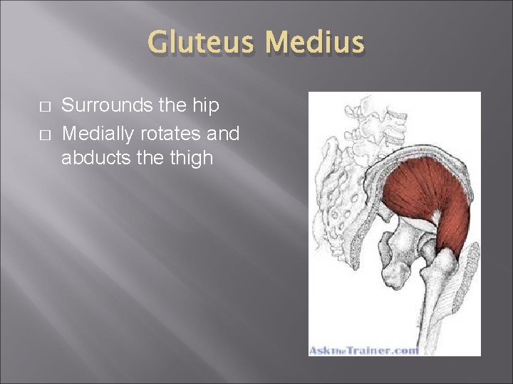
Gluteus Medius � � Surrounds the hip Medially rotates and abducts the thigh
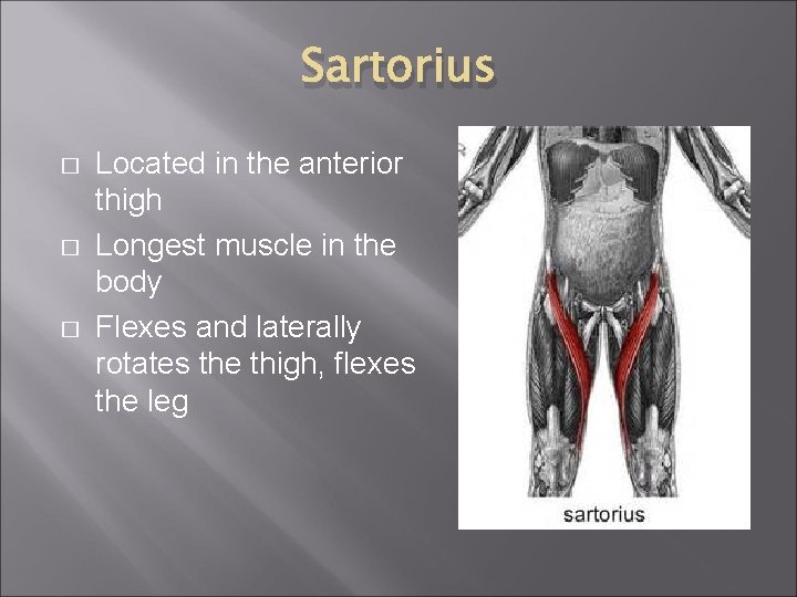
Sartorius � � � Located in the anterior thigh Longest muscle in the body Flexes and laterally rotates the thigh, flexes the leg
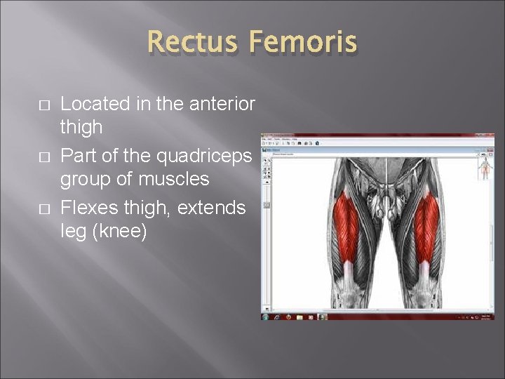
Rectus Femoris � � � Located in the anterior thigh Part of the quadriceps group of muscles Flexes thigh, extends leg (knee)
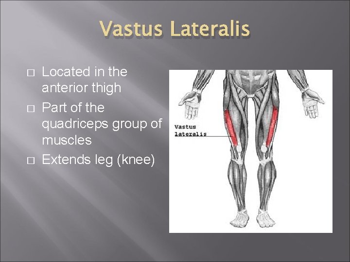
Vastus Lateralis � � � Located in the anterior thigh Part of the quadriceps group of muscles Extends leg (knee)
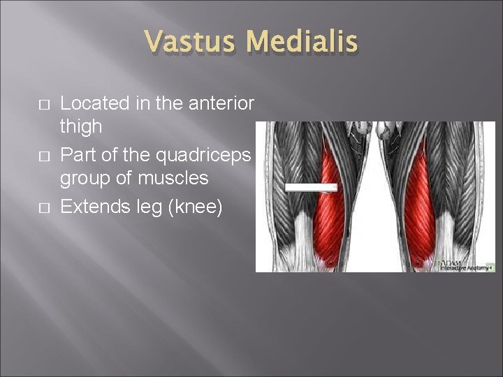
Vastus Medialis � � � Located in the anterior thigh Part of the quadriceps group of muscles Extends leg (knee)
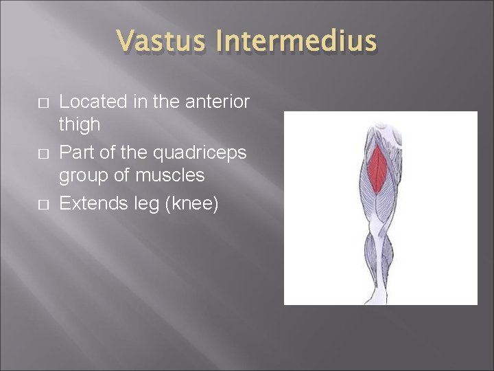
Vastus Intermedius � � � Located in the anterior thigh Part of the quadriceps group of muscles Extends leg (knee)

Adductor muscles � � Located in the inner thigh Adducts and flexes the thigh
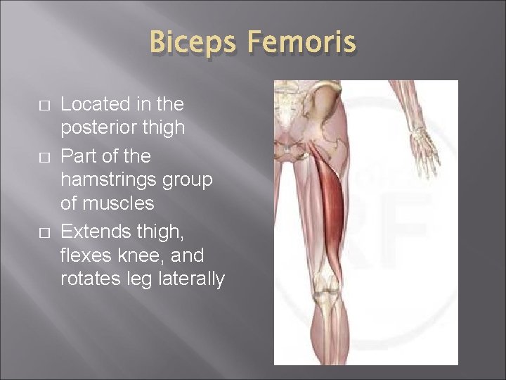
Biceps Femoris � � � Located in the posterior thigh Part of the hamstrings group of muscles Extends thigh, flexes knee, and rotates leg laterally

Semitendinosus � � � Located in the posterior thigh Part of the hamstrings group of muscles Extends thigh, flexes knee, and rotates leg laterally
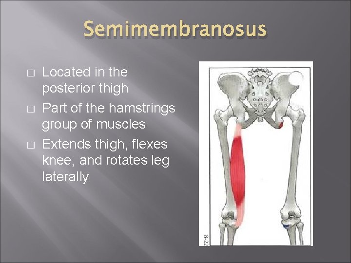
Semimembranosus � � � Located in the posterior thigh Part of the hamstrings group of muscles Extends thigh, flexes knee, and rotates leg laterally
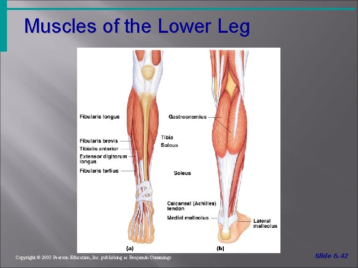
Muscles of the Lower Leg Figure 6. 19 Copyright © 2003 Pearson Education, Inc. publishing as Benjamin Cummings Slide 6. 42
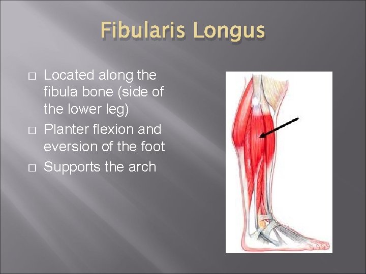
Fibularis Longus � � � Located along the fibula bone (side of the lower leg) Planter flexion and eversion of the foot Supports the arch
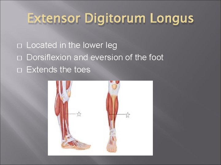
Extensor Digitorum Longus � � � Located in the lower leg Dorsiflexion and eversion of the foot Extends the toes
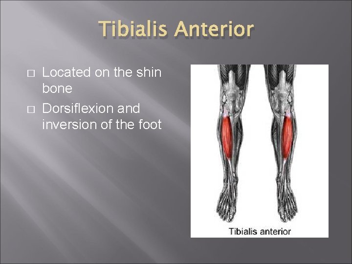
Tibialis Anterior � � Located on the shin bone Dorsiflexion and inversion of the foot
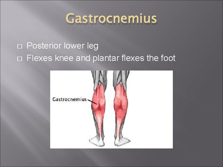
Gastrocnemius � � Posterior lower leg Flexes knee and plantar flexes the foot
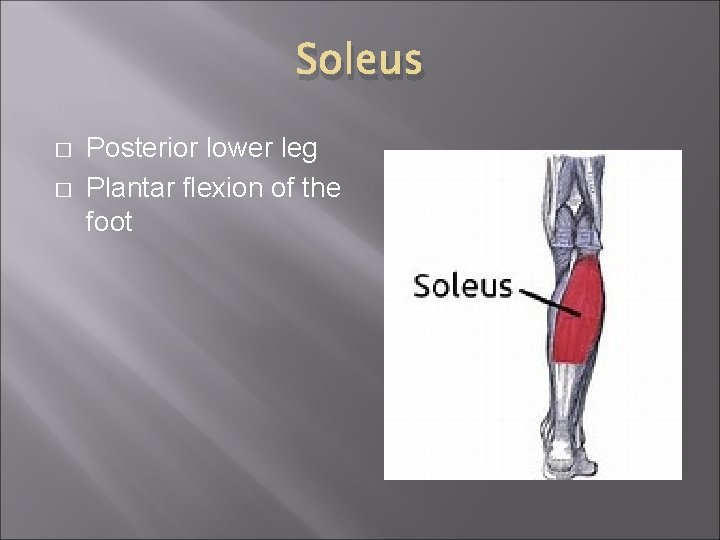
Soleus � � Posterior lower leg Plantar flexion of the foot
Muscle Analysis � � � � Choose a sport or activity where movement is involved (football, yoga, softball, swimming, skate boarding, etc. ) Write a short story about the sport/activity (choose a competition, game, or practice) Include 10 muscles and 5 movements in the story that the participant(s) demonstrates Circle the muscle names & underline the movements in your story You must include one picture with your story Use the “arts and supplies” to complete your final draft The story is a classwork grade of 20 points (1 point for each required term and 5 points for your picture/creativity)
Muscular System Injuries � � � TSWBAT identify injuries of the muscular system. TSWBAT compare a strain and sprain. TSWBAT list the elements of RICE for first aid of muscular system injuries.
Strain vs. Sprain What is a strain? Strains are injuries that involve the stretching or tearing of a musculo-tendinous (muscle and tendon) structure What is a sprain? A sprain is an injury involving the stretching or tearing of a ligament (tissue that connects bone to bone)
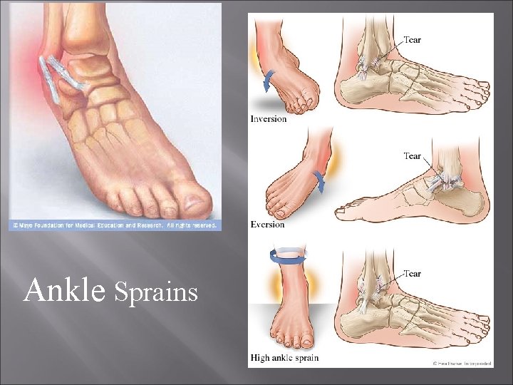
Ankle Sprains
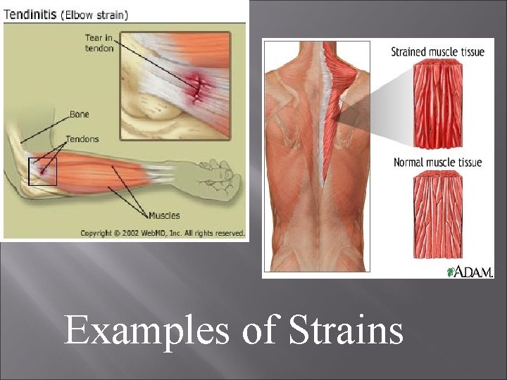
Examples of Strains
Sprains and Strains are categorized according to severity. Grade I (mild) sprain or strain involves some stretching or minor tearing of a ligament or muscle. Grade II (moderate) sprain or strain is a ligament or muscle that is partially torn but still intact. Grade III (severe) sprain or strain means that the ligament or muscle is completely torn, resulting in joint instability.
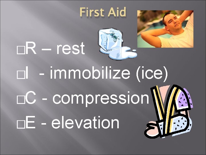
First Aid �R – rest �I - immobilize (ice) �C - compression �E - elevation
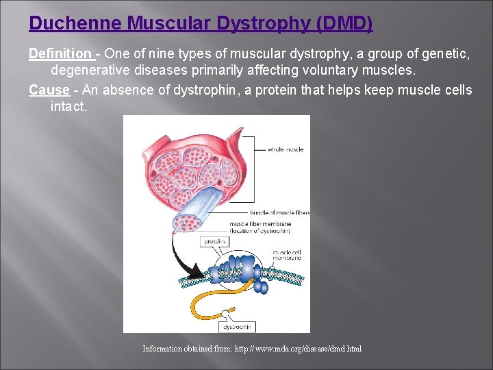
Duchenne Muscular Dystrophy (DMD) Definition - One of nine types of muscular dystrophy, a group of genetic, degenerative diseases primarily affecting voluntary muscles. Cause - An absence of dystrophin, a protein that helps keep muscle cells intact. Information obtained from: http: //www. mda. org/disease/dmd. html
DMD continued. . . Onset - Early childhood - about 2 to 6 years. Symptoms - Generalized weakness first affecting the muscles of the hips, pelvic area, thighs and shoulders. Calves are often enlarged. Progression - DMD eventually affects all voluntary muscles, and the heart and breathing muscles. Inheritance - X-linked recessive. DMD primarily affects boys, who inherit the disease through their mothers. Women can be carriers of DMD but usually exhibit no symptoms. � http: //www. youtube. com/watch? v=_ZAOOHayy 4 I
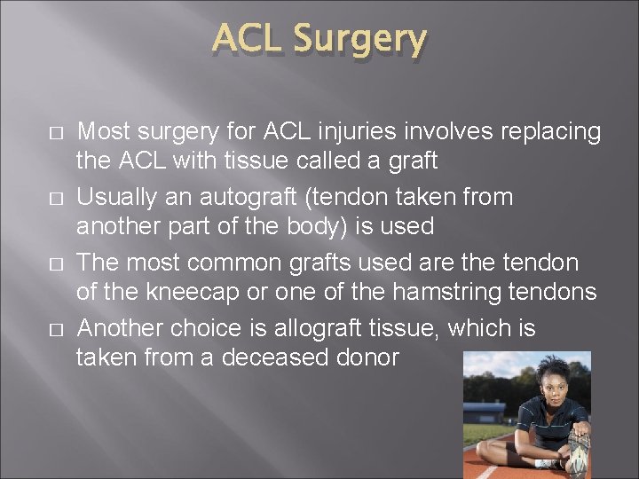
ACL Surgery � � Most surgery for ACL injuries involves replacing the ACL with tissue called a graft Usually an autograft (tendon taken from another part of the body) is used The most common grafts used are the tendon of the kneecap or one of the hamstring tendons Another choice is allograft tissue, which is taken from a deceased donor
Tommy John Surgery � � Repairs an injured elbow ligament (UCL construction) A surgeon replaces the injured UCL with a tendon taken from somewhere else in the patient’s body
Paralysis � � � Loss of muscle function in part of your body Occurs when something goes wrong with the way messages pass between your brain and muscles Can be complete or partial; occur on one or both sides of your body; one area or widespread Paraplegia: lower half of your body Quadriplegia: arms and legs Due to strokes, injuries (spinal cord), or a broken neck
- Slides: 88