
Muscular System Muscles are responsible for all types
Muscular System Muscles are responsible for all types of body movement (Machines of the body) Three basic muscle types are found in the body: Skeletal muscle Cardiac muscle Smooth muscle
Characteristics of Muscle Skeletal and smooth muscle cells are elongated , so called muscle fiber. All can shorten or contract due to the movement of microfilaments All muscles share same terminology ◦ Prefix myo refers to muscle ◦ Prefix mys refers to muscle ◦ Prefix sarco refers to muscle, for example, sarcoplasm.
Types of Muscle
Types of Muscle, cont.
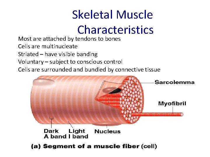
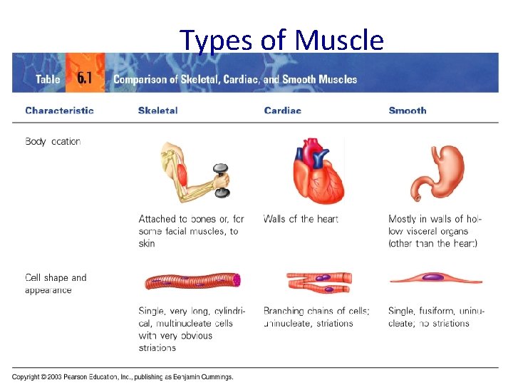
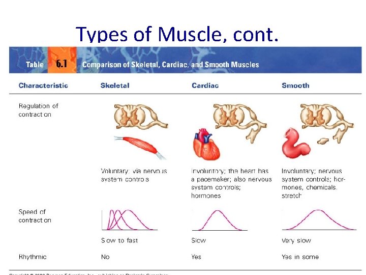
Skeletal Muscle Characteristics Most are attached by tendons to bones Cells are multinucleate Striated – have visible banding Voluntary – subject to conscious control Cells are surrounded and bundled by connective tissue
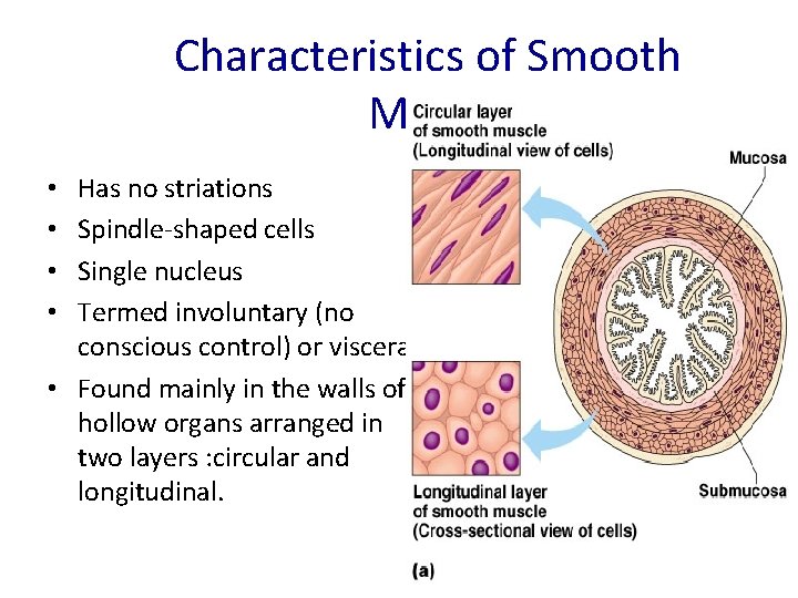
Characteristics of Smooth Muscle Has no striations Spindle-shaped cells Single nucleus Termed involuntary (no conscious control) or visceral. • Found mainly in the walls of hollow organs arranged in two layers : circular and longitudinal. • •
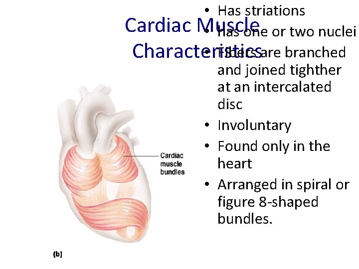
• Has striations Cardiac Muscle • has one or two nuclei • Fibers are branched Characteristics and joined tighther at an intercalated disc • Involuntary • Found only in the heart • Arranged in spiral or figure 8 -shaped bundles.
Functions of Skeletal Muscles 1 -Producing movement 2 - Maintaining posture 3 - Stabilizing joints 4 -Generating heat: is a by-product of muscle activity. As ATP is used to power muscle contraction, nearly threequarters of its energy escapes as heat. This heat is vital in maintaining normal body temperature. Skeletal muscles accounts for at least 40% of body mass , so it is the muscle type most responsible for heat generation.

Connective Tissue Wrappings of Skeletal Muscle · Endomysium – around single muscle fiber · Perimysium – around a fascicle (bundle) of fibers · Epimysium – covers the entire skeletal muscle Copyright © 2003 Pearson Education, Inc. publishing as Benjamin Cummings Figure 6. 1 Slide 6. 4 a
Skeletal Muscle Attachments · Epimysia blends into a connective tissue attachments which are either: · Tendons – cord-like structure which besides anchoring muscles, it provides durability and conserving space. · Aponeuroses – sheet-like structure · Sites of muscle attachment · Bones · Cartilages · Connective tissue coverings Copyright © 2003 Pearson Education, Inc. publishing as Benjamin Cummings Slide 6. 5
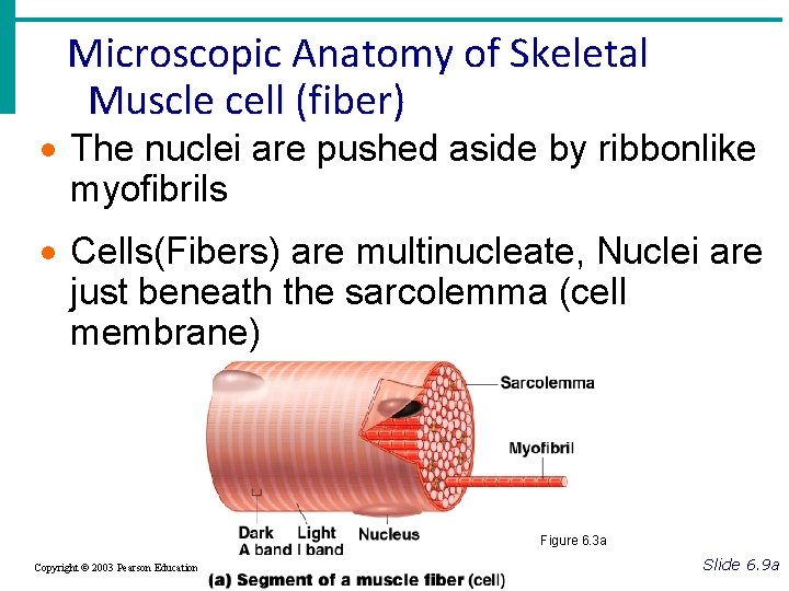
Microscopic Anatomy of Skeletal Muscle cell (fiber) · The nuclei are pushed aside by ribbonlike myofibrils · Cells(Fibers) are multinucleate, Nuclei are just beneath the sarcolemma (cell membrane) Figure 6. 3 a Copyright © 2003 Pearson Education, Inc. publishing as Benjamin Cummings Slide 6. 9 a
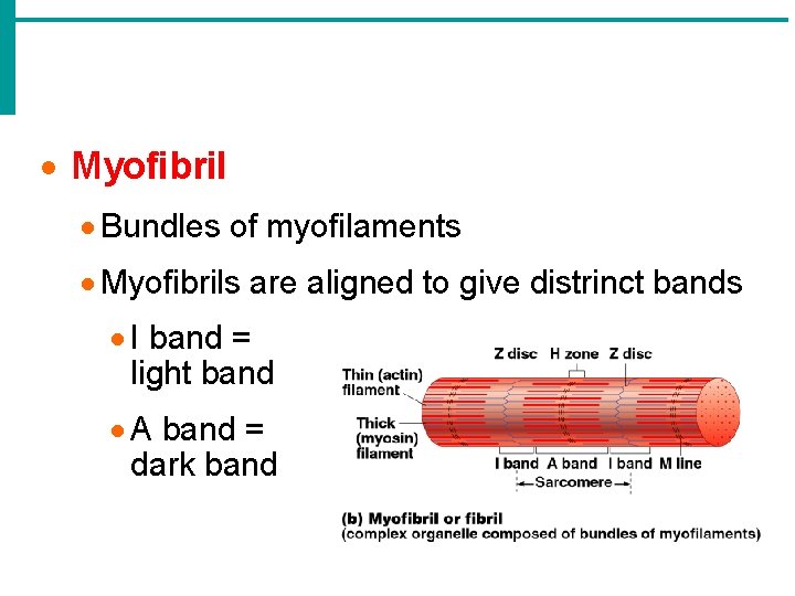
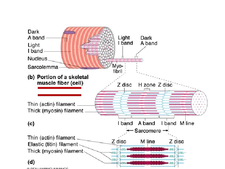
· Myofibril · Bundles of myofilaments · Myofibrils are aligned to give distrinct bands · I band = light band · A band = dark band
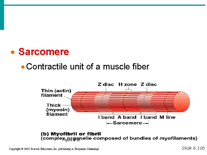
· Sarcomere · Contractile unit of a muscle fiber Figure 6. 3 b Copyright © 2003 Pearson Education, Inc. publishing as Benjamin Cummings Slide 6. 10 b
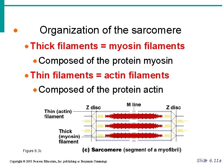
· Organization of the sarcomere · Thick filaments = myosin filaments · Composed of the protein myosin · Thin filaments = actin filaments · Composed of the protein actin Figure 6. 3 c Copyright © 2003 Pearson Education, Inc. publishing as Benjamin Cummings Slide 6. 11 a
Striations in Skeletal Muscle • A- Band- covers the length of the myosin filament • I- Band- covers actin where there is no overlap with myosin • H- Zone- the portion of myosin that does not overlap actin • Z- Line- point where actin filaments anchor
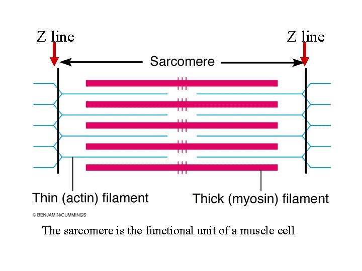
Z line The sarcomere is the functional unit of a muscle cell
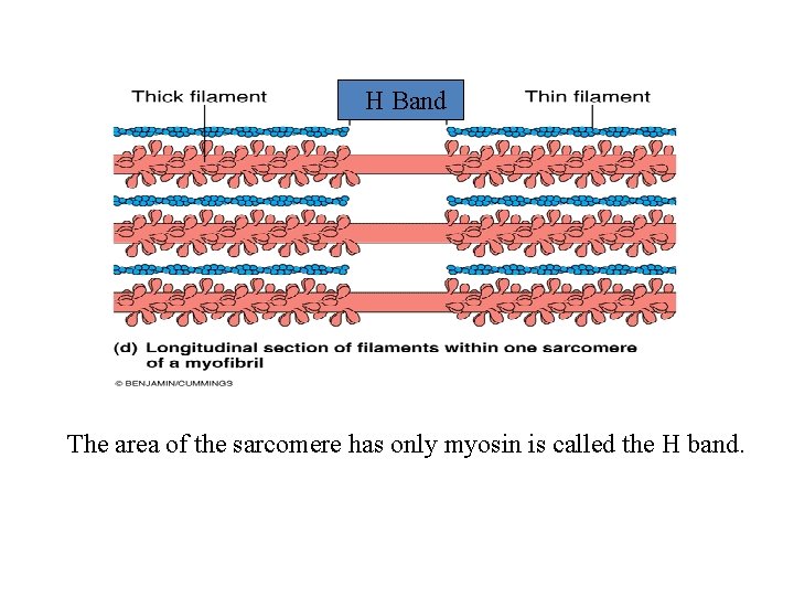
H Band The area of the sarcomere has only myosin is called the H band.
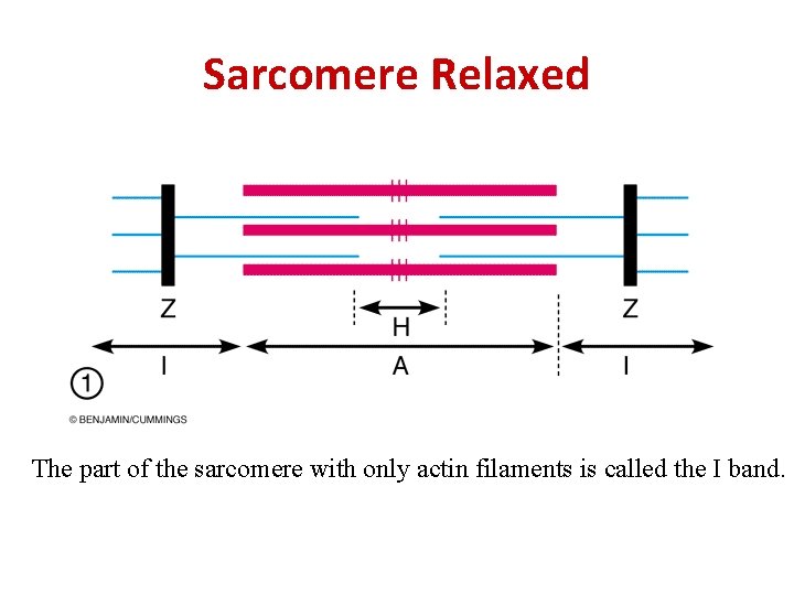
Sarcomere Relaxed The part of the sarcomere with only actin filaments is called the I band.
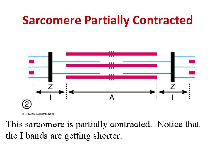
Sarcomere Partially Contracted This sarcomere is partially contracted. Notice that the I bands are getting shorter.
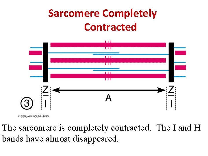
Sarcomere Completely Contracted The sarcomere is completely contracted. The I and H bands have almost disappeared.
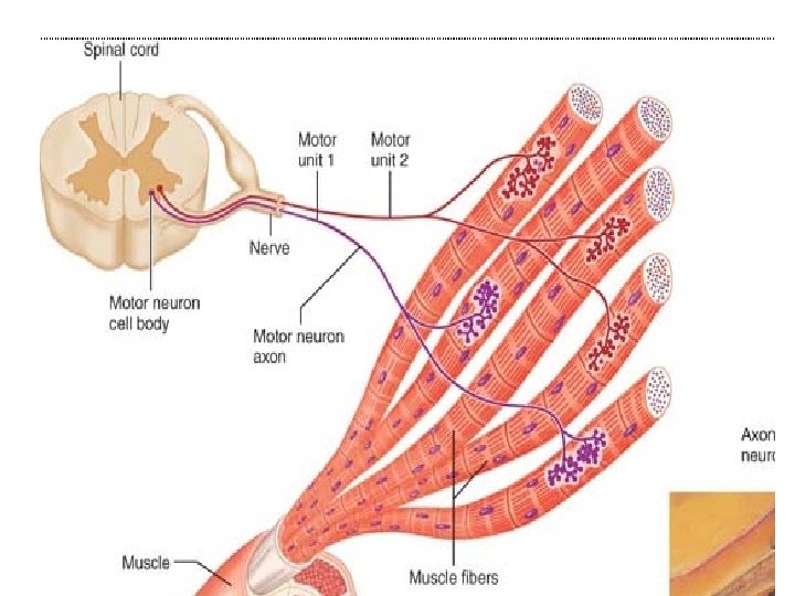
Neuromuscular Components Neuromuscular junction is the point where a motor neuron joins muscle fibers, Motor unit is a motor neuron and all the muscle fibers it innervates, may be few or hundreds. All or none law : with adequate stimulation, a muscle cell will contract to its fullest extent or not at all.
Skeletal muscle activity Muscles have two special functions • 1 -Irritability : the ability to receive and respond to stimulus 2 -Contractility : the ability to shorten • Muscle cell must be stimulated by nerve impulse to contract • There is a gap (synaptic cleft) between the nerve axonal terminal &muscle cell
• This gap is crossed by a chemical transmitter called Acetylcholine (Ach) • Sarcolemma becomes depolarised i. e. , temporarily permeable to sodium • This upset generates an electric current called action potential which is unstoppable leading to contraction of the whole muscle fiber such as lighting a match under a small dry twig.
MUSCLE TONE • Relaxed skeletal muscles are always slightly contracted involantarily. – This state of continuous partial contraction is termed “muscle tone”. • One group of muscle fibers are activated followed by another group and so on. – Spinal reflexes continually activate an alternating subset of motor neurons – No active movement produced – Muscles kept firm, healthy, and ready to respond to stimulation – Helps stabilize joints and maintain posture
The Muscle Twitch The response of a motor unit to a single action potential of its motor neuron is called a muscle twitch. The muscle fibers contract quickly and then relax. Twitch contractions of some muscles are rapid and brief, as with the muscles controlling eye movements. Graded Muscle Responses For proper skeletal movement, contractions must be relatively smooth and vary in strength. These variations are referred to as graded muscle responses. In general, muscle contraction can be graded in two ways: (1) by changing the frequency of stimulation and (2) by changing the strength of the stimulus.
1 - Muscle Response to Changes in Stimulation Frequency The nervous system achieves greater muscular force by increasing the firing rate of motor neurons. the second twitch will be stronger than the first as the second twitch will appear to ride on the shoulders of the first. This phenomenon, called wave summation, occurs because the second contraction occurs before the muscle has completely relaxed. If the muscle is stimulated at an increasingly faster rate, the relaxation time between the twitches becomes shorter and shorter, progressing to a sustained but quivering contraction referred to as unfused or incomplete tetanus
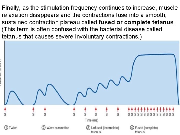
Finally, as the stimulation frequency continues to increase, muscle relaxation disappears and the contractions fuse into a smooth, sustained contraction plateau called fused or complete tetanus. (This term is often confused with the bacterial disease called tetanus that causes severe involuntary contractions. )
2 -Muscle Response to Stronger Stimuli The force of contraction is controlled more precisely by delivering shocks of increasing voltage to the muscle, calling more and more muscle fibers into play. The stimulus at which the first observable contraction occurs is called the threshold stimulus Beyond this point, the muscle contracts more and more vigorously as the stimulus strength is increased until the point at which all the muscle’s motor units are recruited. Increasing the stimulus intensity beyond the maximal stimulus does not produce a stronger contraction.
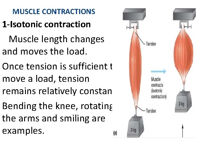
MUSCLE CONTRACTIONS 1 -Isotonic contraction Muscle length changes and moves the load. Once tension is sufficient to move a load, tension remains relatively constant. Bending the knee, rotating the arms and smiling are examples.
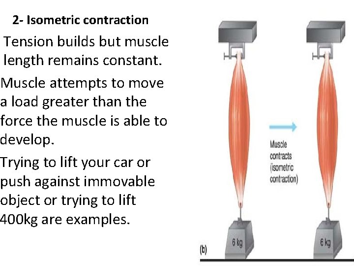
2 - Isometric contraction Tension builds but muscle length remains constant. Muscle attempts to move a load greater than the force the muscle is able to develop. Trying to lift your car or push against immovable object or trying to lift 400 kg are examples.
Effect of exercise on Muscle • Muscles are no exceptions to the saying –use it or lose it • Regular exercise increases muscle size, strength and endurance 1 -Aerobic exercise (isotonic contraction)results in stronger muscle with greater resistance to fatigue (endurance type of exercise). • No increase in size but better heart &lungs functions. • Examples are jogging or biking
2 -Isometric(Resistance) exercise • Require little time • No special equipment • Forcing the muscle to contract with as much as possible leads to enlargement of muscle cell without increase in their number • Examples are weigh lifting and body building. N. B. , The best exercise program for most people is one that include both types of exercise.
Energy for muscle contraction • ATP store in the muscle supplies energy ONLY for 4 -6 seconds • ATP then comes from: 1 - direct phosphorylation of ADP by reaction with creatine phosphate(CP) -No need for oxygen -1 ATP for 1 CP -Provides energy for 15 seconds only
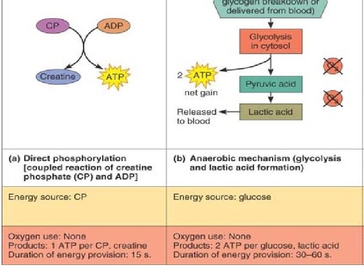
2 -Anaerobic respiration: is the glycolysis and so lactic acid accumulates due to absence of oxygen. • -2 ATP per glucose molecule • -lactic acid accumulates • - so muscle become sluggish or fatigued. • - Provides energy for 30 -60 seconds only
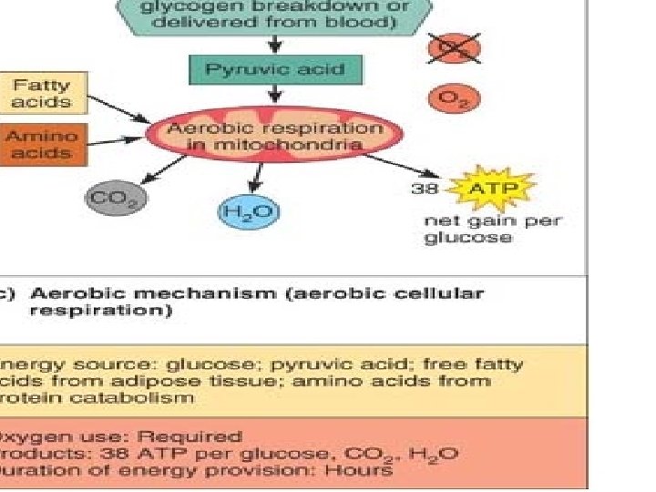
3 -aerobic mechanism (oxidative phosphorylation) • Oxygen is needed to oxidize glucose, pyruvic acid, lactic acid, free fatty acids and aminoacids • 38 ATP for 1 glucose molecule • Duration of energy is HOURS as long as oxygen is available to the muscle.
Muscle fatigue. Muscle fatigue occurs when an exercising muscle can no longer respond to stimulation with the same degree of contraction. Causes for this fatigue is an accumulation of lactic acid, and the depletion of energy reserves. Increased oxygen consumption is needed for a period of time to recover from exercise (paying off an oxygen debt) to get rid of accumulated lactic acid.
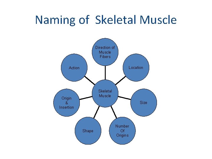
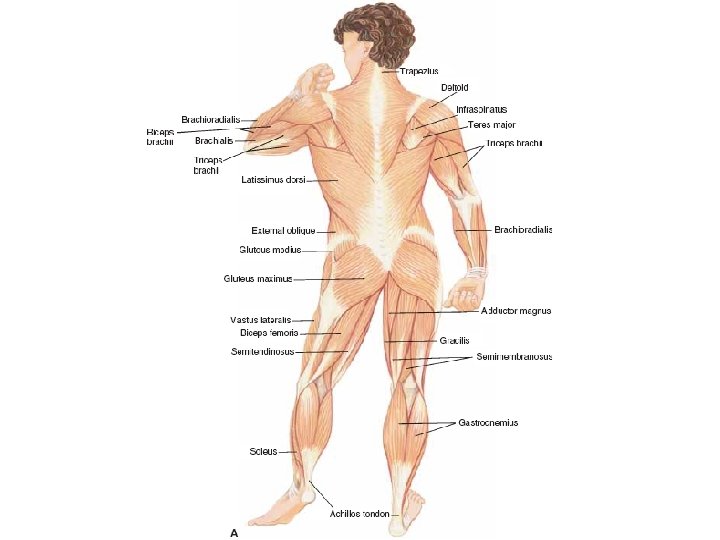
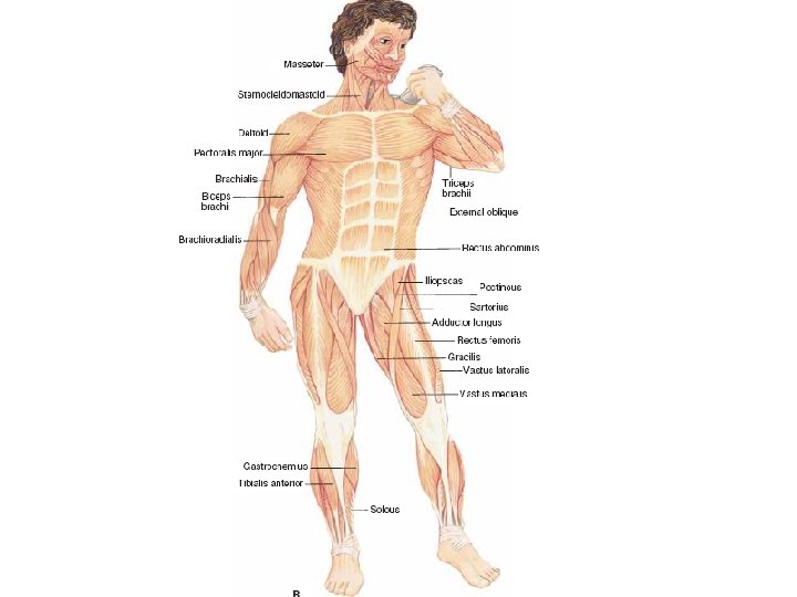
Naming of Skeletal Muscle Direction of Muscle Fibers Location Action Skeletal Muscle Origin & Insertion Size Shape Number Of Origins
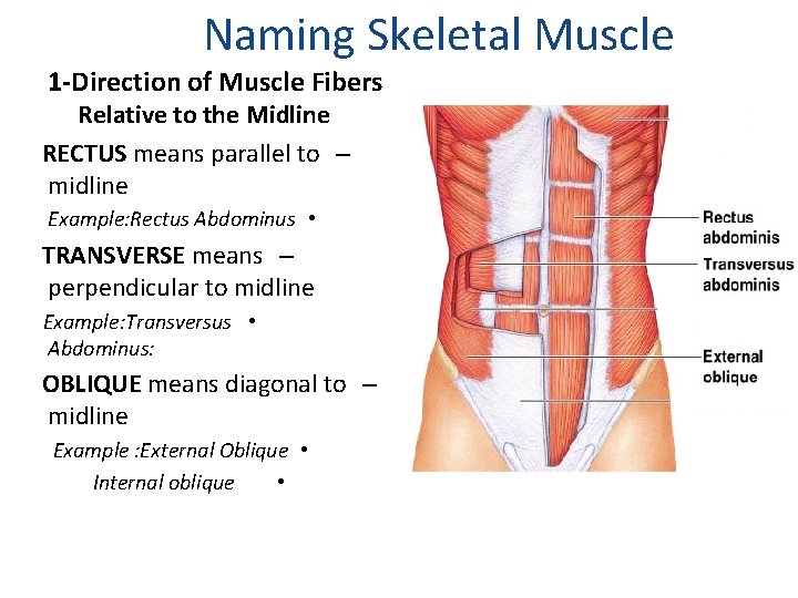
Naming Skeletal Muscle 1 -Direction of Muscle Fibers Relative to the Midline RECTUS means parallel to – midline Example: Rectus Abdominus • TRANSVERSE means – perpendicular to midline Example: Transversus • Abdominus: OBLIQUE means diagonal to – midline Example : External Oblique • Internal oblique •
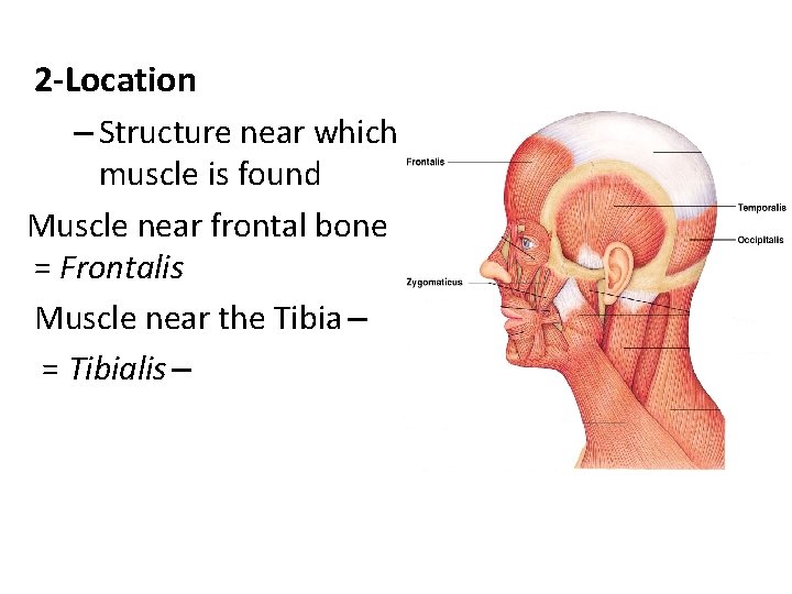
2 -Location – Structure near which a muscle is found Muscle near frontal bone = Frontalis Muscle near the Tibia – = Tibialis –
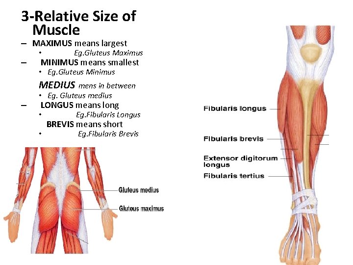
3 -Relative Size of Muscle – MAXIMUS means largest – • Eg. Gluteus Maximus MINIMUS means smallest • Eg. Gluteus Minimus MEDIUS mens in between – • Eg. Gluteus medius LONGUS means long • • Eg. Fibularis Longus BREVIS means short Eg. Fibularis Brevis
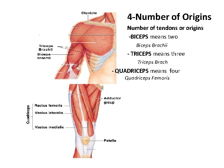
4 -Number of Origins Number of tendons or origins -BICEPS means two Biceps Brachii - TRICEPS means three Triceps Brach - QUADRICEPS means four Quadriceps Femoris
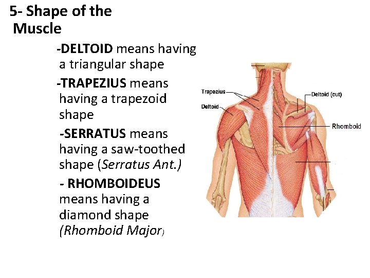
5 - Shape of the Muscle -DELTOID means having a triangular shape -TRAPEZIUS means having a trapezoid shape -SERRATUS means having a saw-toothed shape (Serratus Ant. ) - RHOMBOIDEUS means having a diamond shape (Rhomboid Major)
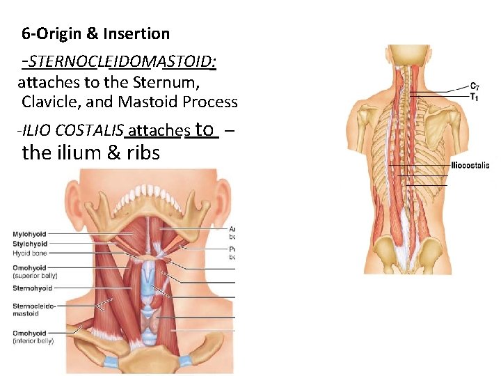
6 -Origin & Insertion -STERNOCLEIDOMASTOID: attaches to the Sternum, Clavicle, and Mastoid Process -ILIO COSTALIS attaches to – the ilium & ribs
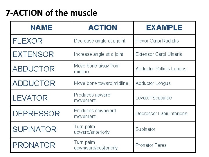
7 -ACTION of the muscle NAME ACTION EXAMPLE FLEXOR Decrease angle at a joint Flexor Carpi Radialis EXTENSOR Increase angle at a joint Extensor Carpi Ulnaris ABDUCTOR Move bone away from midline Abductor Pollicis Longus ADDUCTOR Move bone toward midline Adductor Longus LEVATOR Produces upward movement Levator Scapulae DEPRESSOR Produces downward movement Depressor Labii Inferioris SUPINATOR Turn palm upward/anteriorly Supinator PRONATOR Turn palm downward/posteriorly Pronator Teres
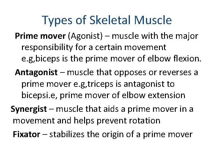
Types of Skeletal Muscle Prime mover (Agonist) – muscle with the major responsibility for a certain movement e. g, biceps is the prime mover of elbow flexion. Antagonist – muscle that opposes or reverses a prime mover e. g, triceps is antagonist to bicepsi. e, prime mover of elbow extension Synergist – muscle that aids a prime mover in a movement and helps prevent rotation Fixator – stabilizes the origin of a prime mover
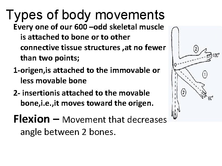
Types of body movements Every one of our 600 –odd skeletal muscle is attached to bone or to other connective tissue structures , at no fewer than two points; 1 -origen, is attached to the immovable or less movable bone 2 - insertionis attached to the movable bone, i. e. , it moves toward the origen. Flexion – Movement that decreases angle between 2 bones.
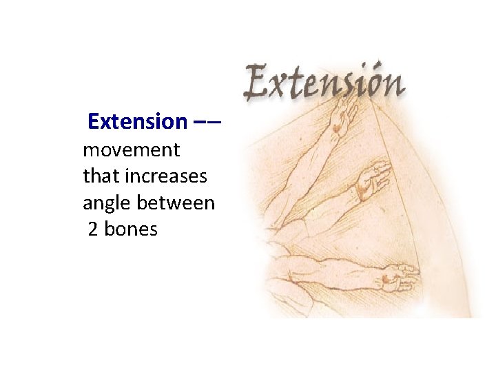
Extension – – movement that increases angle between 2 bones
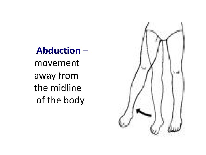
Abduction – movement away from the midline of the body
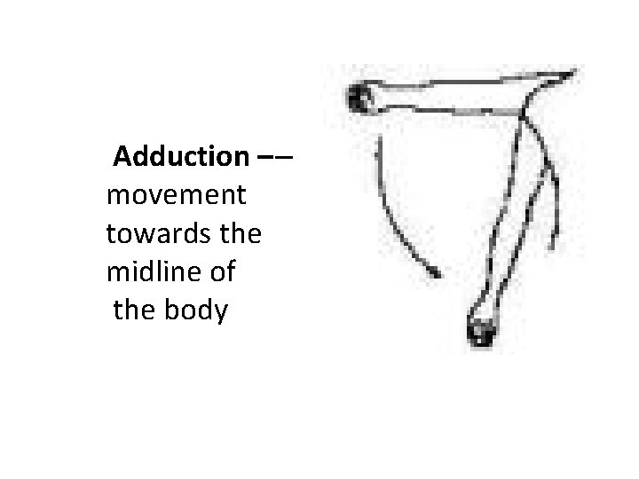
Adduction – – movement towards the midline of the body
Rotation – movement around a longitudinal axis Circumduction-combination of flexion, extension, abduction, and adduction. seen in ball and socket joints as the shoulder
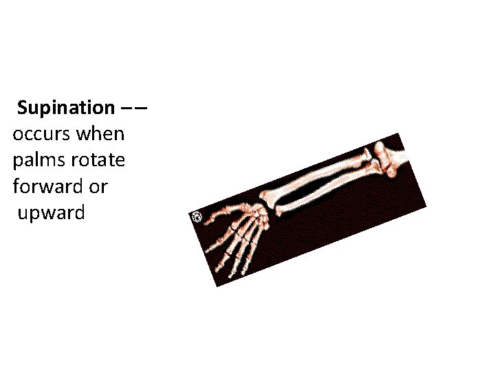
Supination – – occurs when palms rotate forward or upward
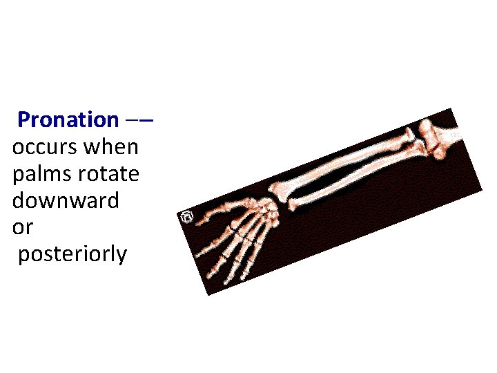
Pronation – – occurs when palms rotate downward or posteriorly
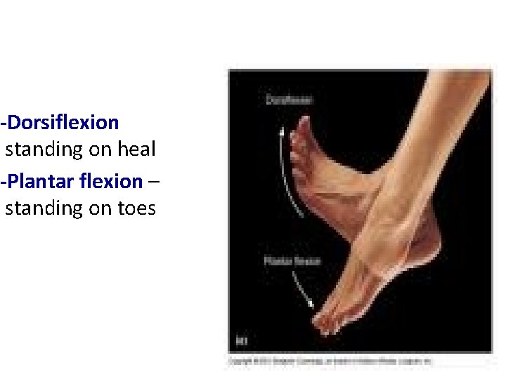
-Dorsiflexion standing on heal -Plantar flexion – standing on toes
Inversion of the foot: turn the sole medially. Eversion of the foot: turn the sole laterally Opposition: moving the thumb to touch the tips of the other fingers
Divisions of the Muscular System Axial muscles: position head and spinal column move rib cage 60% of skeletal muscles Appendicular muscles: support pectoral and pelvic girdles support limbs 40% of skeletal muscles
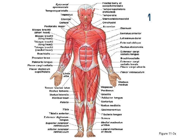
Axial and Appendicular M Figure 11– 3 a
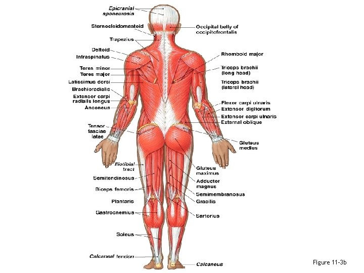
Figure 11– 3 b
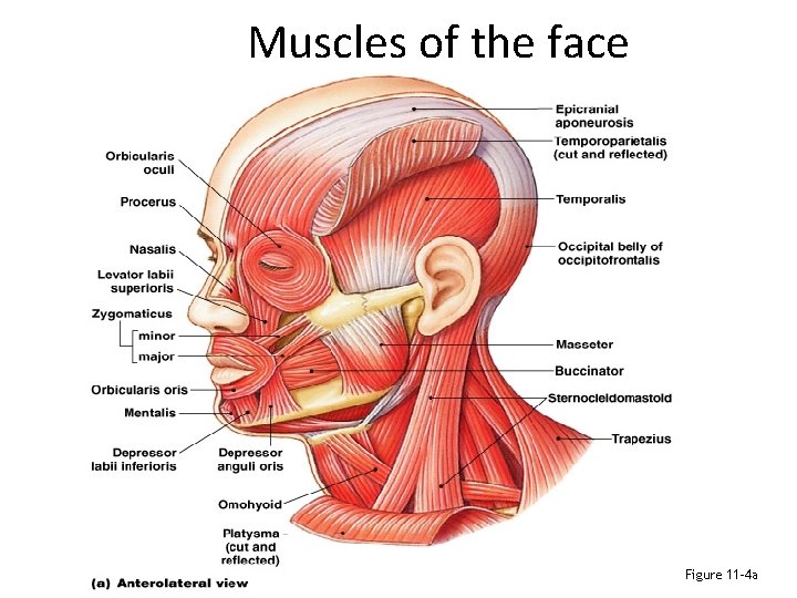
Muscles of the face Figure 11– 4 a
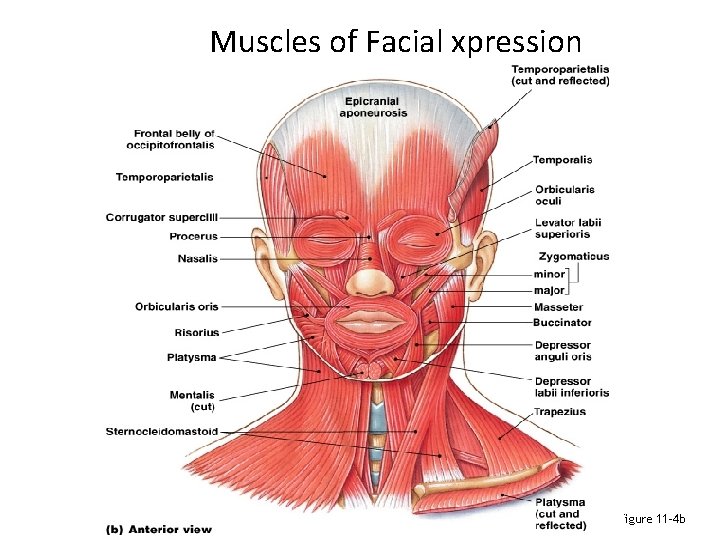
Muscles of Facial xpression Figure 11– 4 b
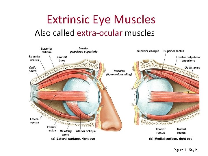
Extrinsic Eye Muscles Also called extra-ocular muscles Figure 11– 5 a, b

Extrinsic Eye Muscles Inferior rectus Superior rectus Medial rectus Lateral rectus Inferior oblique Superior oblique Figure 11– 5 c
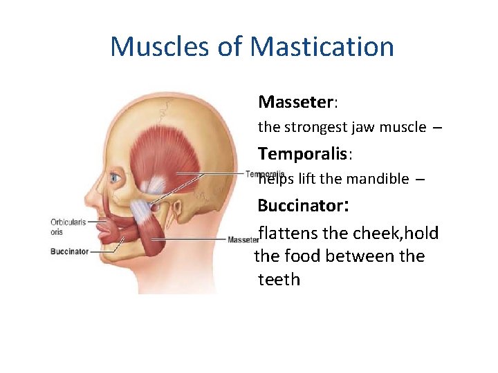
Muscles of Mastication Masseter: the strongest jaw muscle – Temporalis: helps lift the mandible – Buccinator: flattens the cheek, hold the food between the teeth
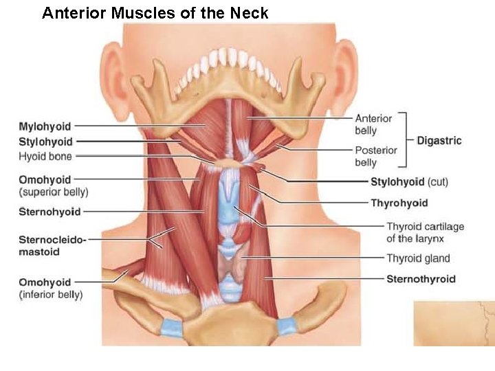
Anterior Muscles of the Neck
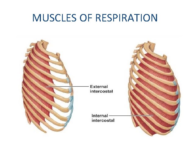
MUSCLES OF RESPIRATION
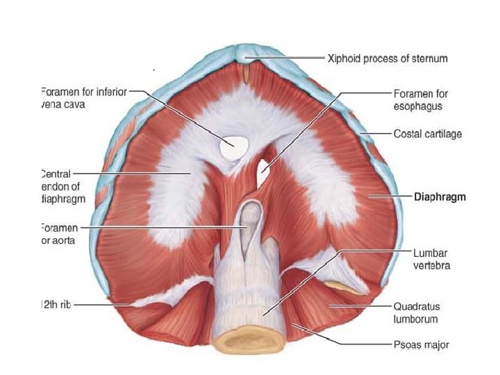
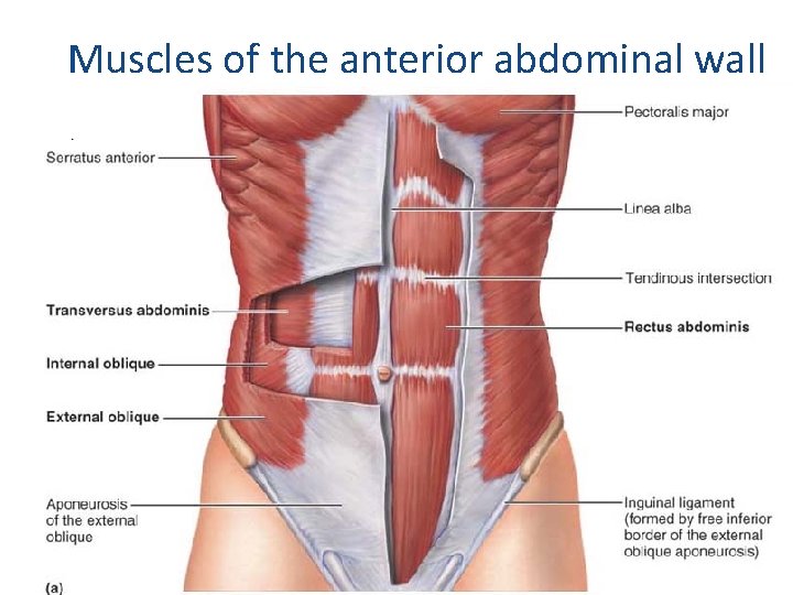
Muscles of the anterior abdominal wall
Functions of Pelvic Floor Muscles -Support organs of pelvic cavity -Flex sacrum and coccyx -Control movement of materials through urethra and anus
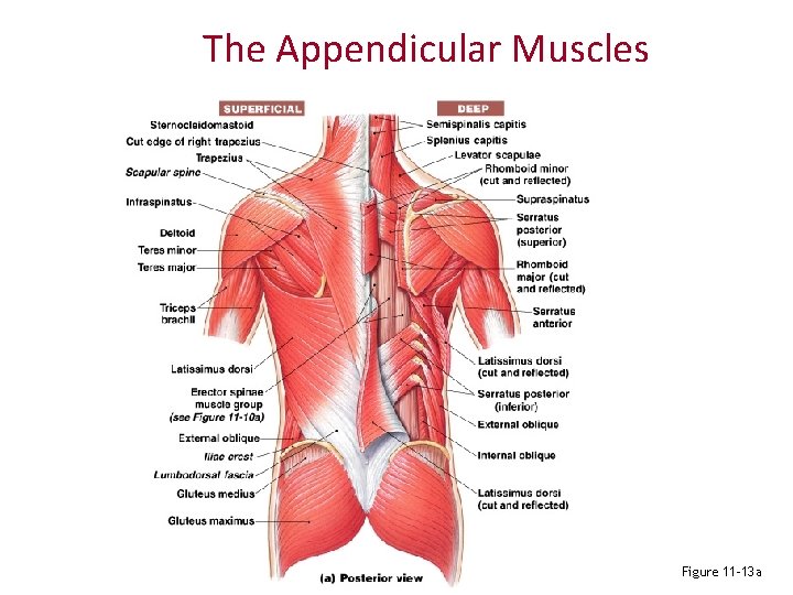
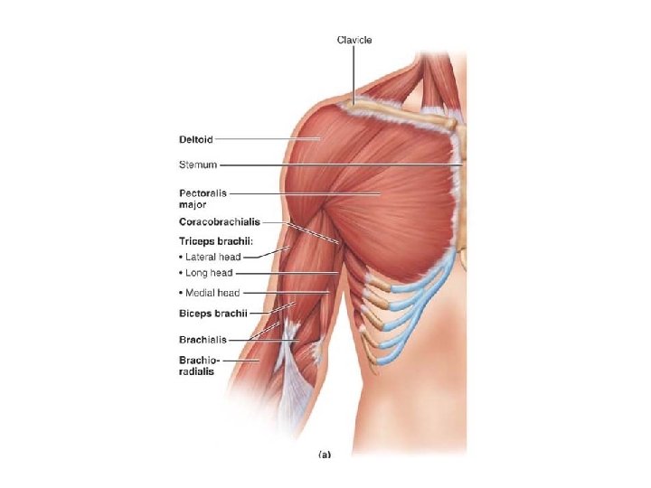
The Appendicular Muscles Figure 11– 13 a
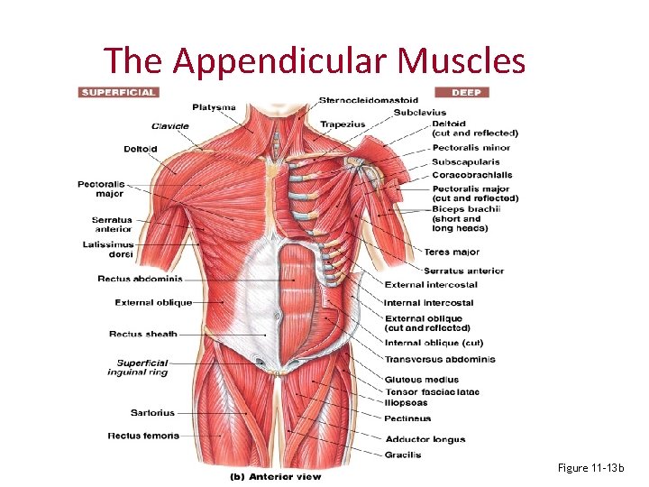
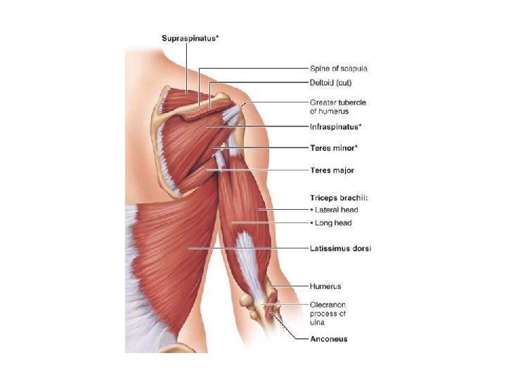
The Appendicular Muscles Figure 11– 13 b
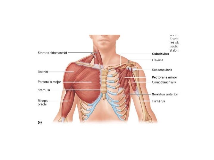
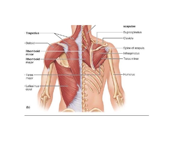
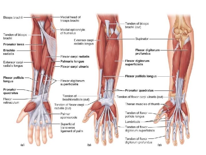
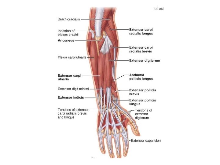
Divisions of Appendicular Muscles 1)Muscles of the shoulders and upper limbs 2) Muscles of the pelvis and lower limbs
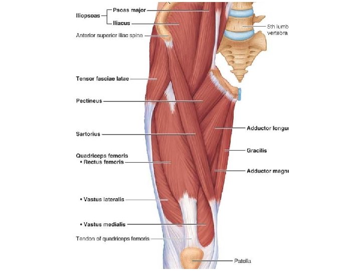
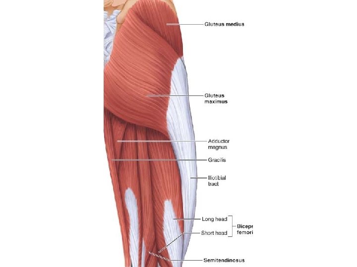
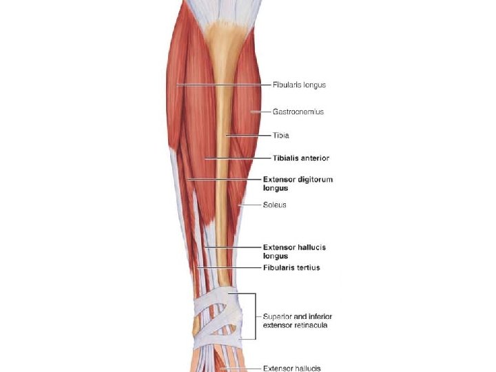
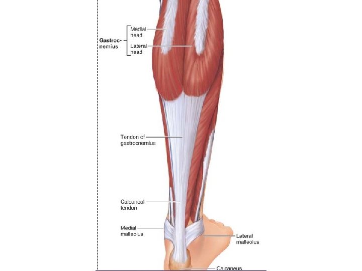
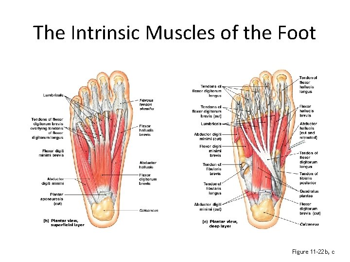
The Intrinsic Muscles of the Foot Figure 11– 22 b, c
Effects of Aging on the Muscular System -Skeletal muscle fibers become smaller in diameter -Skeletal muscles become less elastic: develop increasing amounts of fibrous tissue (fibrosis) -Decreased tolerance for exercise -Decreased ability to recover from muscular injuries
Developmental Aspects of Muscles - The first movement of the fetus (quickening) usually occur by the eixteenth week of pregnancy. - At birth, a baby’s movements are uncoordinated and largely reflexive. - Muscular development reflects the level of neuromuscular coordination, which develops in a headto-toe and proximal-to-distal direction. In other words, a baby can lift its head before it can walk, and gross movements precede fine ones.
- Slides: 89