Muscular System muscle fiber one individual muscle cell

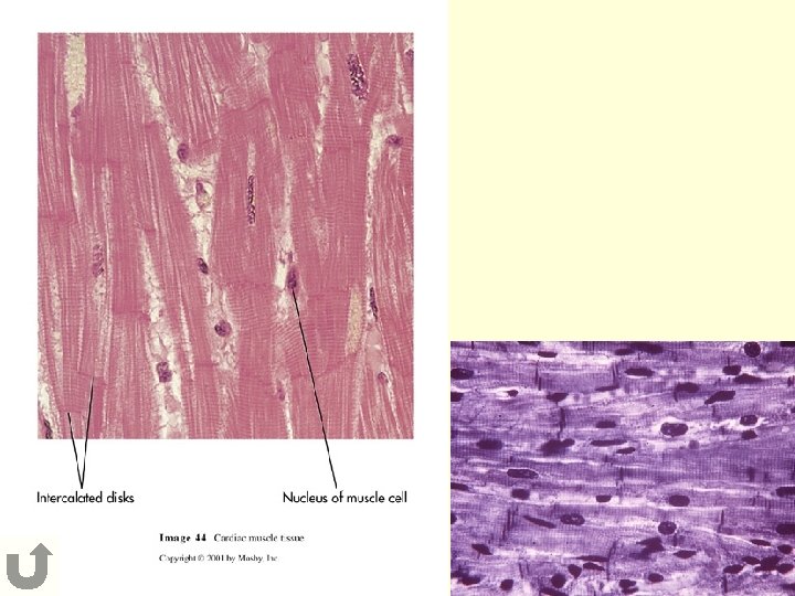
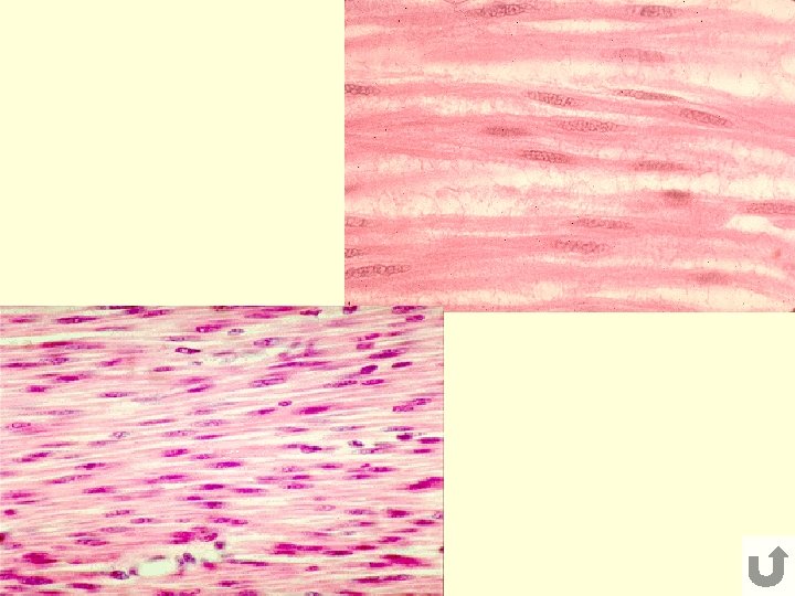
Muscular System muscle fiber = one individual muscle cell Muscle Tissue 1. cardiac -heart • striated • cylindrical, branched -”form follows function” • intercalated disks • involuntary 2. smooth • no striations • fibers tapered at ends • involuntary - bld. vessels, urinary system, digestive system

3. skeletal - attaches to bone • striated • cylindrical • voluntary

- extend from 1 bone, across a joint, to another bone -move bones by pulling on them Functions of Skeletal Muscle 1. Movement • contract (shorten) and relax (lengthen) - insertion - attachment to the stationary bone • origin moves towards origin • insertion - attachment to the movable bone » tendons - attach muscles to bone, made of dense fibrous tissue » bursae - fluid-filled sac b/w tendons and bones • prime mover - mainly responsible for movement • synergists - help produce movement • antagonists - move opposite to synergists - muscles work in teams to produce fluid movements

2. posture - body parts held in a position that favors best function whole muscle doesn’t shorten, only • tonic contraction -few fibers » no movement occurs » hold muscles in position 3. heat production • muscle contraction maintains body temp. - ATP broken down to release energy for contraction - some of that energy is lost as heat

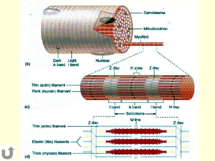
Microscopic Structure of Skel. Muscle muscle bundle of muscle fibers - bicep muscle fiber myofibril myofilaments (myosin and actin filaments) sarcomere – basic functional, contractile unit - myofibril

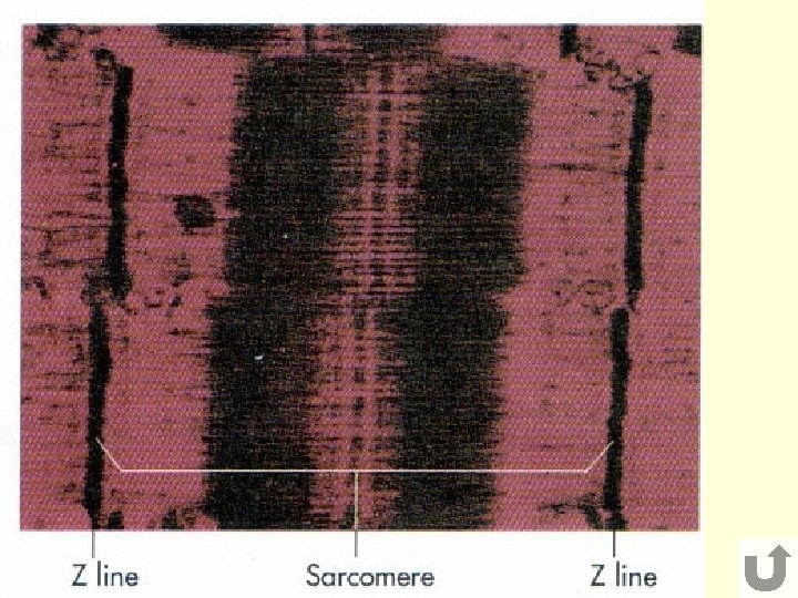
Skeletal Muscle Contraction Sliding Filament Model - when it contracts, the 2 filaments slide towards each other, therefore shortening the sarcomere and also the muscle 1. Z-Disk • connect sarcomere to next sarcomere 2. I-Band • area with ONLY actin filaments 3. A-Band • actin AND myosin present 4. H-Zone • myosin filaments ONLY

Links 1. Sliding Filament Theory 2. Sliding Filament Theory 3. Sliding Filament Theory

synergist prime mover antagonist
- Slides: 12