MUSCULAR SYSTEM Learning Objectives 1 To know basic

MUSCULAR SYSTEM

Learning Objectives 1. To know basic anatomy of muscle 2. Knowledge regarding nomenclature/ classification of muscles 3. Knowledge regarding basic facts of functioning of muscles

Muscles are responsible for all types of body movement – they contract or shorten and are the machine of the body

Three basic muscle types are found in the body ·Skeletal muscle ·Cardiac muscle ·Smooth muscle

Head and Neck Muscles Figure 6. 14 Copyright © 2003 Pearson Education, Inc. publishing as Benjamin Cummings Slide 6. 38

Trunk Muscles Figure 6. 15 Copyright © 2003 Pearson Education, Inc. publishing as Benjamin Cummings Slide 6. 39

Deep Trunk and Arm Muscles Figure 6. 16 Copyright © 2003 Pearson Education, Inc. publishing as Benjamin Cummings Slide 6. 40

Muscles of the Pelvis, Hip, and Thigh Figure 6. 18 c Copyright © 2003 Pearson Education, Inc. publishing as Benjamin Cummings Slide 6. 41

Superficial Muscles: Anterior Figure 6. 20 Copyright © 2003 Pearson Education, Inc. publishing as Benjamin Cummings Slide 6. 43

Superficial Muscles: Posterior Figure 6. 21 Copyright © 2003 Pearson Education, Inc. publishing as Benjamin Cummings Slide 6. 44

Čihák R. , Anatomie 1, Grada Publishing a. s. 2001

Power & Range- Muscle Contraction Maximal power generated by a muscle finally depends on effective mass of contractile tissue i. e number and diamentions of contained fibres Maximal range of contraction depends on length of its fibres Force and range acts at full advantage in parallel fibres
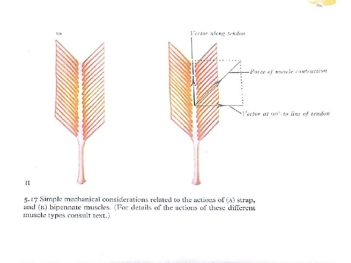

Classification Of Muscles A. By Fascicular Orientation 1. Parallel 2. Pennate 3 Spiral 4 Cruciate
 (a) Quadrilateral- Quadratus lumborum, Thyrohyoid")
1. Parallel ( Relative to muscle direction of pull) (a) Quadrilateral- Quadratus lumborum, Thyrohyoid (b)Long and strap like- Sartorius (c) Strap like with tendinous intersection Rectus abdominis (d ) Fusiform- Biceps brachii
 Unipennate – Flexor Pollicis longus (b)Bipennate- Rectus femoris, Dorsal interossei")
2. Pennate muscles (a) Unipennate – Flexor Pollicis longus (b)Bipennate- Rectus femoris, Dorsal interossei of hand (c )Multipennate - Deltoid (d)Circumpennate- Tibialis anterior

Classification Of Muscles 3. Spiral Supinator 4. Cruciate Sternocledomastoid, Masseter

Classification Of Muscles B. By Type Of Skeletal Muscle Fibre 1. Slow or Red fibres or type I fibres 2. Fast or White fibres or type II fibres

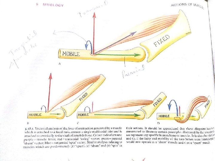
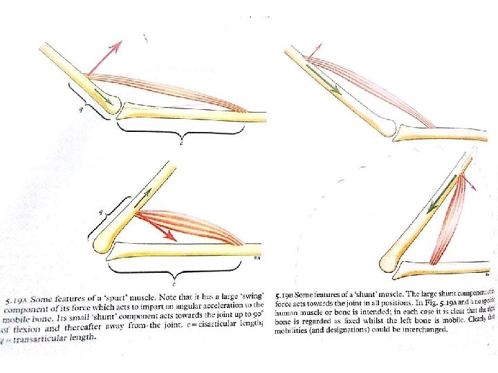
Classification Of Muscles C. By Insertion near or away from joint 1. Shunt Muscle( Away from Joint ) 2. Spurt Muscle ( Near Joint )

Nomenclature of Muscles On Basis of : 1. Shape of muscle Deltoid, Quadratus, Rhomboid, Lumbricals 2. Size Major , minor , longus , brevis 3. Number Of Head Biceps , triceps, Quadriceps femoris, Digastric

Nomenclature 4. Position Supraspinatus, Infraspinatus, Abdominis, Oculi, oris 5. Depth External oblique, Internal oblique Flexor D. Superficialis, Flexor D. Profundus

Nomenclature 6. Attachment : Sternocledomastoid, coracobrachialis 7. Action : Flexor, Extensor, Abductor

Connective Tissue Wrappings of Skeletal Muscle · Endomysium – around single muscle fiber · Perimysium – around a fascicle (bundle) of fibers Copyright © 2003 Pearson Education, Inc. publishing as Benjamin Cummings Figure 6. 1 Slide 6. 4 a

Connective Tissue Wrappings of Skeletal Muscle · Epimysium – covers the entire skeletal muscle · Fascia – on the outside of the epimysium Figure 6. 1 Copyright © 2003 Pearson Education, Inc. publishing as Benjamin Cummings Slide 6. 4 b

Skeletal Muscle Attachments · Epimysium blends into a connective tissue attachment · Tendon – cord-like structure · Aponeuroses – sheet-like structure · Sites of muscle attachment · Bones · Cartilages · Connective tissue coverings Copyright © 2003 Pearson Education, Inc. publishing as Benjamin Cummings Slide 6. 5

Function of Muscles · Produce movement · Maintain posture · Stabilize joints · Generate heat Copyright © 2003 Pearson Education, Inc. publishing as Benjamin Cummings Slide 6. 8

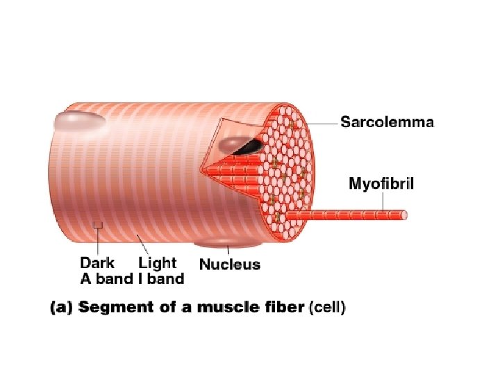
Microscopic Anatomy of Skeletal Muscle · Cells are multinucleate · Nuclei are just beneath the sarcolemma Figure 6. 3 a Copyright © 2003 Pearson Education, Inc. publishing as Benjamin Cummings Slide 6. 9 a

Microscopic Anatomy of Skeletal Muscle · Sarcolemma – specialized plasma membrane · Sarcoplasmic reticulum – specialized smooth endoplasmic reticulum Figure 6. 3 a Copyright © 2003 Pearson Education, Inc. publishing as Benjamin Cummings Slide 6. 9 b

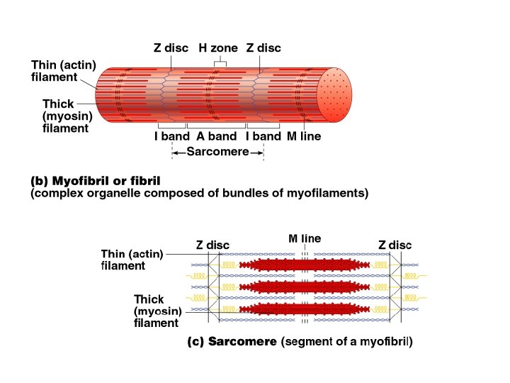
Microscopic Anatomy of Skeletal Muscle · Myofibril · Bundles of myofilaments · Myofibrils are aligned to give distrinct bands · I band = light band · A band = dark band Figure 6. 3 b Copyright © 2003 Pearson Education, Inc. publishing as Benjamin Cummings Slide

Microscopic Anatomy of Skeletal Muscle · Sarcomere · Contractile unit of a muscle fiber Figure 6. 3 b Copyright © 2003 Pearson Education, Inc. publishing as Benjamin Cummings Slide

Microscopic Anatomy of Skeletal Muscle · Organization of the sarcomere · Thick filaments = myosin filaments · Composed of the protein myosin · Has ATPase enzymes Figure 6. 3 c Copyright © 2003 Pearson Education, Inc. publishing as Benjamin Cummings Slide

Microscopic Anatomy of Skeletal Muscle · Organization of the sarcomere · Thin filaments = actin filaments · Composed of the protein actin Figure 6. 3 c Copyright © 2003 Pearson Education, Inc. publishing as Benjamin Cummings Slide
")
Microscopic Anatomy of Skeletal Muscle · Myosin filaments have heads (extensions, or cross bridges) · Myosin and actin overlap somewhat Figure 6. 3 d Copyright © 2003 Pearson Education, Inc. publishing as Benjamin Cummings Slide

Nerve Stimulus to Muscles · Skeletal muscles must be stimulated by a nerve to contract (motor neruron) · Motor unit · One neuron · Muscle cells stimulated by that neuron Copyright © 2003 Pearson Education, Inc. publishing as Benjamin Cummings Figure 6. 4 a Slide 6. 14

Muscle Response to Strong Stimuli · Muscle force depends upon the number of fibers stimulated · More fibers contracting results in greater muscle tension · Muscles can continue to contract unless they run out of energy Copyright © 2003 Pearson Education, Inc. publishing as Benjamin Cummings Slide 6. 22

Muscles and Body Movements · Movement is attained due to a muscle moving an attached bone Figure 6. 12 Copyright © 2003 Pearson Education, Inc. publishing as Benjamin Cummings Slide

Muscles and Body Movements · Muscles are attached to at least two points · Origin – attachment to a immoveable bone · Insertion – attachment to an movable bone Slide

Types of Muscle Contractions · Isotonic contractions · Myofilaments are able to slide past each other during contractions · The muscle shortens · Isometric contractions · Tension in the muscles increases · The muscle is unable to shorten Copyright © 2003 Pearson Education, Inc. publishing as Benjamin Cummings Slide 6. 28


Muscle Tone · Some fibers are contracted even in a relaxed muscle · Different fibers contract at different times to provide muscle tone · The process of stimulating various fibers is under involuntary control Copyright © 2003 Pearson Education, Inc. publishing as Benjamin Cummings Slide 6. 29

Effects of Exercise on Muscle · Results of increased muscle use · Increase in muscle size · Increase in muscle strength · Increase in muscle efficiency · Muscle becomes more fatigue resistant Copyright © 2003 Pearson Education, Inc. publishing as Benjamin Cummings Slide 6. 31

Types of Ordinary Body Movements · Flexion – decreases angle of joint and brings two bones closer together · Extension- opposite of flexion · Rotation- movement of a bone in longitudinal axis, shaking head “no” · Abduction/Adduction (see slides) · Circumduction (see slides) Copyright © 2003 Pearson Education, Inc. publishing as Benjamin Cummings Slide 6. 32

Types of Muscles · Prime mover – muscle with the major responsibility for a certain movement · Antagonist – muscle that opposes or reverses a prime mover · Synergist – muscle that aids a prime mover in a movement and helps prevent rotation · Fixators Copyright © 2003 Pearson Education, Inc. publishing as Benjamin Cummings Slide 6. 35
 ·")
Naming of Skeletal Muscles · Direction of muscle fibers · Example: rectus (straight) · Relative size of the muscle · Example: maximus (largest) Copyright © 2003 Pearson Education, Inc. publishing as Benjamin Cummings Slide

Naming of Skeletal Muscles · Location of the muscle · Example: many muscles are named for bones (e. g. , temporalis) · Number of origins · Example: triceps (three heads) Copyright © 2003 Pearson Education, Inc. publishing as Benjamin Cummings Slide

Naming of Skeletal Muscles · Location of the muscles origin and insertion · Example: sterno (on the sternum) · Shape of the muscle · Example: deltoid (triangular) · Action of the muscle · Example: flexor and extensor (flexes or extends a bone) Copyright © 2003 Pearson Education, Inc. publishing as Benjamin Cummings Slide 6. 37

Smooth Muscle Characteristics · Has no striations · Spindle-shaped cells · Single nucleus · Involuntary – no conscious control · Found mainly in the walls of hollow organs · Slow, sustained and tireless Copyright © 2003 Pearson Education, Inc. publishing as Benjamin Cummings Figure 6. 2 a Slide 6. 6

Cardiac Muscle Characteristics · Has striations · Usually has a single nucleus · Joined to another muscle cell at an intercalated disc · Involuntary · Found only in the heart · Steady pace! Copyright © 2003 Pearson Education, Inc. publishing as Benjamin Cummings Figure 6. 2 b Slide 6. 7

Disorders relating to the Muscular System • Muscular Dystrophy: inherited, muscle enlarge due to increased fat and connective tissue, but fibers degenerate and atrophy • Duchenne MD: lacking a protein to maintain the sarcolemma • Myasthemia Gravis: progressive weakness due to a shortage of acetylcholine receptors

Cardiac muscle tissue intercalated disc Eis, Jelínek, Špaček, Histopatologický atlas, Praha 2006

Abnormal contraction • spasm – involuntary contraction of one muscle • cramp – painful spasm • tetanus – multiple spasms of skeletal muscles • tic – involuntary twiches of muscles, usually under voluntary control • tremor – rhythmical, involuntary contractions of opposite groups of muscles • fasciculations – involuntary, short twiches on motor unit visible under the skin • fibrilace – spontaneous contractions of fibres of one muscle that aren´t visible under the skin
 – fibrous envelope of muscle")
Special muscle structures I • fascia (= perimysium externum) – fibrous envelope of muscle or muscle group – barrier for spreading of inflammation in that specific area • osteofascial septum (= septum osteofasciale) – fascial divider from the superficial fascia to the periosteum – separates the space for muscle groups – compartment (compartimentum) https: //www 2. aofoundation. org/wps/portal/!ut/p/c 0/

Growing old and musle tissue • skeletal muscle tissue starts to be replaced by fibrous and fatty tissue around the age of 30 • reflexes slowdown, loss of flexibility and decrease of strength • change of muscle fibres from quick to slow

Enthesopathy • illness of muscle and tendinous insertions • usually caused by repeated overstraining • e. g. tennis elbow http: //www. fyzioterapie-stepankavojtova. cz/bolestivyloket. html http: //compex. zdravi-cz. eu/tenisovy-loket. php

• Select the trait that does not characterize muscle tissue in general. • A) irritability • B) contractility • C) extensibility • D) All of these are traits of muscle.

• Individual fibers of skeletal muscle have fine sheath of connective tissue called a(n) ________. • A) epimysium • B) perimysium • C) endomysium • D) fascia
 A band to A band •")
• Sarcomeres run from _________. • A) A band to A band • B) Z line to Z line • C) H zone to H zone • D) I band to I band

• What muscle has its origin on the sternum and inserts on the mastoid process of the temporal bone? • A) sternocleiodomastoid • B) splenius capitis • C) semispinalis capitis • D) trapezius
 rectus")
• What is the deepest of the four abdominal muscles? • A) rectus abdominis • B) external abdominal oblique • C) transversus abdominis • D) internal abdominal oblique

• The _______ muscle is a deep, lateral muscle of the forearm that flexes the thumb joints and assists in grasping. • A) flexor pollicis longus • B) flexor carpi ulnaris • C) superficial digital flexor • D) deep digital flexor
 gluteus medius •")
• Which of these muscles is an adductor? • A) gluteus medius • B) tensor fascia lata • C) pectineus • D) iliacus

• Choose the muscle that does not belong to the quadriceps femoris group of the anterior thigh. • A) rectus femoris • B) vastus lateralis • C) vastus medialis • D) biceps femoris
 in the")
• The thenar and hypothenar muscles are located where? • A) in the foot • B) within the hand • C) in the forearm • D) in the lower leg
- Slides: 68