Muscular System Latin Root Words Latin Root Word


")

- Slides: 27
Muscular System
Latin Root Words Latin Root Word Definition Sarco Muscle Myo Muscle Epi- Above Peri- Around Endo- Inside -Um Structure Fascia Band Mere Part Reticulum Net -ase Enzyme Synergy working together Agonist Prime mover Antagonist Working against
Tissue Review Primary purpose of Muscles? Movement
Tissue Review Cardiac Skeletal Smooth
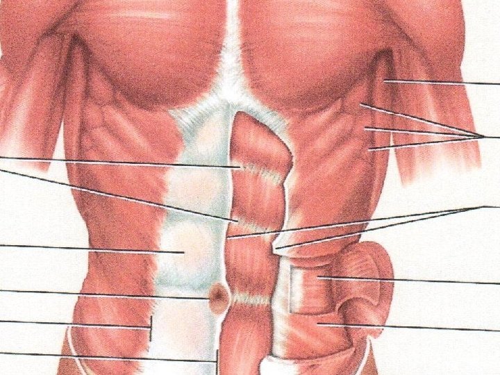
The Muscles: Each muscle is an organ comprised of skeletal Muscle Connective tissue coverings, _____tissue, several ______ Nervous Blood to _______ tissue to cause it to contract, and ______ nourish it. Example Question: Muscles are organs that are composed of what types of tissues?
Muscle Structure Quiz Muscles are organs that are made up of multiple tissues. The tissue that contracts and creates movement is the 1. ______ tissue. Word Bank: Blood Connective Muscle Surrounding the muscle are many different layers of 2. _______ tissue. 3. ______ tissue sends electrical impulses to the muscles that tell them to contract. Finally, 4. _____ tissue supplies the muscle with the resources it needs to contract. Nervous
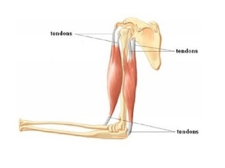
• Connective Tissue Coverings: A muscle has several dense connective coverings. Layers of dense connective tissue, called ____, Fascia surround and separate each muscle. This connective tissue extends beyond the ends of the muscle and gives Tendon that are fused to the periosteum rise to cord like ____ of bones. Sometimes muscles are connected to each other by broad aponeurosis sheets of connective tissue called __________
Aponeurosis
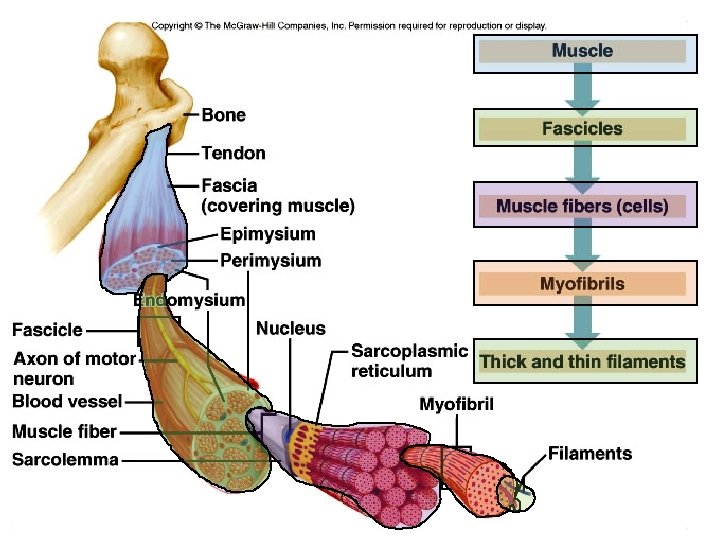
• Connective Tissue Coverings: Under the outer layer another layer of connective tissue around each whole muscle is called the epimysium (above the muscle) ______________ Perimysium (around the muscle) surrounds The ______________ individual bundles of muscle fibers called ______ Fascicles within each muscle Each muscles cell (fiber) is covered by a connective tissue Endomysium (inside the muscle) layer called ____________
Bone Epimysium Perimysium Tendon Endomysium Fascicle Perimysium Endomysium Muscle Fiber Fascicle Perimysium
• Skeletal Muscle Fibers Structure The muscle fiber membrane is called the Sarcolemma __________ which contains the cytoplasm sarcoplasm called _________. Within the sarcoplasm are many parallel Myofibrils __________composed of smaller filaments and Thin Filaments called Thick __________. These myofilaments are actually two types of filaments, a thicker filament composed of the protein myosin _______ and a thinner mostly made of the actin protein _______.
Muscles are attached to bones by 5. _____. The entire muscle is covered by a connective tissue called 6. ______. Directly underneath 6. is the 7. _________. Muscles are composed of many 8. _______ which are wrapped in a connective tissue called the 9. ________. Each 8. is made up of many 10. ______ that are wrapped up in the 11. ________. Word Bank Actin Endomysium Epimysium Fascia Fascicles Myofibrils Myosin Perimysium A 10. is filled with many parallel running fibers called 12. _______. These 12 are composed of thick and thin fibers. The thick fibers are known as 13. _______ and the thin fibers are known as 14. _______ Skeletal Muscle Fibers Tendon
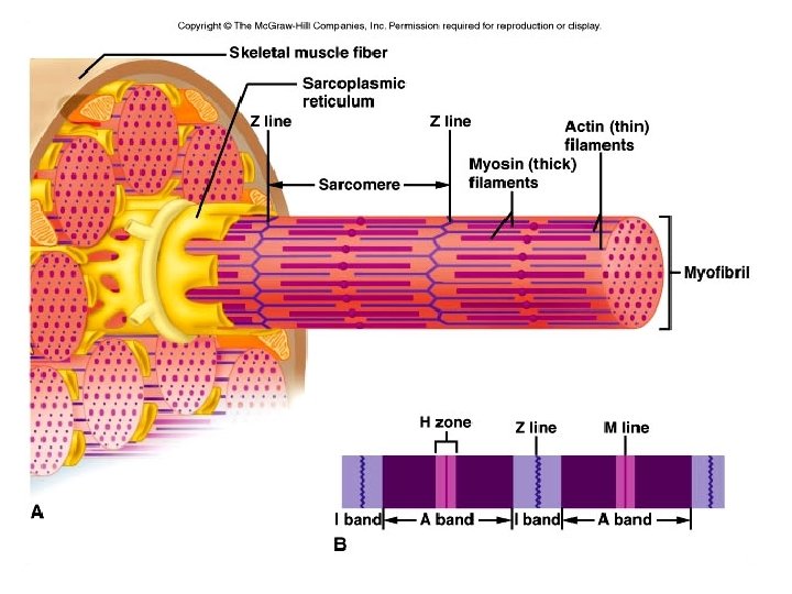
• Skeletal Muscle Fibers Structure A bands and the light The dark stripes are called ___ I bands are called _______. Sarcomere A ________ is defined as a unit extending from Z line one _____ line to the next (center of the light band. )
H Zone Z line I Band M line A Band Z line I Band
Sarcomere Anatomy • I Band = Light, Thin Actin Filaments only • A Band = Dark, Thick Myosin Filaments and Thin Actin Filaments • Z line = I band (actin filaments) connect to Z line • H Zone = Thick Myosin Filaments only • M Line = Thickening in the H zone
Muscle Identification • Use the text book to label the muscles in your packet. • You will need to begin memorizing these muscles. • Page 188 - 199
Daily Warm Up 01/22/2016 – 01/25/2016 • Match the connective tissue with the structure that it surrounds. 1. 2. 3. 4. D ___Epimysium a. Surrounds fascicles C ___Endomysium b. covers entire muscles and bones B ___Fascia c. surrounds individual muscle cells/ fibers A ___Perimysium d. surrounds muscle tissue • List the structures of a muscle from smallest(1) to largest(5). Myofibril, Muscle, Myosin and Actin Filaments, Muscle Cell/Fiber, Fascicle 1. Myosin and Actin Filaments 2. Myofibril 3. Muscle Cell/Fiber 4. Fascicle 5. Muscle
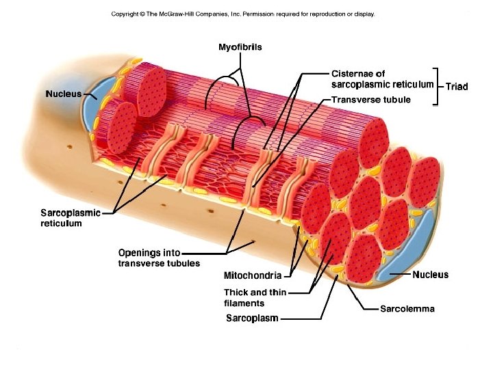
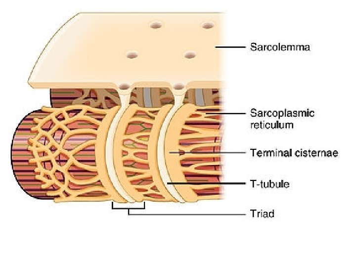
T Tubules • Where are they located? Around the myofibrils • Are they open or close to the outside? Open • What other tubular structure are they associated with? Sarcoplasmic Reticulum • What ion does this tubular structure contain? Calcium Ions (Ca 2+)
Skeletal Muscle Cell Parts • Sarcolemma: Skeletal muscle cell/fiber cell membrane • Transverse (T) Tubules • Invaginations of the sarcolemma • Used to communicate with sarcoplasmic reticulum • Sarcoplasm • Skeletal muscle cell/fiber cytoplasm • Sarcoplasm Reticulum: Skeletal muscle cell/fiber cell membrane that surrounds each myofibril • Terminal Cisternae • Enlarged areas of the sarcoplasmic reticulum that surround the T Tubules
4. 3. 5. 2. 6. 1. 7. 8. 9. 10. 1 1.