Muscular System Functions Movement Posture Stabilize Joints Generate

Muscular System

Functions: • Movement • Posture • Stabilize Joints • Generate Heat

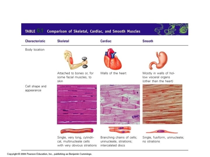
Types: • Skeletal- voluntary, striated, provides movement • Cardiac- involuntary, striated, in the heart • Smooth- involuntary, not striated, visceral (such as the digestive system)
 is wrapped by endomysium • Several")
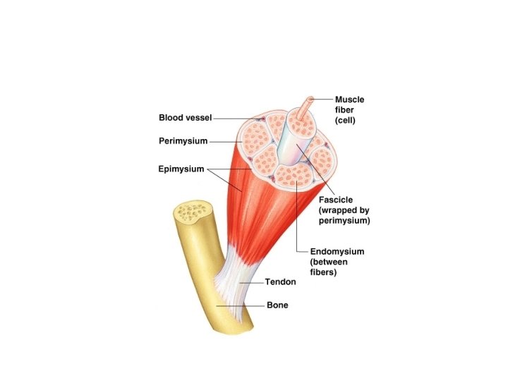
Anatomy of Skeletal Muscle • Muscle fiber (cell) is wrapped by endomysium • Several wrapped fibers are bundled and wrapped by perimysium • Each bundle is called a fascicle • Many fascicles are wrapped by tough epimysium • Epimysium ends in a cord like tendon or a sheet like aponeuroses
 • Made of many myofibrils covered by a plasma membrane called")
Muscle Fiber (Cell) • Made of many myofibrils covered by a plasma membrane called the sarcolema • Each myofibril has contractile units called a sarcomere

mm
 bands and light (I) bands • Z disc- midway")
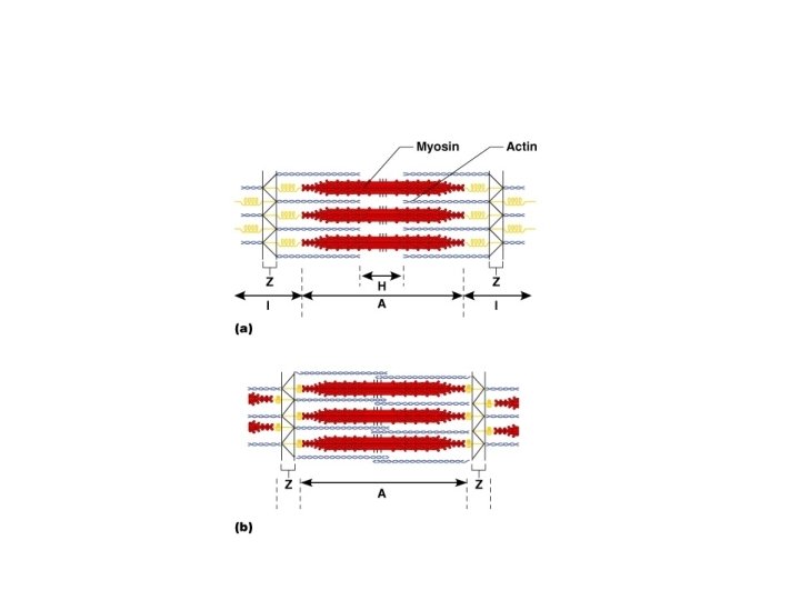
Sarcomere • Alternating dark (A) bands and light (I) bands • Z disc- midway in (I) band • H zone midway in (A) band • M line- center of H zone
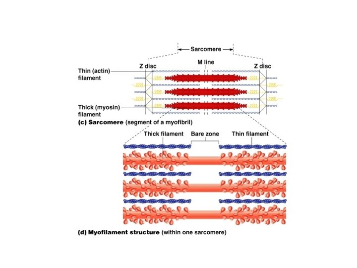

Sarcomere • 2 types of protein filaments: –Thick filaments= myosin –Thin filaments= actin • Cross bridges on myosin filaments connect the two

Sliding Filaments • Thin fibers are pulled to the center of the sarcomere by myosin head cross bridges • Triggered by rise in Ca+ which attaches to the myosin heads to pull it back • ATP releases the head and it springs forward and pushes the thin filaments over the thick filaments = Muscle Shortens
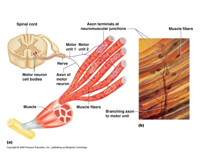

Skeletal muscle must be stimulated by a nerve • Motor Unit = one neuron and all the muscle cells it stimulates • More fibers stimulated by one neuron, the greater the force • Less fibers stimulated by one neuron, the finer the movement

Glucose is necessary to provide ATP • Muscles can store glucose as glycogen • Muscles contain myoglobin which will bind oxygen
 good for minutes •")
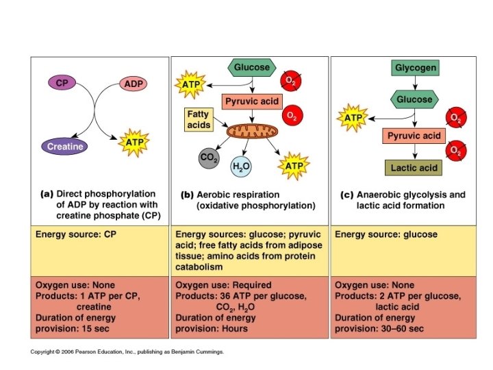
3 pathways to ATP production • 1. Creatine phosphate (CP) good for minutes • 2. Aerobic respiration= C 6 H 12 O 6 + O 2 CO 2 + H 2 O + ATP 95% of all ATP produced (36 -38 ATP/glucose) • 3. Anaerobic glycolysis= without oxygen, glucose is broken into 2 pyruvate acid, releases 2 ATP and a byproduct-lactic acid (causes muscle soreness)

Types of muscle contraction • Isotonic – contraction occurs and results in movement • Isometric- the myosin filaments try to contract but the muscle is against something immovable • Muscle tone- even when the muscle is relaxed, few of the muscle fibers continue to contract and make the muscle firm

Exercise effect • Aerobic- endurance – does NOT increase size – DOES resist fatigue – more blood hemoglobin, more mitochondria, more glycogen • more myoglobin- holds oxygen

Exercise effect continued • Isometric- resistance – Does increase size and strength – more myofilaments – more connective tissue • larger cells

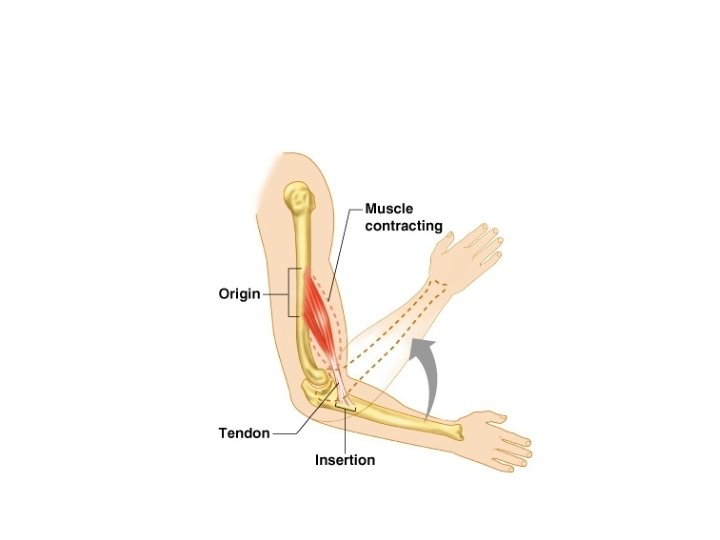
5 Golden Rules • 1. muscles cross at least one joint • 2. the bulk of the muscle lies proximal to the joint • 3. all muscles have at least two attachments: --origin and insertion • 4. muscles can only pull; never push • 5. during contraction, the insertion moves towards the origin
, oblique 2. Relative size: Maximus, Minimus")
Naming Muscles 1. Directions of fibers: Rectus (parallel), oblique 2. Relative size: Maximus, Minimus 3. Location over bone: Frontalis, Temporalis 4. Number of origins: Biceps, Triceps

5. Location of origin and insertion: sternocleidomastoid 6. Shape: Deltoid 7. Action: adductor, flexor

8. Prime Mover- major responsibility for movement 9. Antagonist- when prime mover is contracted, it is relaxed 10. Synergist- helper to prime mover 11. Fixator- special synergists that hold a muscle stable

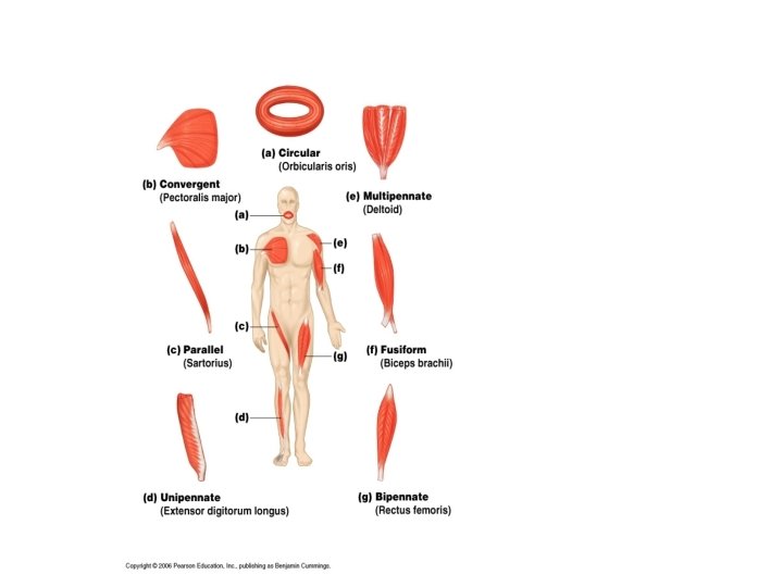
Shapes of Muscles Circular- sphincters Convergent- fan shaped Parallel- strap like Fusiform- special parallel shape with a big body • Pennate- feather like • •

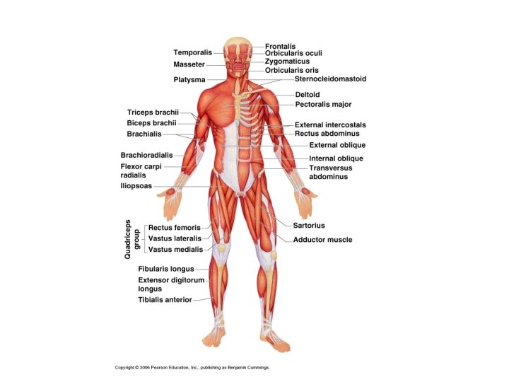
Muscles of the Head Frontalis- raises eyebrows Orbicularis oculi- blinking, squinting Zygomaticus- smile Depressor anguli oris- frown, (antagonists= smile/frown) • Lavator labii superioris- disgust • Depressor labii- pout • •
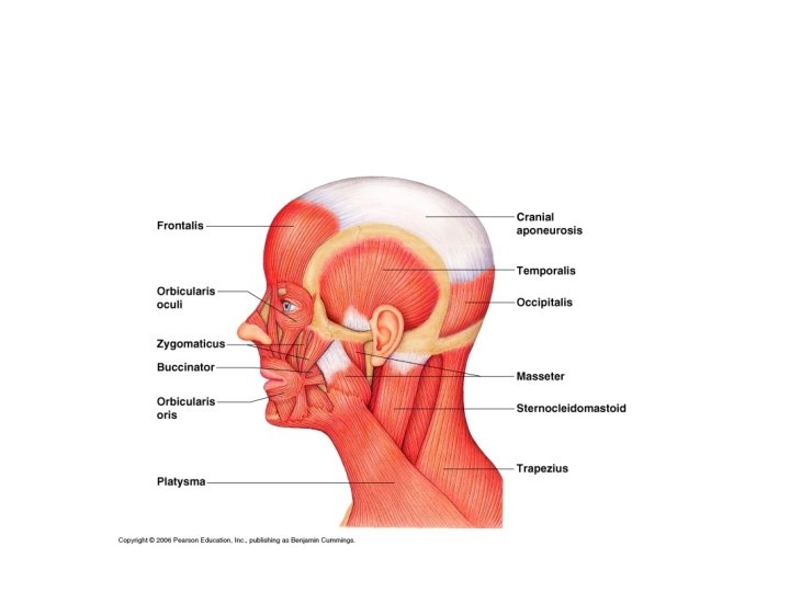
 • Masseter- prime mover (from zygomatic to ramus of mandible) • Temporalis-")
Chewing (Mastication) • Masseter- prime mover (from zygomatic to ramus of mandible) • Temporalis- synergist • Buccinator- holds food between teeth/sucking • Platysma- tenses skin of neck during shaving

Swallowing • Digastric- open mouth- depresses mandible • Mylohyoid- forces bolus into pharynx • Pharyngeal constrictors- propels bolus to esophagus

Neck • Sternocleidomastoid- head flexion, neck flex, rotate head towards opposite shoulder • Scalenes- flex and rotate neck

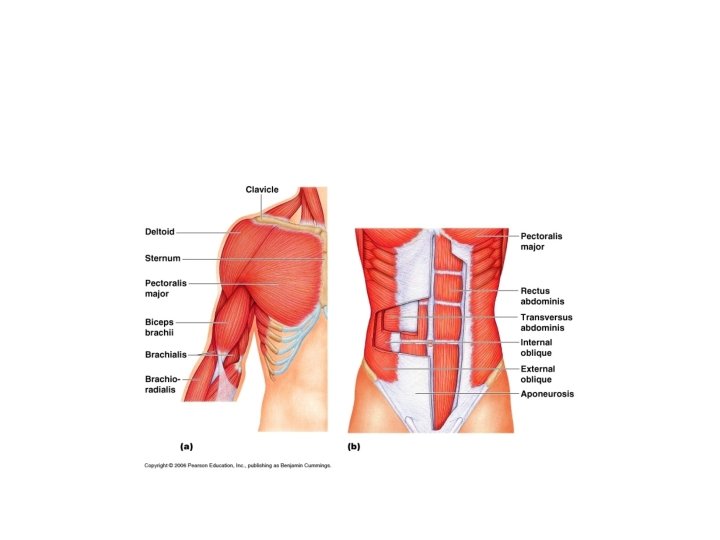
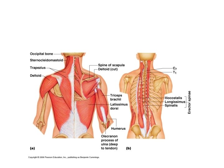
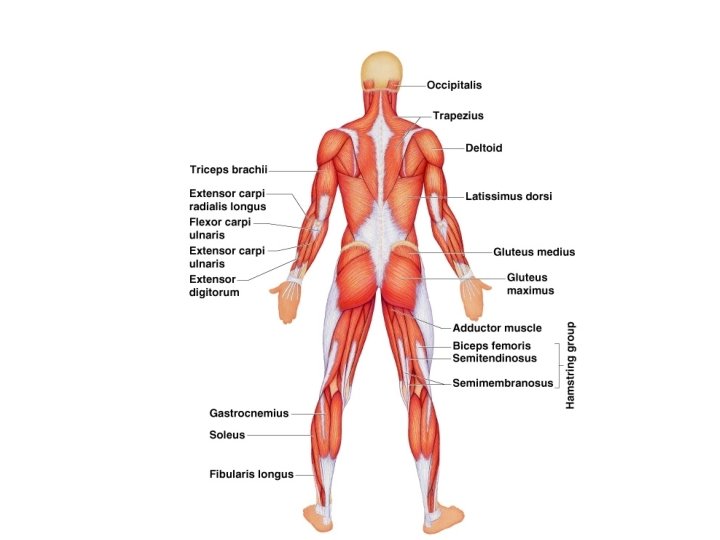
Shoulder Joint • Trapezius- depresses shoulder/adducts scapula • Pectoralis major- prime mover of arm flexion, adducts arm, rotates medial
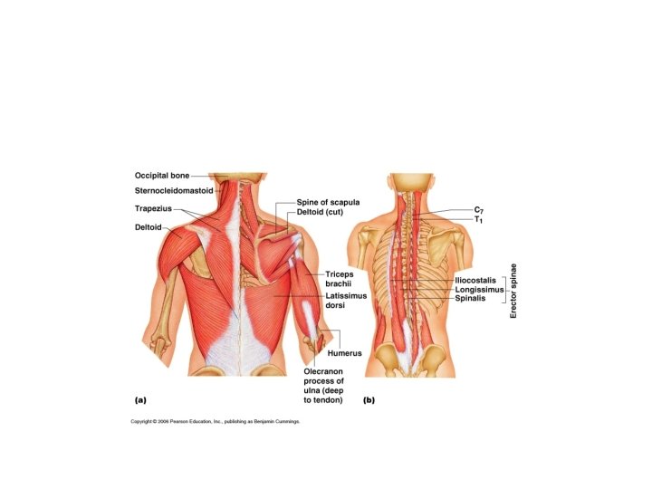

Shoulder continued • Latissimus dorsi- prime mover of arm extension, arm adductor brings arm down in hammering, swimming, rowing • Deltoid- antagonist of pectoralis major, and latissimus dorsi, prime mover of arm abduction, swinging arm movement while walking
 • Supraspinatus- prevents downward dislocation, assists in abduction • Infraspinatus- rotates")
Shoulder (rotator cuff) • Supraspinatus- prevents downward dislocation, assists in abduction • Infraspinatus- rotates humerus laterally • Teres major- medial rotation of humerus, extends • Also: Coracobrachialis • coracoid – humerus, flexion and adduction of humerus

Elbow Joint • Triceps brachii- antagonist to forearm flexors, strong forearm extensor, stabilizes shoulder • Biceps brachii- flexes elbow, supinates forearm, lifts radius • Brachialis- forearm flexor, lifts ulna • Brachioradialis- synergist to forearm flexion

Back Trunk • Erector Spinae- prime mover of back extension- made of 3 muscles: Iliocostalis, Longissimus, and Spinalis • Bending forward at the waist touching fingers to the floor they are relaxed (held together by ligaments). That is why lifting can be a problem
 • External intercostals- synergist to diaphragm in inspiration, elevates rib cage •")
Thorax (breathing) • External intercostals- synergist to diaphragm in inspiration, elevates rib cage • Diaphragm- prime mover of inspiration • Internal intercostals- aid in expiration, depresses the rib cage

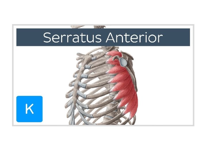
Thorax continued • Serratus anterior- “boxer’s muscle” raises the point of the shoulder, abduction and raising the arm, pushing, punching

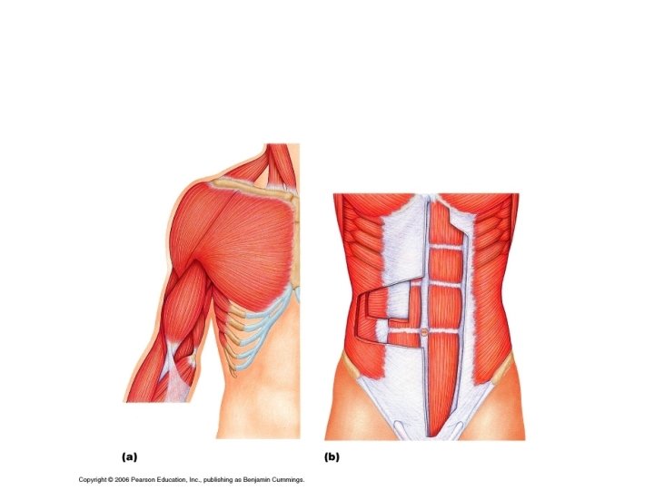
Abdominal Girdle • Rectus Abdominus- pubic crest to xiphoid process, flex and rotate lumbar vertebrae • External oblique- diagonal fibers, “sit-up”, flex abdomen • Internal oblique- under external oblique • Transverse abdominus- runs across and under obliques

Hip and Knee • Iliopsoas- prime mover of hip flexion • Sartorius- strap like, superficial, flexes and laterally rotates thigh, flexes knee, “cross leg position” • Adductor magnus: • Adductor longus: adducts thigh • Adductor brevis: • Gracilis- adducts thigh, medial rotation when walking
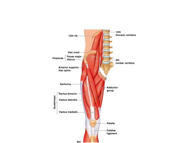
 • All extend knee: • Rectus Femoris: also flexes hip")
Quadriceps group (ant. Thigh) • All extend knee: • Rectus Femoris: also flexes hip on thigh • Vastus lateralis • Vastus intermedius • Vastus medialis

Hip and Thigh posterior • Gluteus Maximus- major, powerful, extensor of the thigh, (rising from chair, stairs, running) • Gluteus medius- injection site • Gluteus minimus- adducts thigh

Hamstring group • Posterior thigh, sciatic nerve runs through, powerful knee flexors, extends thigh: • Biceps Femoris- extends thigh, flexes knee, lateral rotation of leg when knee is flexed • Semitendinosus • Semimembranosus
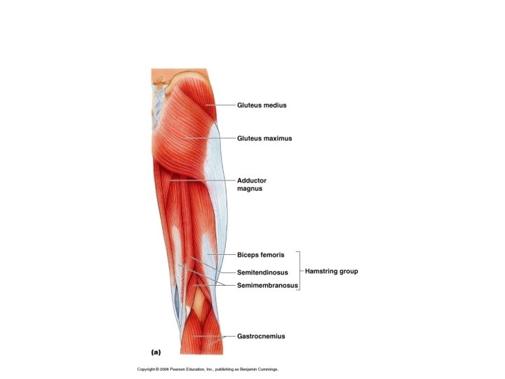

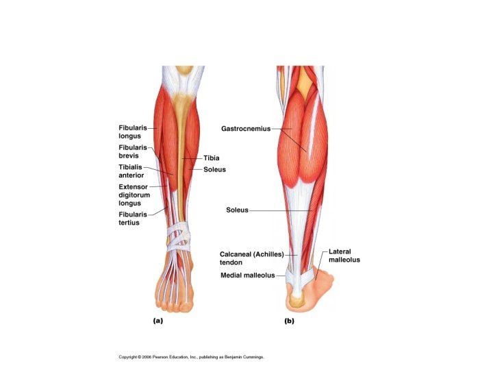
Lower Leg • Gastrocnemius- prominent belly forms proximal curve of the calf, plantar flexion, large Achilles Tendon (Calcaneal) • Soleus- lies under gastrocnemius, also plantar flexor
- Slides: 58