MUSCULAR SYSTEM Figure 50 37 Ballandsocket joint Head

MUSCULAR SYSTEM

Figure 50. 37 Ball-and-socket joint Head of humerus Hinge joint Pivot joint Humerus Scapula Ulna Radius

")
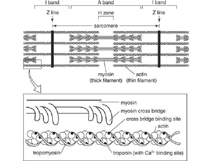
Figure 50. 26 a Muscle Bundle of muscle fibers Nuclei Single muscle fiber (cell) Plasma membrane Myofibril Z lines Sarcomere
 Thin filaments (actin) TEM")
Figure 50. 26 b Z lines Sarcomere Thick filaments (myosin) Thin filaments (actin) TEM Z line M line Sarcomere 0. 5 m Z line
 Myofibril Plasma membrane")
Figure 50. 30 a Synaptic terminal T tubule Sarcoplasmic reticulum (SR) Myofibril Plasma membrane of muscle fiber Axon of motor neuron Mitochondrion Sarcomere Ca 2 released from SR
 Myosin-binding sites")
Figure 50. 29 Ca 2 -binding sites Tropomyosin Actin Troponin complex (a) Myosin-binding sites blocked Ca 2 Myosinbinding site (b) Myosin-binding sites exposed
 ATP Thick filament")
Figure 50. 28 a-5 1 Thin filament Myosin head (lowenergy configuration) ATP Thick filament 5 Thin filament moves toward center of sarcomere. ADP Pi 4 Myosinbinding sites Actin Low-energy configuration ADP 2 Pi Cross-bridge High-energy configuration 3
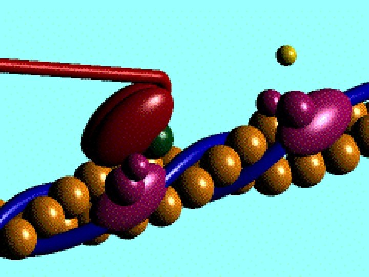
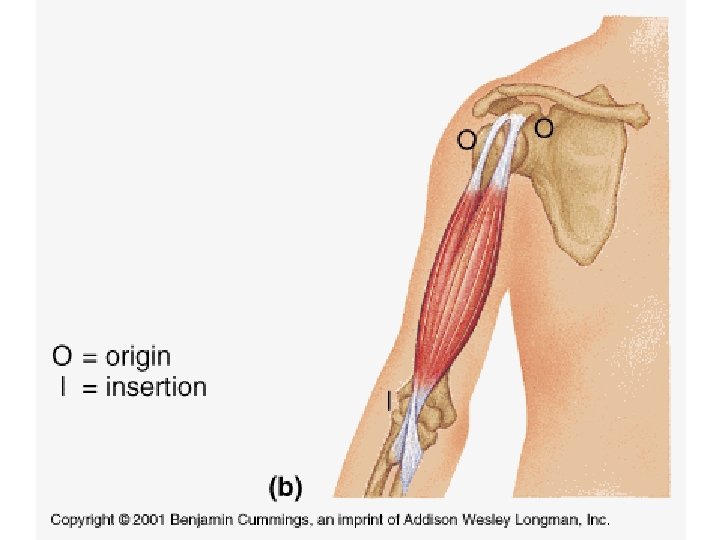

Figure 50. 31 Spinal cord Motor unit 1 Motor unit 2 Synaptic terminals Nerve Motor neuron cell body Motor neuron axon Muscle fibers Tendon
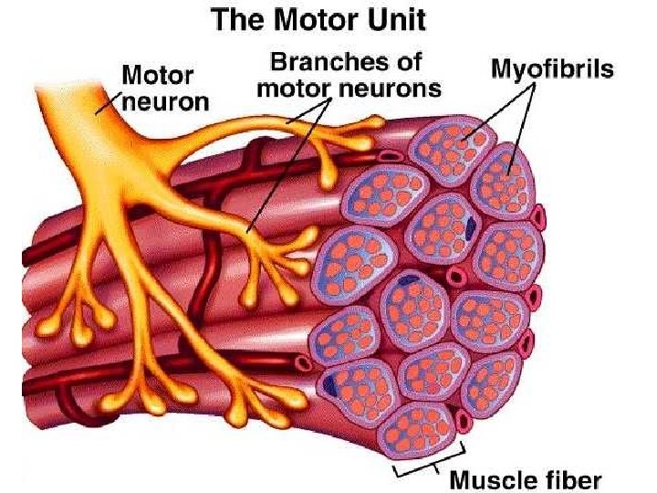

Figure 50. 30 b 1 Synaptic terminal of motor neuron T tubule Plasma membrane Synaptic cleft 2 ACh Sarcoplasmic reticulum (SR) Ca 2 pump 3 Ca 2 ATP 6 7 4 CYTOSOL Ca 2 5

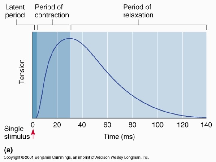

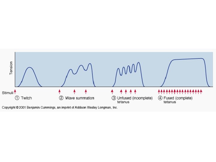
Figure 50. 32 Tension Tetanus Summation of two twitches Single twitch Action potential Time Pair of action potentials Series of action potentials at high frequency
- Slides: 29