Muscular System Anatomy Physiology II Tony Serino Ph


.")


 • A motor neuron is stimulated to fire an")

 • Calcium ions from SR flood the myofibrils")
")


")


- Slides: 53
Muscular System Anatomy & Physiology II Tony Serino, Ph. D. Biology Dept. Misericordia University
Muscular System • Functions: § Movement –generation of force and/or shortening § Maintenance of posture § Joint stabilization § Heat Generation § Attributes: § contractility, irritability, extensibility, and elasticity
Types of Muscle Cells Skeletal Muscle –voluntary, striated Cardiac Muscle –involuntary, striated Smooth Muscle –involuntary, no striations
Muscles wrapped with CT, that is continuous with tendon and periosteum
The elasticity of the CT sheaths, tendon and the muscle cells = the Series Elastic Component
Antagonistic Muscle Arrangement This arrangement plus the series elastic component allows the muscle to return to its original length.
Develop as a fusion of myoblasts, which accounts for multinucleated cells, extra myoblasts remain as satellite cells.
Skeletal Muscle Cells • Long, cylindrical, non-branching, multinucleated • 10 -100 mcm wide and up to 35 cm long • Voluntary, no spontaneous depolarization normally • Contractile proteins (myosin & actin) arranged in bundles called myofibrils
Unique Muscle Cell Structures Sarcomere
Each skeletal muscle cell must be innervated by a motor neuron to begin contracting.
Neuronal AP triggers release of ACh at neuromuscular junction (motor end plate).
ACh is released and diffuses across gap
ACh bind to the nicotinic receptor and triggers a MEPP
The MEPP triggers an AP that races along the sarcolemma and down the T-tubules. The depolarization affects the SR cisternae which releases Ca++ into the cytoplasm. The rise of intracellular Ca++ triggers the mechanical events of contraction.
Muscle Cell Contraction (Excitation-Contraction Coupling) • A motor neuron is stimulated to fire an AP • AP reaches synaptic terminal triggering an influx of Ca++ • The Ca++ stimulates the release of ACh • ACh diffuses across cleft and binds to nicotinic receptors in motor end plate • This causes Na+ channels to open; causing the generation of a MEPP • The MEPP triggers an AP along sarcolemma and into T-tubules • This deplorarizes the SR cisternae which releases stored Ca++ into the cytoplasm
Each myofibril consists of overlapping thick and thin filaments arranged in units called sarcomeres.
Muscle Contraction: Mechanical Events (Sliding Filaments) • Calcium ions from SR flood the myofibrils • This causes the thick and thin filaments to bind to each other (generates tension) and may cause them to slide past each other • This causes the sarcomere to shorten
Myofibril Anatomy H Band M Line Z Line (Cross section)
Overlapping Tick and Thin Filaments
Thick Filament Structure
Thin Filament Structure: Twisted bead chain of actin proteins Thin Filament: Actin, Tropomyosin and Troponin
Calcium is trigger
Contraction Events Detachment
Contraction Events Detachment Reset: energize myosin head
Contraction Events Detachment Reset Attachment
Contraction Events Detachment Reset Power Stroke Attachment
The H Band shrinks as the filaments slide past each other.
Muscle Contraction Review
Muscles are arranged as Motor Unit = 1 motor neuron + all the muscle fibers it controls (innervates) The size of the motor unit depends on the degree of control needed in that particular whole muscle.
Single Muscle Twitch
Treppe –an increase in tension development with no summation present; due to enzyme warming, increase blood flow, more Ca 2+ availability, etc. treppe
Biomechanics of Force Production • • Tension = force exerted on an object by a muscle Load = force exerted on muscle by the weight of an object Twitch = the mechanical response of a muscle to an AP Types of Contractions: • • Isometric = muscle increases tension without shortening Isotonic = muscle shortens with no further increase in tension Bicep Tension Load Fulcrum (pivot point) Weight of arm + object
Isotonic Contraction
Isometric Contraction
Factors Affecting Muscle Fiber Performance § Load –affects duration, degree and velocity of contraction • Increasing load decreases velocity § Frequency of stimulation § Initial Length of muscle fiber § Type of muscle fiber –fibers differ in strength, size, ATP splitting rate, and resistance to fatigue
Load Effect on Degree and Duration of Contraction
Load vs. Velocity of Contraction As load increases, velocity decreases.
Factors Affecting Muscle Fiber Performance § Load –affects velocity of contraction • Increasing load decreases velocity § Frequency of stimulation § Initial Length of muscle fiber § Type of muscle fiber –fibers differ in strength, size, ATP splitting rate, and resistance to fatigue
Increase frequency of stimulation allows tension to Mechanical add to previous contraction’s tension (Wave) Summation
Factors Affecting Muscle Fiber Performance § Load –affects velocity of contraction • Increasing load decreases velocity § Frequency of stimulation § Initial Length of muscle fiber § Type of muscle fiber –fibers differ in strength, size, ATP splitting rate, and resistance to fatigue
Initial Length of Muscle Fiber: affects the maximum tension that can be developed due to degree of overlap between thick and thin filaments
Factors Affecting Muscle Fiber Performance § Load –affects velocity of contraction • Increasing load decreases velocity § Frequency of stimulation § Initial Length of muscle fiber § Type of muscle fiber –fibers differ in strength, size, ATP splitting rate, and resistance to fatigue
Types of Muscle Fiber: each motor unit consists of only one type of muscle fiber • Slow twitch, red (oxidative) fibers (SO) –small diameter, weakest, slow ATPase, much myoglobin and mitochondria, abundant blood supply, fatigue resistant • Fast twitch, red (oxidative) fibers (FO) –medium diameter, moderate strength, fast ATPase, abundant mitochondria and myoglobin, good blood supply, moderate fatigue resistance • Fast twitch, white (glycolytic) fibers (FG) –largest diameter, great strength, fast ATPase, low amount of myoglobin or mitochondria, decreased blood supply, high in glycolytic enzymes, tire quickly
Control of Whole Muscle Tension dependent on: • Tension developed by each fiber – Dependent on fiber type, initial length and degree of wave summation • Amount of fibers stimulated to contract – The number of motor units responding is directly related to amount of tension produced – If the body needs more power, it recruits more motor units to respond – Known as recruitment (motor unit summation)
Energy Use: stored ATP in muscle used quickly so re-supply is crucial to function 1. Creatine Phosphate –quick re-supply, allowing time for aerobic respiration to gear up 2. Aerobic Respiration –oxidative phosphorylation dependent on adequate blood supply of oxygen, uses different sources for energy: a) b) c) 3. Stored glycogen Glucose and fatty acids from blood Fatty acids from blood Anaerobic Respiration -becomes dominant as need for oxygen exceeds ability of blood to transport it into muscles After exercise, energy continues to be consumed at increased levels to re-build reserves, etc. , this is part of the oxygen debt incurred during the exercise (defined as the amount of energy required to rebuild supplies used during the exercise (glycogen, creatine, proteins, etc. ))
Anaerobic threshold Aerobic threshold exceeded when the delivery of Oxygen is not enough to maintain aerobic metabolism. The perfusion of oxygen maximum (VO 2 max) is exceeded.
Lactic Acid Cycle
Fatigue –inability to maintain contraction tension even while being stimulated. Two kinds: • Primary Fatigue –due to accumulation of lactic acid in sarcoplasm, this changes the cytoplasm p. H and begins to change protein configurations which ends contraction. • Secondary Fatigue –related to the loss of energy reserves in the body, as seen in day after soreness. Why this triggers a low intensity pain signal (a dull ache) is unknown.
Cardiac Muscle Striated, single nucleus, branched cells, connected together by intercalated discs (with many gap junctions) Spontaneously contracts, needs no innervation, involuntary
Smooth Muscle No sarcomeres, therefore, no striations, single nucleated, small spindle shaped cells Spontaneously contracts, involuntary control, can remain contracted for long periods of time without fatiguing Two types: Visceral (single unit) –united by gap junctions Multi-unit –needs innervations, behaves like skeletal muscle (Ex. Iris)
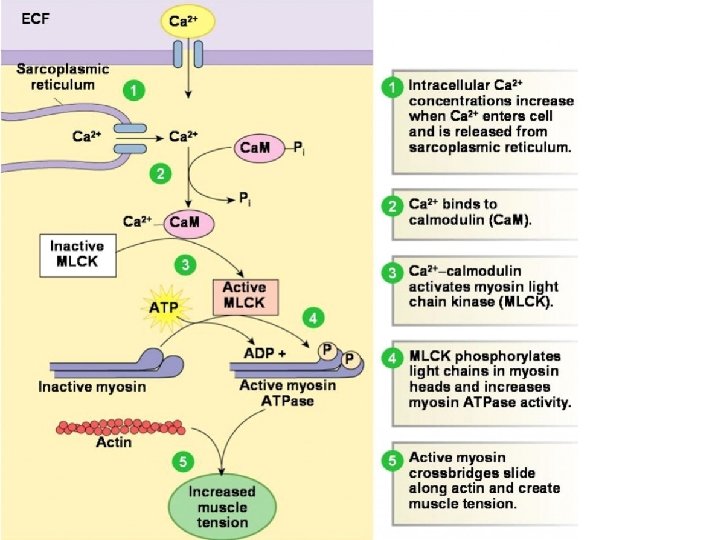
Smooth Muscle Contraction Control Contraction: increased cytosolic Ca++, and activation of MLCK State of contraction can be maintained without further use of ATP Relaxation: due to decrease cytosolic Ca++, deactivation of MLCK, and activation of MLCP
Visceral Smooth Muscle