Muscular and Skeletal System Powerpoint 2 Unit 8


 – Outer portion of")


 Muscle � Connects the various parts of the skeleton through one")


 types of protein")


- Slides: 23
Muscular and Skeletal System Powerpoint #2 Unit 8 – Chapters 35/36 Working together to create movement
Skeletal System Structures: Bones Cartilage Ligaments Tendons
Skeletal System Function: Supports body Protects internal organs Allows for movement Stores mineral reserves Provides a site for blood cell formation
Bones: 206 bones in the human body
Bones: 3 Parts Spongy bone – Not soft or spongy – Very strong – Structure resembles the supporting structure of bridges. – Strong but lightweight
Compact bone: – Very dense (no spaces like spongy bone) – Outer portion of bone – Contains Haversian canal for veins and arteries to run though
Bone Marrow: – Soft tissue – Found in bone cavities – Yellow Marrow: fat cells – Red marrow: makes red blood cells
8
Types of Muscle � � The human body is comprised of 324 muscles Muscle makes up 30 -35% (in women) and 42 -47% (in men) of body mass. Three types of muscle: Skeletal muscle Cardiac muscle Smooth muscle 9
A. Skeletal (Striated) Muscle � Connects the various parts of the skeleton through one or more connective tissue tendons � During muscle contraction, skeletal muscle shortens and moves various parts of the skeleton � Activated through signals carried to the muscles via nerves voluntary control � Repeated activation of a skeletal muscle can lead to fatigue � Can have many nuclei 10
Skeletal Muscles work in PAIRS Bending or straightening of elbow requires the coordinated movement of biceps and triceps muscles 11
B. Smooth Muscle � Located in the blood vessels, the respiratory tract, the iris of the eye, the gastro-intestinal tract � The � Is contractions are slow and uniform fatigue resistant � Activation � Has 12 is involuntary one nucleus
C. Cardiac Muscle � Has characteristics of both skeletal and smooth muscle � Functions to provide the contractile activity of the heart � Is very fatigue resistant � Activation of cardiac muscle is involuntary (like smooth muscle) � Can 13 have 2 nuclei, usually has 1
Components of skeletal muscle myofibril 14 muscle fiber bundle
Muscle Fibers � Cylinder-shaped � Each fiber is made up of a number of myofilaments � Diameter � Each of fiber activated via same nerve: motor unit fiber has capillaries that supply nutrients and eliminate waste � Divided 15 of fiber (0. 05 -0. 10 mm) Length of fiber (appr. 15 cm) fiber contains contractile machinery and cell organelles � Group � Each cells that make up skeletal muscle into functional units called sarcomeres
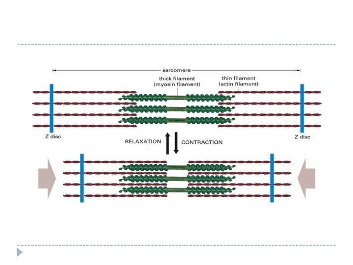
High microscope magnification of sarcomeres within a myofibril 16
Muscle Contraction �Organized �Two in series ( attached end to end) types of protein myofilaments: - Actin: thin filament - Myosin: thick filament �Projecting from each myosin are tiny contractile myosin bridges 17
Muscle Contraction �During muscle contraction the myofilaments myosin and actin slide toward each other and overlap. This shortens the sarcomere and the entire muscle. Muscle cells are "shocked" by nerve impulses from motor neurons.
Muscle Contraction The filaments slide together because myosin attaches to actin and pulls on it. Myosin head (H) attaches to actin filament (A), forming a cross bridge. After the cross bridge is formed the myosin head bends, pulling on the actin filaments and causing them to slide: Muscle contraction is a little like climbing a rope. The cross bridge cycle is: grab -> pull -> release, repeated over and over (a) At rest b) Contraction
Cartilage Tough, elastic, connective tissue Found in: ears, between bones, larynx, and other various places.
Tendons Connect Muscle to Bone
Ligaments Connect Bone to Bone