Muscles SAURABH MARU ASSISTANT PROFESSOR SCHOOL OF PHARMACY

Muscles SAURABH MARU ASSISTANT PROFESSOR SCHOOL OF PHARMACY & TECHNOLOGY MANAGEMENT, SVKM’S NMIMS, SHIRPUR

TYPES OF MUSCLES

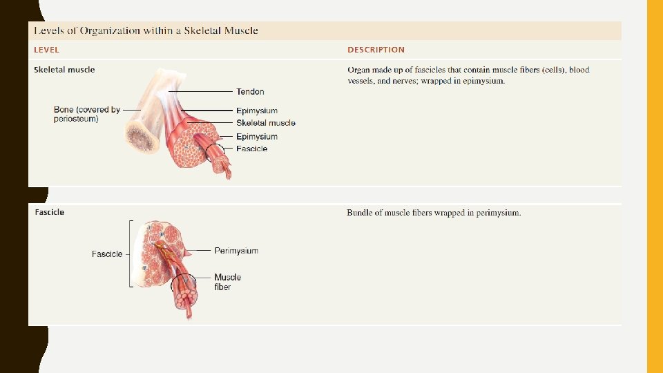
MICROSCOPY OF A SKELETAL MUSCLE FIBRE • Diameter of a mature skeletal muscle fiber: 10 to 100 micro. M avg. 10 cm (4 in. ) • Each skeletal muscle fiber arises during embryonic development from the fusion of a hundred or more small mesodermal cells called myoblasts • Each mature skeletal muscle fiber has a hundred or more nuclei. • Once fusion has occurred, the muscle fiber loses its ability to undergo cell division • Number of skeletal muscle fibers is set before birth

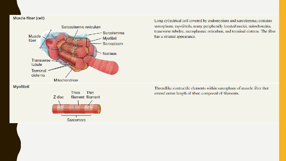
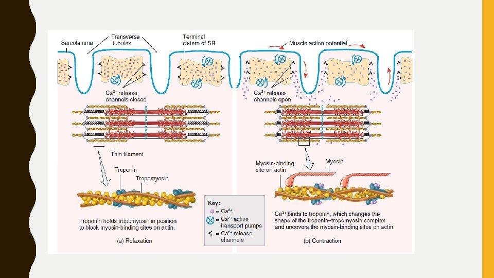
SARCOLEMMA, TRANSVERSE TUBULES, AND SARCOPLASM • Plasma membrane of a muscle cell: Sarcolemma • Thousands of tiny invaginations of sarcolemma: transverse (T) tubules (tunnel) • Muscle action potentials travel along the sarcolemma and through the T tubules • Within sarcolemma is sarcoplasm- cytoplasm of a muscle fiber. • Sarcoplasm have glycogen- used for synthesis of ATP. • Contains a red-coloured protein called myoglobin - binds oxygen molecules that diffuse into muscle fibers from interstitial fluid.

• Myoglobin releases oxygen when it is needed by the mitochondria for ATP production. • mitochondria lie in rows throughout the muscle fiber, close to contractile muscle proteins that use ATP during contraction
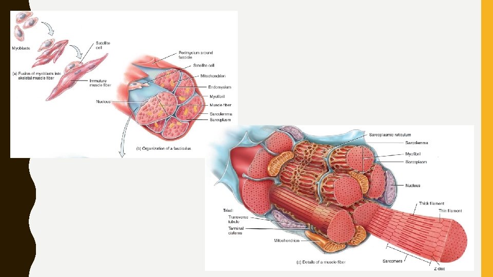

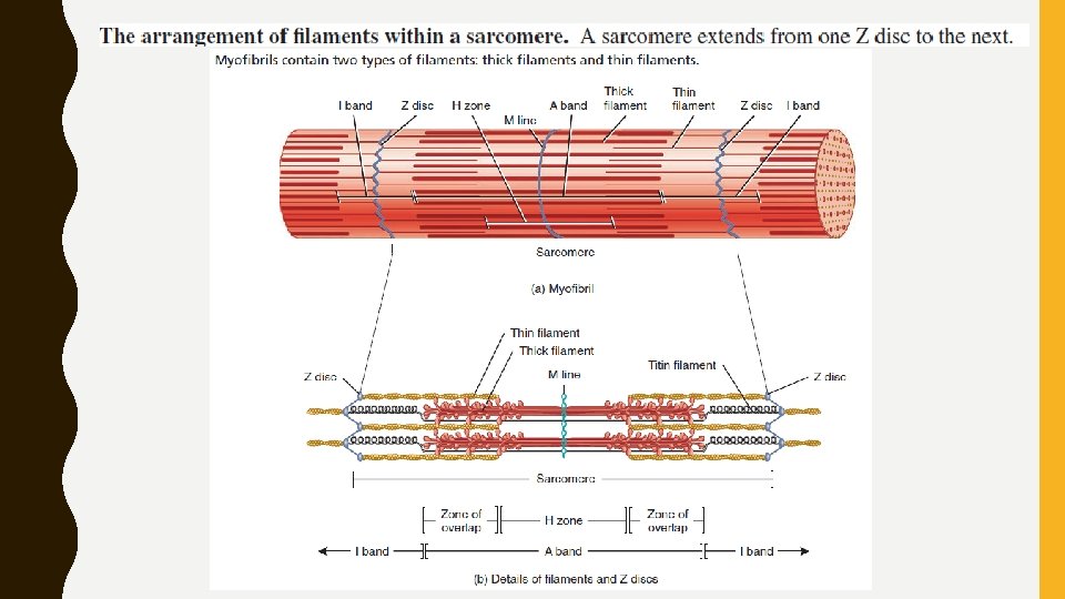
Myofibrils and Sarcoplasmic Reticulum • Sarcoplasm appears stuffed with little threads –myofibrils contractile organelles of skeletal muscle • Extend the entire length of a muscle fiber- skeletal muscle fiber appear striped (striated) • A fluid-filled system of membranous sacs –sarcoplasmic reticulum encircles each myofibril - similar to smooth endoplasmic reticulum in non-muscular cells. • Stores calcium ions (Ca 2++).
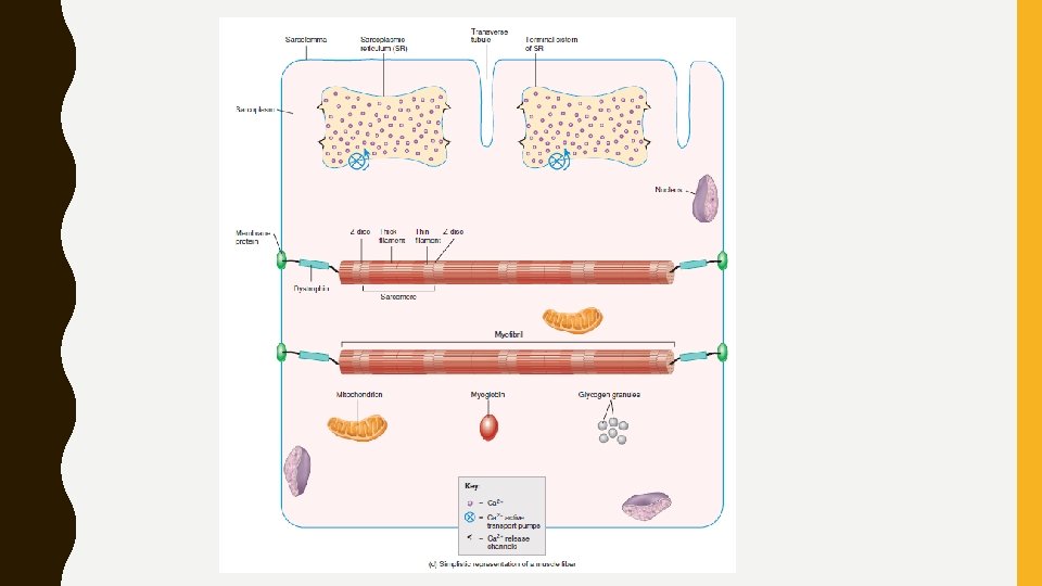

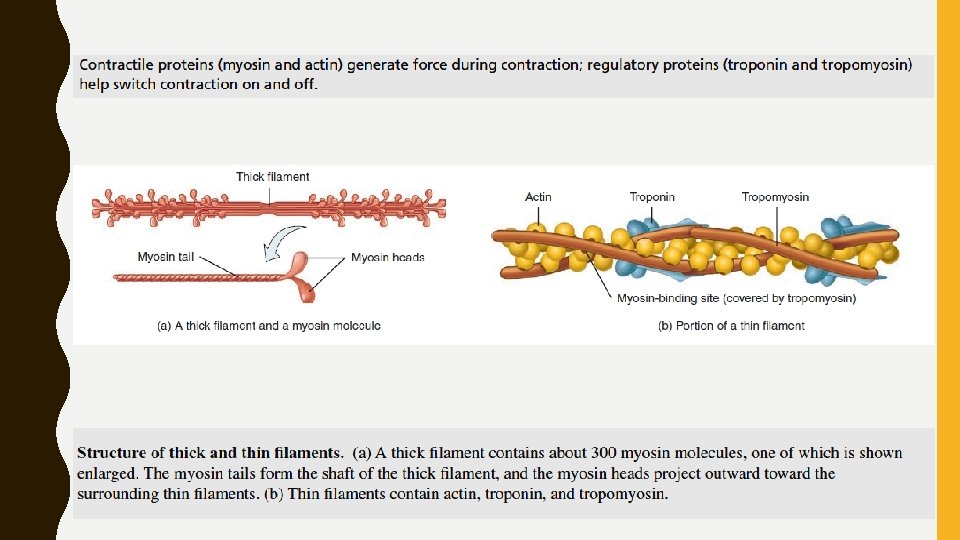
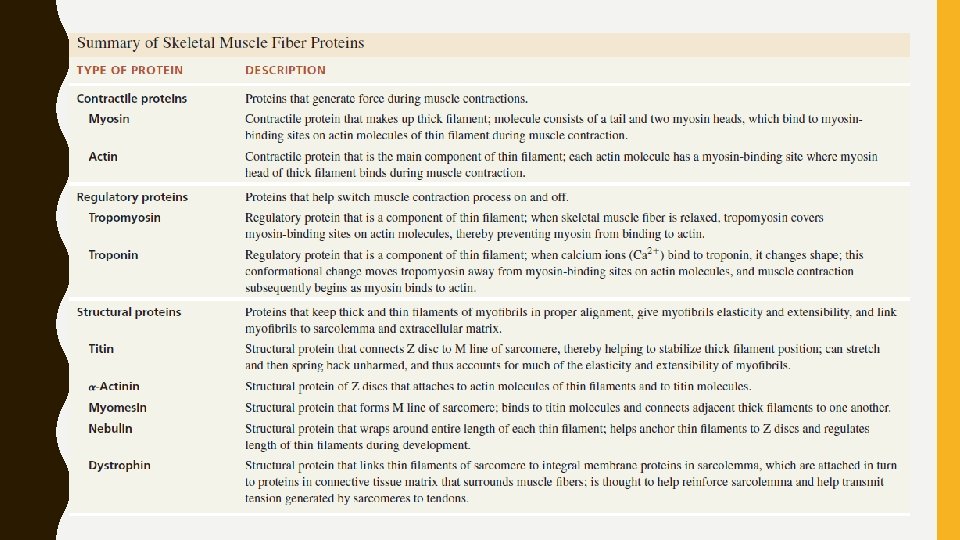
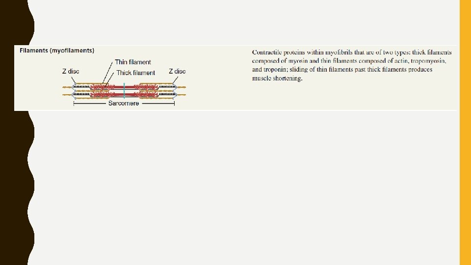
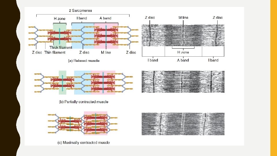
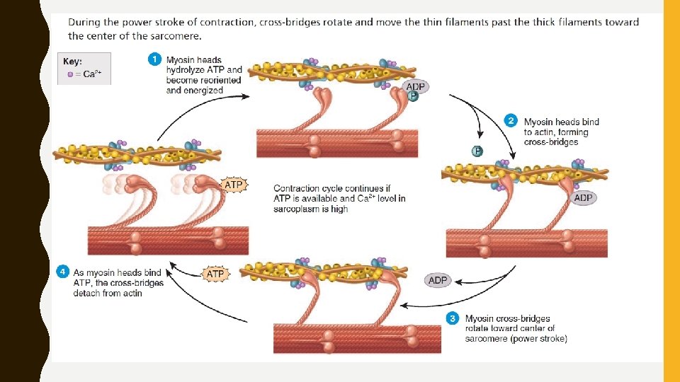
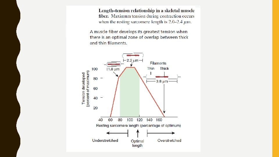
Filaments and the Sarcomere • Smaller protein structures within myofibrils: filaments or myofilaments • Thin filaments: 8 nm in diameter and 1– 2 m long – composed of protein actin, • Thick filaments: 16 nm in diameter and 1– 2 m long – composed of protein myosin. • Both involved in contractile process. • There are two thin filaments for every thick filament in regions of filament overlap. • Arranged in compartments called sarcomeres - basic functional units of a myofibril

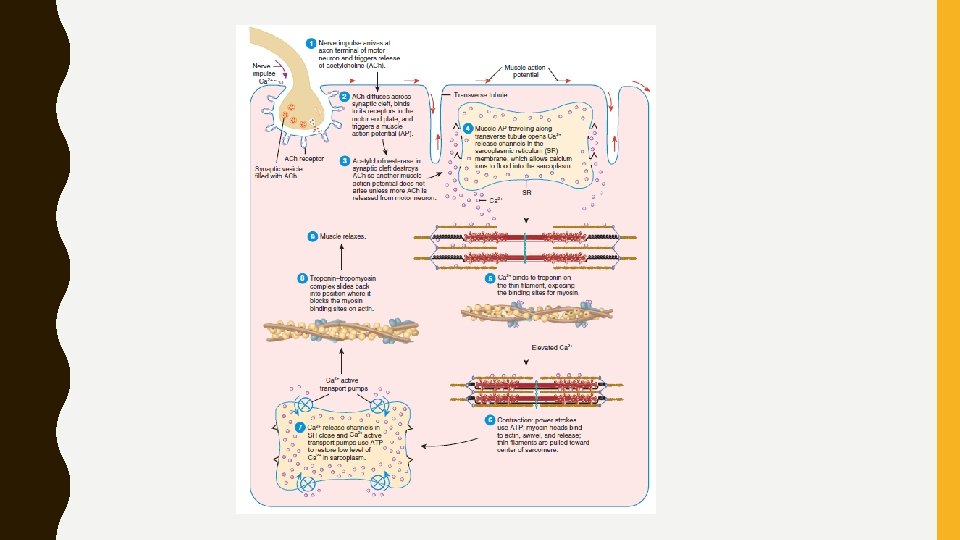
Neuromuscular Junction i. iii. iv. Release of acetylcholine Activation of ACh receptors Production of muscle action potential Termination of ACh activity: By ACh. E

Thank you
- Slides: 22