Muscles of Thigh Lecture Objectives List the muscles

Muscles of Thigh

Lecture Objectives • List the muscles of the thigh. • Describe the attachments of the thigh muscles and their nerve supply. • Describe the femoral triangle. • Describe the femoral sheath and its contents.

Thigh compartments

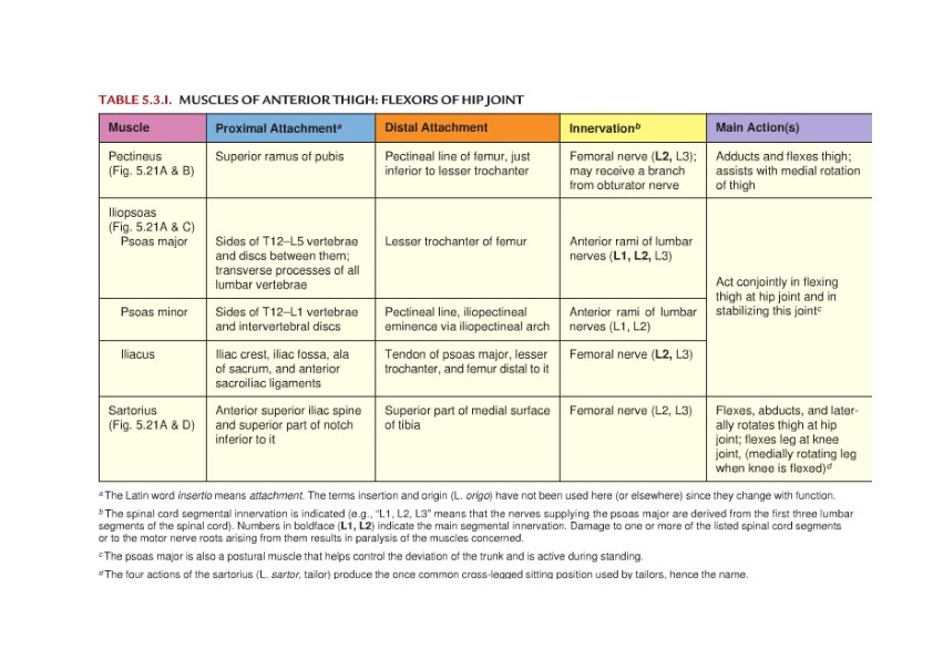
Anterior Thigh Muscles Mainly femoral nerve • Flexors of the hip • Pectineus m. • Adduction & flexion • Iliopsoas m. • Iliacus m. • Psoas major m. Major flexor • Sartorius m. • Longest • Flex hip & knee

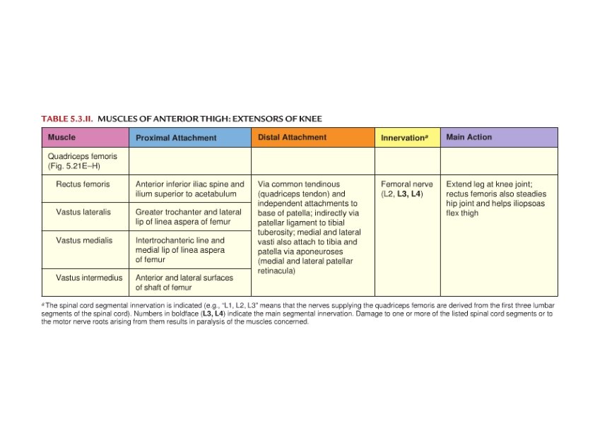
Anterior Thigh Muscles • Extensors of the knee • Quadriceps femoris m. • Rectus femoris m. • Can flex the hip • Vastus medialis m. • Vastus intermedius m. • Articularis genu • Vastus lateralis m.

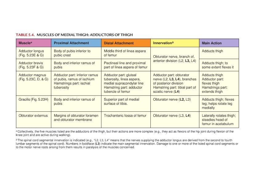
Medial Thigh Muscles Mainly obturator nerve Adductor group • Adductor longus m. • Most anterior • Adductor brevis m. • Deep to pectineus & adductor longus

Medial Thigh Muscles • Adductor magnus m. • Most posterior • Adductor hiatus Adductor part Hamstring part Sciatic nerve

Medial Thigh Muscles • Gracilis m. • Most medial and superficial • Crosses hip & knee joints • Obturator externus m. • Lateral rotation

Femoral Triangle: Boundaries • Superior • Lateral • Medial Floor Iliopsoas Pectineus

Femoral Triangle: Content • Lateral Medial • Nerve – artery – vein – lymphatics • Femoral sheath • Continuation of • Trnasversalis fascia – anteriorly • Iliacus fascia ‐ posteriorly • Content • artery – vein – lymphatics • Femoral canal • Femoral ring
 Canal • Location • Boundaries • Anteromedial – Sartorius • Posterior –")
Adductor (Subsartorial) Canal • Location • Boundaries • Anteromedial – Sartorius • Posterior – longus & magnus • Lateral – Vastus medialis • Content

Femoral Artery • Its entrance to the thigh • Position • Midway between ASIS and pubic symphysis • Femoral sheath • Relations • Sartorius • Iliopsoas & adductor muscles • Femoral vein and nerve • Exit to popliteal region • Adductor hiatus • Profunda femoris a. (deep a. of thigh) (thigh region) • Deep to the adductor longus

Femoral Nerve • Largest branch of the lumbar plexus • Relations • • Psoas m. Iliacus m. Inguinal ligament Femoral sheath

Saphenous Nerve • Cutaneous branch of the femoral nerve • Relations In Femoral triangle Within Adductor canal Cross Femoral a. Between Sartorius & gracilis tendons • Companies great saphenous v. • Anterior to Medial malleolus • •

Obturator Nerve: Divisions • Anterior to obturator externus & adductor brevis mm. • Posterior • Traverse obturator externus m. • Posterior to adductor brevis m. • Anterior to adductor magnus m.

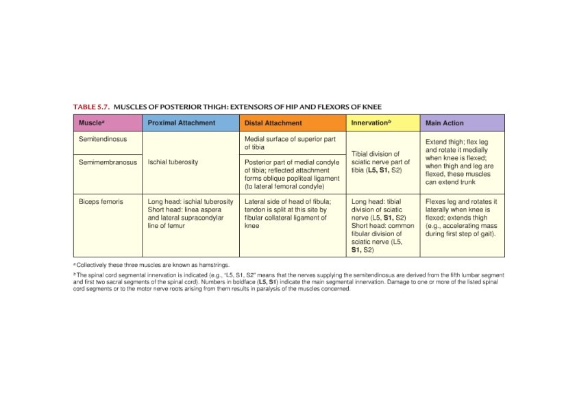
Posterior Thigh Muscles Hamstring muscles • Extend hip & flex knee • Tibial division of sciatic nerve • Semitendinosus m. • Semimembranosus m. • Biceps femoris m. • Long head • Short head • Fibular division of the sciatic nerve

Sciatic Nerve: Relations • Greater sciatic notch • Piriformis m. • Gluteus maximus m. • Biceps femoris m. • At the superior part of the popliteal fossa divides into its terminal branches • Tibial n. • Common peroneal n.

Surface Anatomy of Femoral Triangle

Surface Anatomy of Anterior Thigh • Femoral triangle • Femoral artery • Femoral vein • Femoral nerve • Patellar ligament

Catheterization • Femoral artery catheterization • Midway between the ASIS and the symphysis pubis • Just below the inguinal ligament • Femoral vein catheterization • Just medial to the felt femoral artery • High incidence of thrombosis

Surface Anatomy Sciatic Nerve • Location of sciatic nerve • Line between greater trochanter and ischial tuberosity • Down middle of posterior thigh • Tibial nerve • Middle popliteal fossa • Common fibular nerve • Tendon of biceps femoris muscle

Surface Anatomy of Posterior Thigh
- Slides: 26