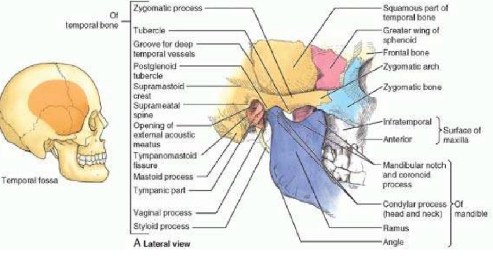
Muscles of mastication Muscles of mastication TEMPOROMANDIBULAR JOINT

Muscles of mastication


Muscles of mastication
 is a modified hinge type of synovial joint, permitting movement")
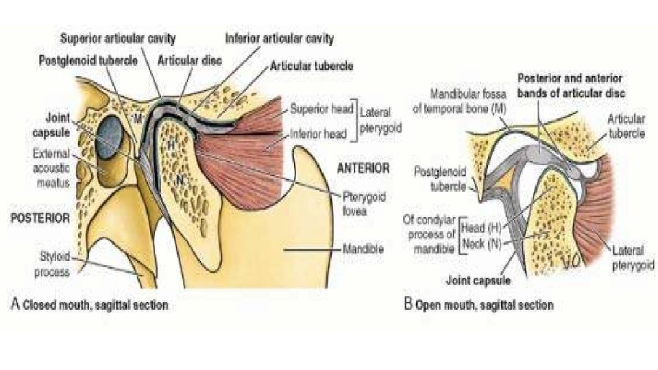
TEMPOROMANDIBULAR JOINT The (TMJ) is a modified hinge type of synovial joint, permitting movement in three planes. The articular surfaces involved are the head of the mandible, the articular tubercle of the temporal bone, and the mandibular fossa The articular surfaces of the TMJ are covered by fibrocartilage rather than hyaline cartilage as in a typical synovial joint. An articular disc divides the joint cavity into two separate synovial compartments. The joint capsule of the TMJ is loose. The fibrous layer of the capsule attaches to the margins of the articular area on the temporal bone and around the neck of the mandible. The thick part of the joint capsule forms the intrinsic lateral ligament (temporomandibular ligament), which strengthens the TMJ laterally and, with the postglenoid tubercle, acts to prevent posterior dislocation of the joint.

Two extrinsic ligaments and the lateral ligament connect the mandible to the cranium. 1. The stylomandibular ligament , a thickening of the fibrous capsule of the parotid gland, runs from the styloid process to the angle of the mandible It does not contribute significantly to the strength of the TMJ. 2. The sphenomandibular ligament runs from the spine of the sphenoid to the lingula of the mandible.
 Temporalis, masseter,")
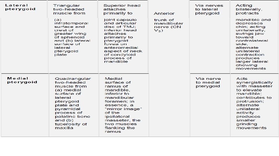
MOVEMENTS AT THE TEMPOROMANDIBULAR JOINT Movements of Mandible Muscles Elevation (close mouth) Temporalis, masseter, and medial pterygoid Depression (open mouth) Lateral pterygoid and suprahyoid and infrahyoid muscles. Protrusion (protrude chin) Lateral pterygoid, masseter, and medial pterygoid Retrusion (retrude chin) Temporalis (posterior oblique and near horizontal fibers) and masseter Lateral movements (grinding and chewing) Temporalis of same side, pterygoids of opposite side, and masseter. . Nerve: Auriculotemporal nerve, masseteric nerve (v 3). . Artery: Superficial temporal artery.

Dislocation of TMJ Sometimes during yawning or taking a large bite, excessive contraction of the lateral pterygoids may cause the heads of the mandibles to dislocate anteriorly, by passing anterior to the articular tubercles. In this position, the mandible remains depressed and the person may not be able to close the mouth.

24 October 2016 Dr. Aiman Q Afar 12
- Slides: 12