
Muscles of Head Neck By Dr Pardeep Kumar
Muscles of Head & Neck By Dr. Pardeep Kumar
Head and Neck Muscles � There are two main groups of head muscles. �Facial muscles Inserted into soft tissues Pull on the skin of the face �Chewing muscles) muscles (Masticatory Responsible for breaking down the foods we eat

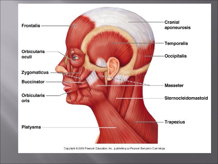
FACIAL epicranial aponeurosis MUSCLES frontalis occipitalis masseter buccinator risorius

Frontalis muscle - Facial Originates at the cranial aponeurosis and inserts to the skin of the eyebrows. � Raises your eyebrows. � Wrinkles your forehead �
Orbicularis Oculi - Facial � � � � It is the sphincter muscle of eyes. Fibers run circles around the eyes. Allows you to close your eyes, squint, blink, and wink. Origin: Frontal bone and maxilla Insertion: Tissue around eyes Action: Blinks and closes eyes Flow of tears by helping to empty the lacrimal sac.
Orbicularis Oris � � � Fibers run circles around the mouth. Closes the mouth and protrudes the lips Often called the “kissing” muscle. Origin: Mandible and Maxilla � Insertion: Skin and muscle around mouth � Action: Closes and protrudes lips. �
Buccinator – Facial and Chewing Runs horizontally across the cheek and inserts into the orbicularis oris. � Flattens the cheek (whistling or blowing a trumpet). � Compresses the cheek to hold the food between the teeth during chewing. �
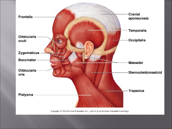
Zygomaticus - Facial Extends from the corner of the mouth to the cheekbone. � Called the smiling muscle because it raises the corners of the mouth upward. �
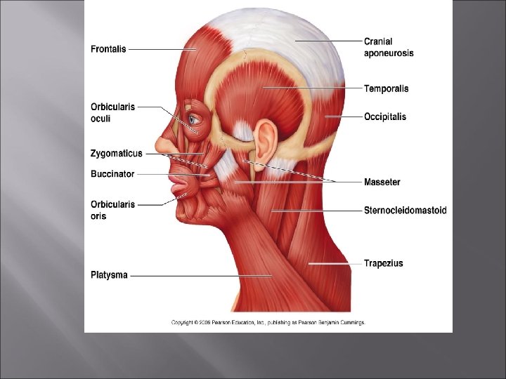
Masseter – Chewing Covers the angle of the lower jaw. � Runs from the temporal bone to the mandible. � Closes the jaw by elevating the mandible. �
Temporalis – Chewing � � � Fan-shaped muscle overlying the temporal bone. Inserts into the mandible. Acts as synergist of the masseter in closing the jaw. Origin: Floor of Temporal fossa Insert: Coronoid process of Mandible Action: Closes jaw
Medial pterygoid � � Origin : - pterygoid process of sphenoid bone Insertion : -medical surface near angle of mandible. Action : -pulls chin at opporite side Produce grinding motion
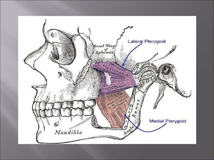
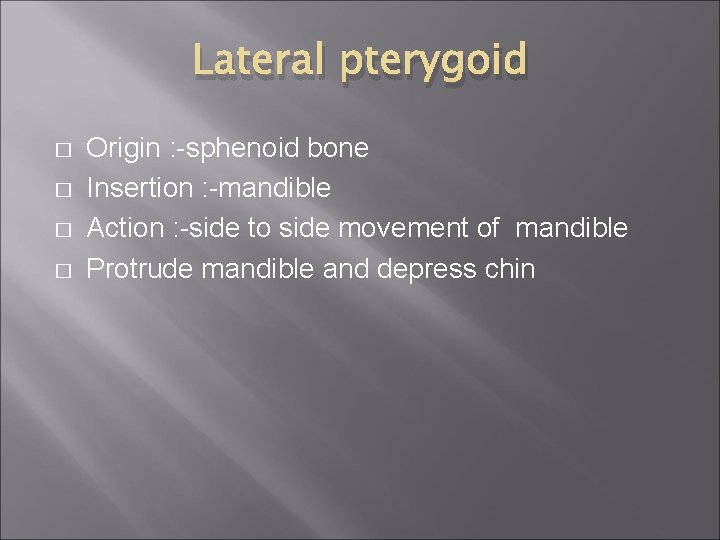
Lateral pterygoid � � Origin : -sphenoid bone Insertion : -mandible Action : -side to side movement of mandible Protrude mandible and depress chin
Muscles of Neck are divided into 4 groups 1 - superficial 2 - Suprahyoid 3 - Infrahyoid 4 - Deep �
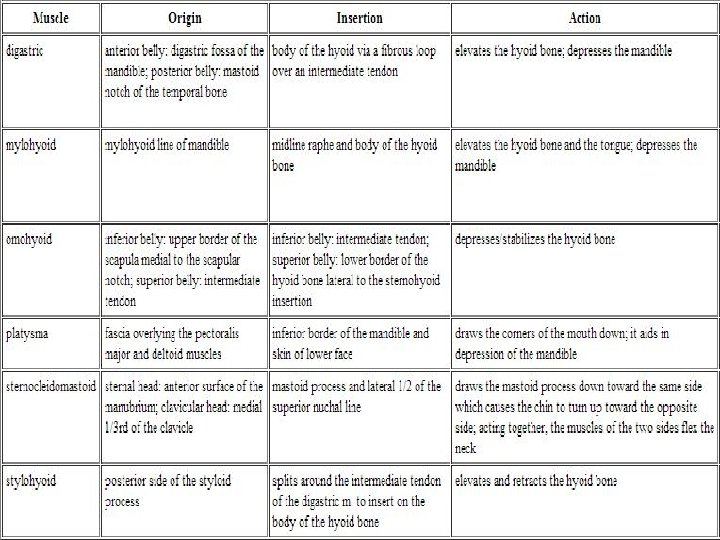
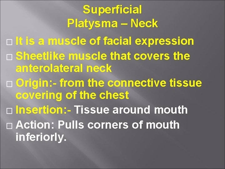
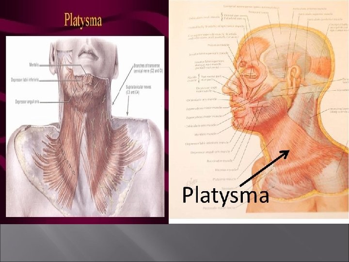
Superficial Platysma – Neck � It is a muscle of facial expression � Sheetlike muscle that covers the anterolateral neck � Origin: - from the connective tissue covering of the chest � Insertion: - Tissue around mouth � Action: Pulls corners of mouth inferiorly.

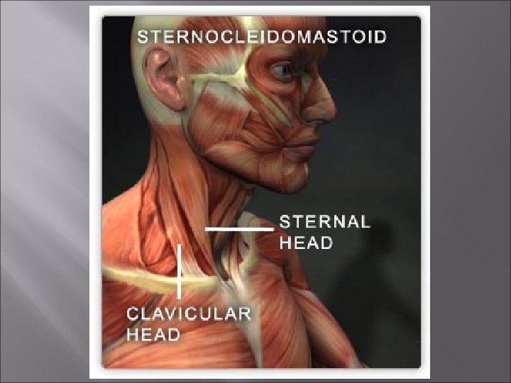
Sternocleidomastoid – Neck � � � Two-headed paired muscle. One head originates at the sternum and the second head originates at the clavicle. Heads fuse before inserting into the mastoid process of the temporal bone. When both are contracted, they flex the neck (often called the prayer muscle). When one contracts, it turns the head the opposite direction.
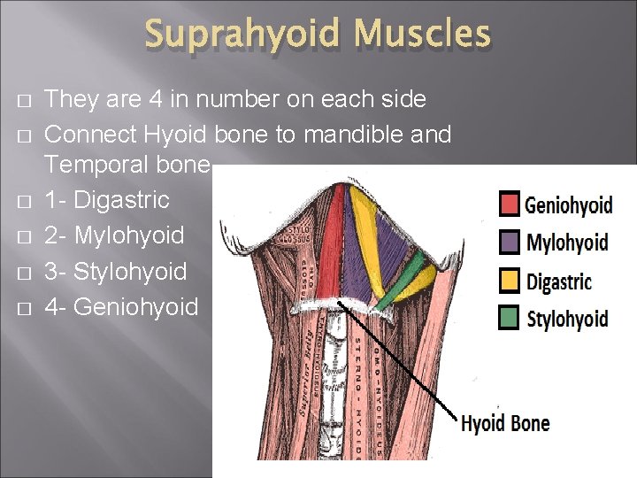
Suprahyoid Muscles � � � They are 4 in number on each side Connect Hyoid bone to mandible and Temporal bone. 1 - Digastric 2 - Mylohyoid 3 - Stylohyoid 4 - Geniohyoid
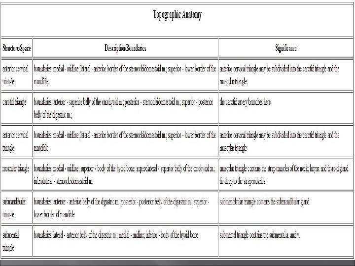
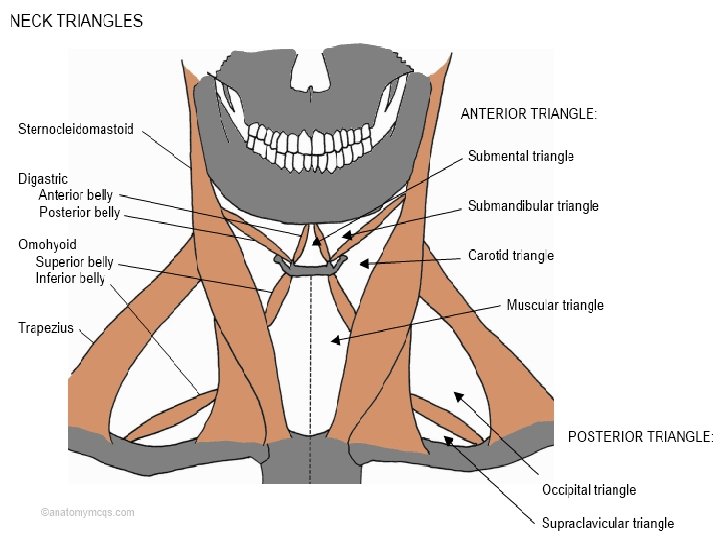
Digastric � � It has 2 bellies Origin : - anterior belly lower border of mandible and posterior belly arise from mastoid process Insertion : -hyoid bone Two bellies of digastric and lower border of mandible form the Submandibular Triangle

Mylohyoid � � � Origin : -body of mandible Insertion : - hyoid bone Action : - supports the floor of mouth

Stylohyoid � � � Origin : - styloid process of temporal bone Insertion : -hyoid bone Geniohyoid Muscle The pair of muscles lie superior to mylohoid Action : - action of suprahyoid muscles are to pull the hyoid upward , backward and forward in swallowing and speaking
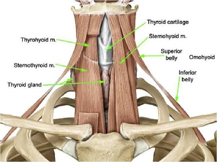
Infrahyoid Muscles � � � � Ribbon like shape so often called the Strap Muscles. 4 muscles anchors the hyoid bone to sternum the clavicle and scapula. 2 Groups (A) Superficial (strnohyoid and Omohyoid ) (B) Deep (sternothyroid and thyrohyoid) Origin and insertion is according to their names Action : -depress , retract and steady of hyoid bone and larynx in swallowing and speaking
Deep muscles � � � � It includes Scalenus anterior Scalenus posterior Location: - front and side of cervical vertebra Origin : -transerse process of C- vertebra Insertion : scalenus anterior inserted into upper surface of first rib and scalenus posterior is inserted into second rib Action : -elevate first rib , elevate the second rib or bending of cervical column.

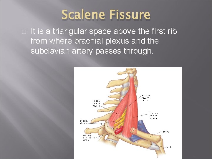
Scalene Fissure � It is a triangular space above the first rib from where brachial plexus and the subclavian artery passes through.

- Slides: 34