Muscles of arm and forearm and cubital fossa

Muscles of arm and forearm and cubital fossa Prof. Abdulameer Al-Nuaimi E-mail: a. al-nuaimi@sheffield. ac. uk abdulameerh@yahoo. com

Cubital Fossa

cubital fossa The cubital fossa is triangular in shape, and thus has three borders: Lateral border – medial border of the brachioradialis muscle. Medial border – lateral border of the pronator teres muscle. Superior border – hypothetical line between the epicondyles of the humerus. The floor of the cubital fossa is formed proximally by the brachialis, and distally by the supinator muscle. The roof consists of skin and fascia, and is reinforced by the bicipital aponeurosis. Within the roof runs the median cubital vein, which can be accessed for venepuncture

Contents of cubital fossa The contents of the cubital fossa include vessels, nerves and the biceps tendon (lateral to medial): Radial nerve –passing underneath the brachioradialis muscle. It divides into its deep and superficial branches. Biceps tendon – It runs through the cubital fossa, attaching to the radial tuberosity. Brachial artery –It bifurcates into the radial and ulnar arteries at the apex of the cubital fossa. Median nerve – It leaves the cubital fossa between the two heads of the pronator teres.

Brachioradialis M. Pronator teres muscle Supinator M. Median cubital vein Cubital Fossa

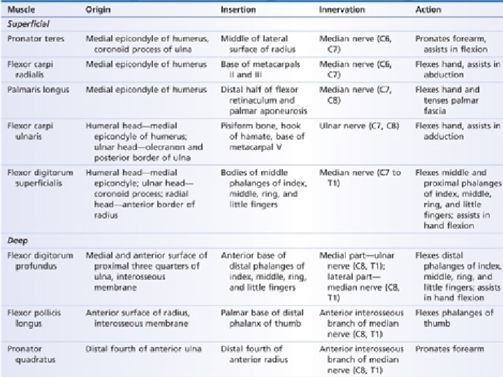
Anterior compartment of the forearm Superficial group Flexor digitorum superficialis

Brachioradialis Deep muscles of anterior compartment Of the forearm

Extensor Compartment Forearm

Posterior Forearm

Abductor pollicis longus Extensor indicis Extensor pollicis longus Extensor pollicis brevis

On ulna

Thank You
- Slides: 13