Muscles fasciae and trigons on the neck Semmelweis


 m. mylohyoid N. V/3 Mylohyoid line of mandibule Mylohyoid")
 m. sternohyoid Inner side of sternum and capsule of")


 Fom the base of the mandible Sheathes the sternocleidomastoid")
 Sheathes the infrahyoid muscles In the midline forms a")
 In the midline behind the pharynx is atteched strongly")


- Slides: 30
Muscles, fasciae and trigons on the neck Semmelweis Egyetem ÁOK Anatómiai Intézet Dr. Csáki Ágnes 2018. 02. 06.
Deep, prevertebral muscles of the neck M. rectus capitis anterior M. rectus capitis lateralis M. longus capitis M. longus colli Scalenus muscles Actions: flex the head and neck anteriorly Innervation: ventral rami of spinal nerves
Scalene muscles posterior anterior Name Origin Insertion m. anterior scalene tubercle on the 1. rib anterior tubercles of the transverse processes of C 3 -C 6 m. middle scalene behind posterior tubercles of the subclavian groove on the transverse processes of C 2 -C 7 1. rib m. posterior scalene 2. rib posterior tubercles of the transverse processes of C 5 -C 7 innervation: ventral rami of spinal nerves C 3 -C 8 Function The three muscles: ipsilateral contraction causes ipsilateral flexion of the neck, and bilateral contraction causes anterior flexion of the neck in case of forced respiration act as accessory muscles of respiration
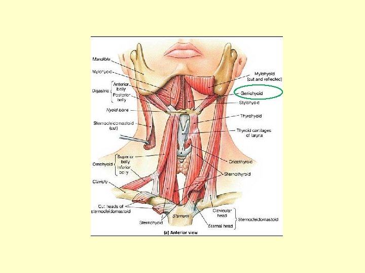
Suprahyoid muscles origin insertion (1) m. mylohyoid N. V/3 Mylohyoid line of mandibule Mylohyoid raphe and body of hyoid bone (2) m. digastric Anterior belly - inner side of the mentum Tendon is connected to the hyoid bone /N. V/3 Posterior belly - mastoid process /N. V II (3) m. stylohyoid N. VII Styloid process (4) m. geniohyoid N. XII Hyoid bone Body and greater horn of the hyoid bone around the tendon of the digastric Body of hyoid bone Inner side of the mentum (genu of mandible) The suprahyoid muscles are a group of four muscles, located superiorly to the hyoid bone of the neck. They all act to elevate the hyoid bone – an action involved in swallowing, sucking, drinking, opening of mouth.
Infrahyoid muscles origin insertion (1) m. sternohyoid Inner side of sternum and capsule of the sternoclavicular joint Body of the hyoid bone (2) m. sternothyroid Inner side of sternum and I. rib cartilage Linea obliqua of the thyroid cartilage (3) m. thyrohyoid linea obliqua of the thyroid cartilage Body and greater horn of the hyoid bone (4) m. omohyoid Inferior belly: sup. transverse scapular lig. Tendon is connected to the sternoclavicular superior belly: body and greater horn of the joint and veins hyoid bone The infrahyoid muscles are a group of four muscles that are located inferiorly to the hyoid bone in the neck Depress the hyoid bone take parts in swallowing, sucking, drinking, opening of mouth Innervation: Ansa cervicalis profunda (Cervical plexus C 1 - C 3)
Lateral view of supra- and infrahyoid muscles
Superficial neck muscles Sternocleidomastoid m. Originates from the manubrium sterni and clavicle, inserts to the mastoid process of the skull ipsilateral contraction causes ipsilateral flexion and rotation of the head („ear comes near to the shoulder”, bilateral contraction causes protraction of the head Innervation: IX cranial nerve (with the trapezius m. )
Triangles of the neck Posterior area antero-lateral area ANTERIOR AREA: (according to the scm m. ) 1. Submandibular triangle: two bellies of the digastric and base of mandibule Base of the trigon: the mylohyoid m. Content: submandibular gland 2. Submentale triangle: anterior bellies of the two digastric m. , hyoid bone Base of the trigon: the mylohyoid m. Content: lymph nodes 3. Carotid triangle: sternocleidomastoid, omohyoid and posterior belly of digastric m. Content: carotid sheeth (bifurcation of common carotid, internal jugular vein, vagus nerve) Puls!
LATERAL AREA: supraclavicular triangle divided by the omohyoid m. into 1. Omotrapezoid triangle : bordered by sternocleidomastoid, omohyoid and trapezius Base of the triangle: deep neck muscles covered by the prevertebral fascia Content: cervical plexus (Erb point-cervical pl. ) (and brachial plexus trunks) 2. Omoclavicular triangle: sternocleidomastoid, omohyoid and clavicle Content: scalene hiatus (gap) with the subclavian artery and trunks of the brachial plexus
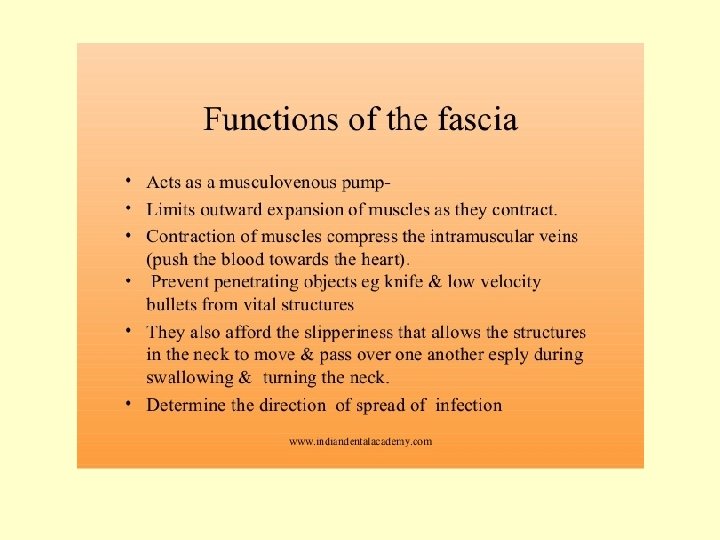
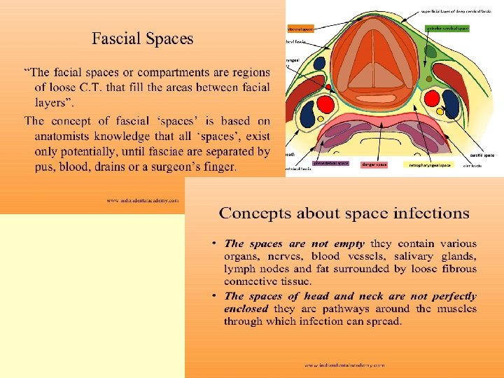
Cervical fascias Fascia is a layer of fibrous tissue that surrounds muscles, vessels and nerves. In the neck, there are several layers of fascias, which act to support and compartmentalise the structures present
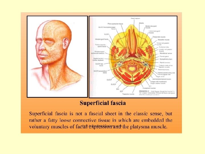
https: //www. slideshare. net/indiandentalacademy/compartments-of-the-head-and-neck-kiran-nxpowerlite
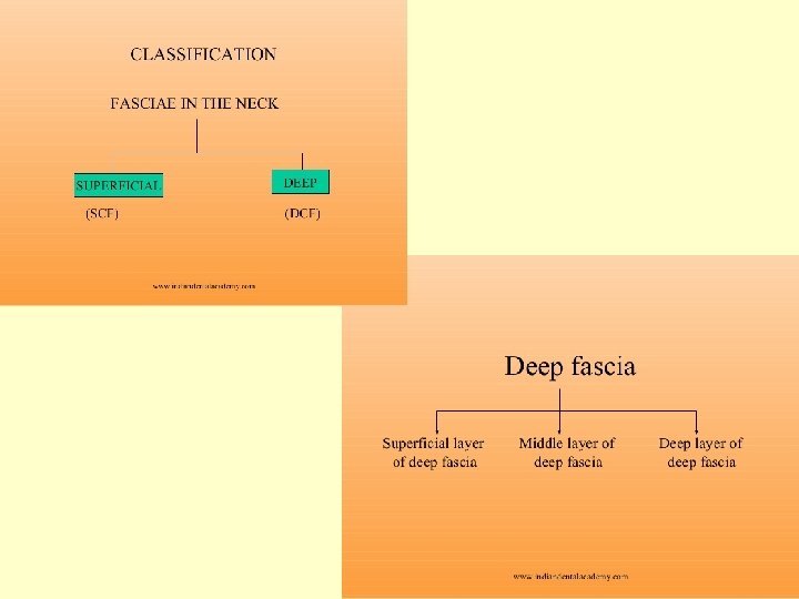
SUPERFICIAL CERVICAL FASCIA Subcutan connective tissue and the platysma only DEEP CERVICAL FASCIA : spf layer (investing layer) pretracheal layer prevertebral layer
Superficial or investing layer (yellow) Fom the base of the mandible Sheathes the sternocleidomastoid and the trapezius At the angle of the mandibule fuses with the parotideomasseteric fascia („parotid nest”) Superficial layer
Middle or pretracheal layer (green) Sheathes the infrahyoid muscles In the midline forms a strong, aponeurosis like layer Connects the tendon of the omohyoid to the sternoclavicular joint Forms the carotid sheath! Pretracheal layer
Deep or prevertebral layer (violet) In the midline behind the pharynx is atteched strongly to the longus capitis and longus colli Laterally forms the scalene „tent” Carotid sheath Prevertebral layer
Transverse section of the neck. The investing layer of fascia in highlighted in blue. The fascia completely envelopes the sternocleidomastoid and trapezius. Deep cevical fascia - Spf. layer or investing layer - pretracheal layer (visceral part) (carotid sheath) - prevertebral layer
The trachea, esophagus, and infrahyoid muscles are enclosed by the pretracheal layer. It can be anatomically divided into two parts: Muscular – encloses the infrahyoid muscles(purple) Visceral – encloses the trachea and oesophagus and forms the carotid sheath Deep cevical fascia - Spf. Layer or investing layer -pretracheal layer (muscular) (visceral) (carotid sheath) - prevertebral layer
The prevertebral layer surrounds the vertebral column and its associated muscles (scalene, longus colli, longus capitis m. , and deep muscles of the back) Deep cevical fascia - Spf. Layer or investing layer - pretracheal layer (visceral part) (carotid sheath) - prevertebral layer
Scalene tent Lower section – scalene tent
FASCIAL SPACES Pretracheal-Buccopharyngeal fascia Alar fascia Superficial Retropharyngeal Danger Prevertebral Parapharyngeal space
The prevertebral fascia consists two layers: prevertebral and alar fascia The alar fascia fuses anteriorly with the buccopharyngeal fascia at the level of the 4 th thoracic vertebra. Behind the alar fascia the danger space continuos downward to the thorax.
The superficial layer and muscular part of pretracheal layer end at the sternum, but the visceral layer and the prevertebral layer continous downward to the thorax!! - inflammation
Superficial space - anterior jugular veins Numb. 2 spf layer Numb. 3 pretracheal layer
Irodalom: images. MD, 2006 Current Medicine LLC Kahle W, Leonhardt H, Platzer W: Color Atlas/Text of Human Anatomy, 1992, Thieme, Stuttgart Putz R, Pabst R (editors): Sobotta Atlas of Human Anatomy, 1993, Urban & Schwarzenberg, München Romanes GJ (editor): Cunningham’s Textbook of Anatomy, 1991, Oxford University Press, Oxford Szentágothai J, Réthelyi M: Funkcionális anatómia, 2002, Medicina, Budapest Vízkelety T: Az ortopédia tankönyve, 1995, Semmelweis Kiadó, Budapest http: //teachmeanatomy. info/the-basics/ https: //www. youtube. com/watch? v=F 2 s. SVWEdj. WQ https: //www. slideshare. net/indiandentalacademy/compartments-of-the-head-and-neck-kirannx-powerlite Thank you!