Muscles Bone Cartilage Muscles Skeletal Smooth Cardiac 4

Muscles Bone, Cartilage

Muscles Skeletal Smooth Cardiac

4 Unique Characteristics of Muscle Tissue • Excitability is equated with responsiveness. • Contractility causes the fiber to shorten resulting in either a pull on bones or the movement of specific body parts. • Elasticity is the muscle’s ability to return to its original length when tension is released. • Extensibility is capability of extending in length in response to the contraction of opposing muscle fibers.

Skeletal Muscles T endon Skeletal muscle Epimysium Vessels Nerves Perimysium Muscle fascicle Endomysium Muscle f iber
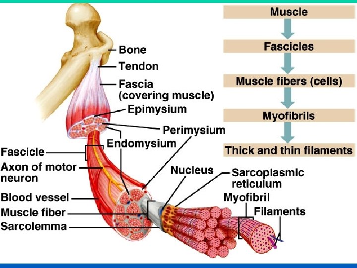

Skeletal Muscles Voluntary muscles head : The original of a muscle belly. The contractile part Muscle Origin: attachment that moves the least Insertion: attachment that moves the most Under varying circumstances the degree of mobility of the attachments may be reversed; therefore, the terms origin and insertion are interchangeable

Skeletal Muscles Tendon a cord of connective tissue into which muscles fibers end by which a muscle is attached to bone or other structures

Nerve Supply of Skeletal Muscle ØThe nerve trunk to a muscle is a mixed nerve Øabout 60% is motor and 40% is sensory Øit also contains some sympathetic autonomic fibers. ØMotor Point ØThe place of entrance of the nerve into the muscle Øat about the midpoint on its deep surface, often near the margin ØThis arrangement allows the muscle to move with minimum interference with the nerve trunk Mo t o r n e u ro n Motor point

Skeletal Muscle Action • Prime mover: – The chief muscle or member of a chief group of muscles responsible for a particular movement. • Antagonist: – Any muscle that opposes the action of the prime mover • Before a prime mover can contract, the antagonist muscle must be equally relaxed – this is brought about by nervous reflex inhibition Prime mover Antagonist Extending knee Flexion knee

Skeletal Muscle Action • Fixator: – contracts isometrically to stabilize the origin of the prime mover so that it can act efficiently. • Synergists • assist the prime mover in performing its action. • may also assist an agonist by preventing movement at a joint and thereby stabilizing the origin of the agonist Prime mover Fixator

Skeletal Muscles many muscles can act as a prime mover, an antagonist, a fixator, or a synergist, depending on the movement to be accomplished P ri me mover Synergist P ri me mover

Naming Muscles I. Skeletal muscles are named according to certain criteria A. Location- may indicate bone or body region that muscle is associated with B. Shape- Muscles often have a definitive shape, after which they are name Ex. Deltoid means triangle (and the deltoid muscle is triangular) C. Relative Size 1. Maximus= largest 2. Minimus= smallest 3. Longus= long 4. Brevis= short Ex. Gluteus maximus (larger) and minimus (smaller)

Naming Muscle D. Direction of Muscle Fibers - may reflect the direction of the fibers in relation to midline or other axis 1. Rectus= straight (runs parallel) 2. Transversus/oblique ( right angles)/ obliquely Ex. Rectus femoris- muscle that runs parallel with the femur E. Number of Origins 1. Biceps= two origins 2. Triceps= three origins 3. Quadriceps= four origins Ex. Biceps Brachii F. Location of origin and insertions 1. may be named according to the attachment points 2. Origin is always named first Ex. Sternocleidomastoid (dual origin on sternum and clavicle; insertion on mastoid process

Naming Muscle G. Action 1. Uses words such as flexor, extensor, or adductor Ex. Adductor longus on thigh adducts the thigh H. Sometimes several criteria are combined in a name. Extensor carpi radialis longus 1. muscle’s action (extensor) 2. joint it acts on (carpi= wrist) 3. Where it is (radialis = radius of forearm) 4. size (long relative to other wrist muscles

Skeletal Muscles Naming Of Skeletal Muscle Shape Name Shape Deltoid Teres Rectus Triangular Round Straight Name Size Major Latissimus Longissimus Large Broadest Longest Size

Skeletal Muscles Naming Of Skeletal Muscle Number of heads or bellies Name Number of heads or bellies Biceps Quadriceps Digastric Two heads Four heads Two bellies Position Name Position Pectoralis Supraspinatus Brachii Of the chest Above spine of scapula Of the arm

Skeletal Muscles Depth Naming Of Skeletal Muscle Name Depth Profundus Superficialis Externus Deep Superficial External Attachments Name Attachment Sternocleidomastoid From sternum and clavicle to mastoid process From coracoid process to arm Corachobrachialis

Skeletal Muscles Naming Of Skeletal Muscle Action Name Action Extensor Flexor Constrictor Extend Flex Constrict

Smooth Muscles • Composed of short muscle fibers that have a fusiform shape and single centrally located nucleus • Contraction is slow, resistant to fatigue, and usually sustained for an extended period of time. • Takes longer than skeletal muscle to contract and relax. Contraction is under involuntary control. ØPeristalsis: propulsion of contents

Cardiac Muscles Striated muscle fibers that branch and unite with each other ØConducting system of the heart: specialized cardiac fibers

Bone a special form of connective tissue in which calcium salts are deposited and which provides a framework, or skeleton, for the other tissues of the body. q. Compact bone: Solid mass q. Cancellous bone: Branching network of trabecula Function of bones ØThe rigid supporting framework of the body ØLevers for muscles ØProtection to certain viscera (e. g. , brain, spinal cord, heart, lung, liver, bladder) ØContain marrow, which is factory for blood cells ØStorehouse of calcium and phosphate

Classification Of bones Regionally General shape

Bone Regionally Region of skeleton Number of bones Axial skeleton Skull Cranium 8 Face 14 Auditory ossicles 6 Hyoid 1 Vertebrae 26 Sternum 1 Ribs 24 Appendicular skeleton Shoulder girdles Clavicle 2 Scapula 2

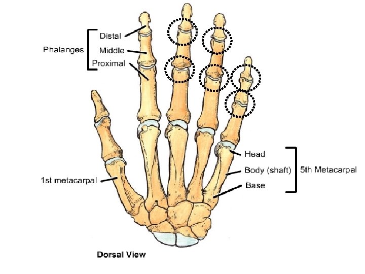
Region of skeleton Number of bones Upper extremities Humerus 2 Radius 2 Ulna 2 Carpals 16 Metacarpals 10 Phalanges 28 Hip bone 2 Pelvic girdle Lower extremities Femur 2 Patella 2 Fibula 2 Tibia 2 Tarsals 14 Metatarsals 10 Phalanges 28 206

Bone General Shape Long bones Short bones Flat bones Irregular bones Sesamoid bones

Bone General Shape Long bones ØTheir length is greater than their breadth ØFound in the limbs (e. g. , the humerus, femur, metacarpals, metatarsal, and phalanges)

Bone General Shape Short bones ØThey are roughly cuboidal shape ØFound in hand foot (e. g. , the scaphoid, launate, talus, and calcaneum).

Bone General Shape Flat bones ØThey are composed of thin inner and outer layers of compact bones, the tables, separated by a layer of cancellous bone , the diploe ØFound in the vault of the skull (e. g. , the frontal and parietal bones)

Bone General Shape Irregular bones ØThin shell of compact bone with an interior made up of cancellous bone ØE. g. , bones of skull, the vertebra, and the pelvic bones)

Bone General Shape Sesamoid bones ØSmall nodules of bone that are found in certain tendons where they rub over bony surfaces ØE. g. , patella. .

Bone q. Bone marrow q. Periosteum q. Development of bone Periosteum the fibrous covering of a bone.

BONE MARKINGS Every bump, groove, and hole has a name on your bones
 that grow")
Bone Markings • Two types of bone markings: – Projections (aka processes) that grow out from the bone – Depressions (cavities) that indent the bone
 Condyle: Rounded articular projection Condyle")
Joint Projections • 1) Condyle: Rounded articular projection Condyle
 Head: bony expansion on a narrow neck • 3) Facet:")
Joint Projections • 2) Head: bony expansion on a narrow neck • 3) Facet: smooth, nearly flat articular surface
 Ramus: Armlike bar of bone")
Joint Projections • 4) Ramus: Armlike bar of bone
 Crest: Narrow ridge of bone (Line: smaller than a crest) 2)")
Ligament/Tendon Projections 1) Crest: Narrow ridge of bone (Line: smaller than a crest) 2) Epicondyle: Raised area on or above a condyle
 Tubercle: Small rounded projection 4) Tuberosity: large rounded or roughened projection 5) Trochanter:")
3) Tubercle: Small rounded projection 4) Tuberosity: large rounded or roughened projection 5) Trochanter: very large, blunt projection (only on femur) Proximal Tibia
 Spine: Sharp, pointed projection Thoracic Vertebrae")
6) Spine: Sharp, pointed projection Thoracic Vertebrae
 Meatus: (me -")
DEPRESSIONS • Allow blood vessels or nerves to pass through. 1) Meatus: (me - A- tus) Canal or tube
 Fossa: shallow basin 3) Fissure: narrow, slitlike opening")
Depressions 2) Fossa: shallow basin 3) Fissure: narrow, slitlike opening
 Sinus: Cavity within a bone; filled with air and lined with mucous")
Depressions 4) Sinus: Cavity within a bone; filled with air and lined with mucous membranes 5) Foramen: Round or oval opening Foramen Magnum
 Sulcus, Groove or Furrow: a shallow depression")
Depressions 6) Sulcus, Groove or Furrow: a shallow depression
 Condyle 2) Head 3) Facet 4) Ramus 5) Crest 6) Epicondyle")
Review: Projections 1) Condyle 2) Head 3) Facet 4) Ramus 5) Crest 6) Epicondyle 7) Tubercle 8) Tuberosity 9) Trochanter 10) Spine Depressions 1) Meatus 2) Fossa 3) Fissure 4) Sinus 5) Sulcus or Groove or Furrow

Bone Development Osteogenesis and ossification: • The process of bone tissue formation, which leads to: – The formation of the bony skeleton in embryos – Bone growth until early adulthood – Bone thickness, remodeling, and fracture repair
")
Bone Growth - Ossification • Cartilage template laid down. • Osteoblasts (bone building cells) located in Ossification Centers.

Bone Growth Ossification • Primary Ossification Center in diaphasis. • Secondary Ossification Centers in epiphisis.

Bone Growth - Ossification • Grow toward one another, cartilage remains between them. • As long as cartilage remains undamaged, growth can occur.

Endochondral Ossification • Begins in the second month of development • Uses hyaline cartilage “bones” as models for bone construction • Requires breakdown of hyaline cartilage prior to ossification

Stages of Endochondral Ossification • Formation of bone collar • Cavitation of the hyaline cartilage • Invasion of internal cavities by the periosteal bud, and spongy bone formation • Formation of the medullary cavity; appearance of secondary ossification centers in the epiphyses • Ossification of the epiphyses, with hyaline cartilage remaining only in the epiphyseal plates

Stages of Endochondral Ossification Secondary ossification center Epiphyseal blood vessel Deteriorating cartilage matrix Hyaline cartilage Spongy bone formation Primary ossification center Bone collar Articular cartilage Spongy bone Medullary cavity Epiphyseal plate cartilage Blood vessel of periosteal bud 1 Formation of bone collar around hyaline cartilage model. 2 Cavitation of the hyaline cartilage within the cartilage model. 3 Invasion of internal cavities by the periosteal bud and spongy bone formation. 4 Formation of the medullary cavity as ossification continues; appearance of secondary ossification centers in the epiphyses in preparation for stage 5. 5 Ossification of the epiphyses; when completed, hyaline cartilage remains only in the epiphyseal plates and articular cartilages

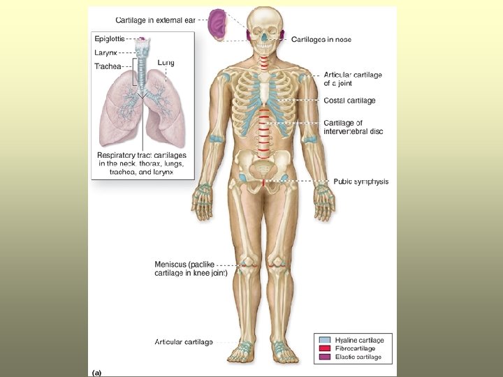
Cartilage Form of connective tissue in which cells and fibers are embedded in gel-like matrix Cartilage Hyaline cartilage Fibro cartilage Elastic cartilage

Hyaline cartilage High proportion of amorphous matrix that has the same refractive index as the fibers embedded in it

Fibro cartilage Fibro Cartilage Has many collagen fibers embedded in a small amount of matrix

Elastic cartilage Elastic Cartilage Possesses large numbers of elastic fibers embedded in matrix

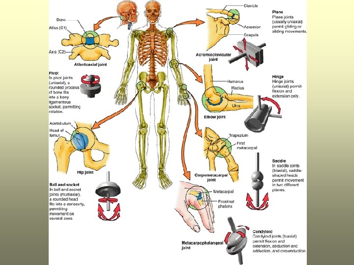
Joints * Fibrous joint Plane Cartilaginous joint Hinge Pivot places where bones meet each other (articulate). Synovial joint Condyloid Ellipsoid Saddle Ball and socket

Classification of Joints Fibrous Joints dense connective tissues connect bones between bones in close contact Cartilaginous Joints hyaline cartilage or fibrocartilage connect bones Synovial Joints most complex allow free movement synarthrotic immovable amphiarthrotic slightly movable diarthrotic freely movable

Fibrous Joints 3 Types Syndesmosis Suture Gomphosis ØSyndesmosis Øa sheet or bundle of fibrous tissue connects bones Ø amphiarthrotic Ø lies between tibia and fibula

Fibrous Joints Suture • between flat bones • synarthrotic • thin layer of connective tissue connects bones Gomphosis • cone-shaped bony process in a socket • tooth in jawbone • synarthrotic

Cartilaginous Joints 2 Types Synchondrosis Symphysis Synchondrosis Ø bands of hyaline cartilage unite bones Ø epiphyseal plate (temporary) Ø between manubrium and first rib Ø synarthrotic

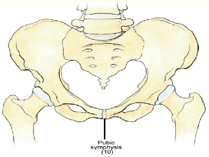
Cartilaginous Joints Symphysis • pad of fibrocartilage between bones • pubis symphysis • joint between bodies of adjacent vertebrae • amphiarthrotic
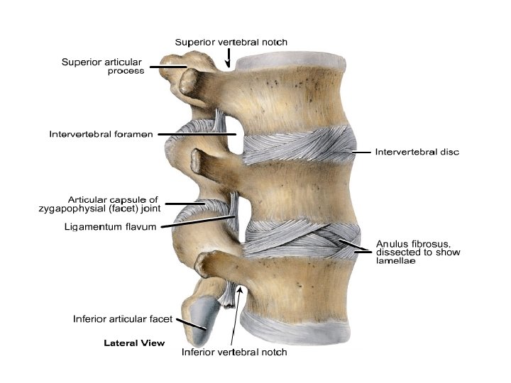

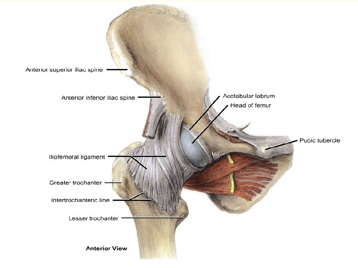
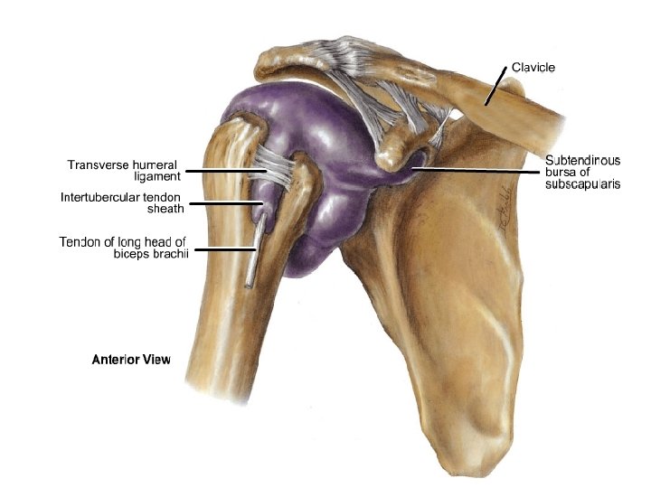
Joints Synovial joint • Where a space intervenes between the articulating ends of bones, the joint is called a synovial joint (i. e. most of the joints of the body. Knee, shoulder…. . ). • articular capsule encloses the joint. • lined by a synovial membrane which secretes a lubricating fluid (synovial fluid) Capsule Øa fibrous or membranous envelope surrounding an organ. ØAn articular capsule surrounds each synovial joint, being attached to the bones just beyond the limits of the joint cavity. Disc a flat round structure usually applied to plates of cartilage in joints. Ligament a band of fibrous connective tissue by which bones are connected to each other.

General Anatomy of Synovial Joints • Basic features: – articular capsule – joint cavity – synovial fluid – articular cartilage – ligaments – nerves – blood vessels

Joints Synovial joint

Joints Synovial joint Plane Hinge Pivot Condyloid Ellipsoid Saddle Ball and socket

Joints Synovial joint Types of joints Articular shape Movement Example Plane Flat *Sternoclavicular *Acromioclavicular Slide one another

Joints Synovial joint Types of joints Articular shape Movement Example Hinge Flat, planar *Interphalangeal joints of hand & foot; *Knee *Elbow Motion in one plane; flexion. extension

Joints Synovial joint Types of joints Articular shape Movement Example Pivot Central bony pivot is surrounded by bony - ligmentous ring Atlantoaxial rotation

Joints Synovial joint Types of joints shape Movement Example Condyloid Flexion, extension, abduction, adduction, small rotation *Metacarpophalangeal Convex or concave

Joints Synovial joint Types of joints Articular shape Movement Example Ellipsoid Flexion, extension, abduction, adduction, *Wrist joint Elliptical convex fits into an elliptical concave

Joints Synovial joint Types of joints Articular shape Movement Example Saddle Reciprocally concavo-convex *Carpometacarpal joint of the thumb Flexion, extension, abduction, adduction, rotation
")
Joints Synovial joint Types of joints Articular shape Movement Example Spheroidal (ball & socket) Convex surface in concave cavity *Shoulder *hip Wide-ranging flexion, extension, abduction, adduction, rotation, circumduction

Joints Stability Of joints Articular surfaces Ligaments Muscle tone

Joints Stability Of joints Articular surfaces

Joints Stability Of joints Ligaments

Joints Stability Of joints Muscle tone

Mobility and Stability in Joints • Motion permitted ranges from none to various extensive motions. • Structure determines both its mobility and its stability. – more mobile = less stable

Ligaments Ligament a band of fibrous connective tissue by which bones are connected to each other. Fibrous Unstrechable under normal condition Iliofemoral ligament of the hip Elastic Regain its original length after Stretching Ligamentum flavum

Ligaments

Ligaments

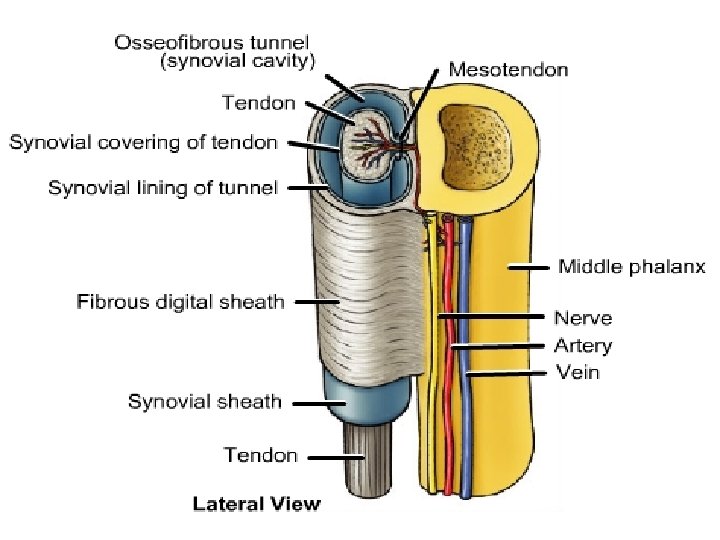
Bursa ØA membranous sac containing a small amount of viscous fluid. ØA bursa is usually found in tissues where friction develops, such as where a tendon crosses a bony prominence. ØA bursa may form synovial sheaths to surround tendons as they cross other tendons or bone.

Synovial sheath §Tubular bursa that surrounds a tendon §tendon become suspended within bursa by a mesotendon

Synovial sheath

Thank you
- Slides: 92