Muscles are responsible for all types of body

Muscles are responsible for all types of body movement Muscles make up about ½ of the body’s mass Three basic muscle types are found in the body 1) Skeletal muscle 2) Cardiac muscle 3) Smooth muscle
 Contraction of muscles is")
Muscle cells are elongated (muscle cell = muscle fiber) Contraction of muscles is due to the movement of microfilaments All muscles share some terminology Prefix myo refers to muscle Prefix mys refers to muscle Prefix sarco refers to flesh

Most are attached by tendons to bones Cells are multinucleated Striated – have visible banding Voluntary – subject to conscious control but also controlled automatically by reflexes Cells are surrounded and bundled by connective tissue

Endomysium – delicate connective tissue sheath that surrounds each single muscle fiber Perimysium – coarser conntective tissue that surrounds many fibers Fascicle – name for the above system of muscle fibers bundled together by perimysium Figure 6. 1

Epimysium – covers the entire skeletal muscle Bundles the fascicles together Figure 6. 1

Epimysium blends into a connective tissue attachment Tendon – cord-like structure Provides durability and conserves space CT holds up to crossing rough bony projections Small size allows more tendons to pass over a joint Aponeuroses – sheet-like structure Sites of muscle attachment 1) Bones 2) Cartilages 3)Connective tissue coverings
 Has no striations 2) Spindle-shaped cells 3) Single nucleus 4) Involuntary –")
1) Has no striations 2) Spindle-shaped cells 3) Single nucleus 4) Involuntary – no conscious control 5) Found mainly in the walls of hollow organs Skeletal muscle would be like a speedy wind up car that loses power quickly Smooth muscle is like a steady heavy duty engine that lumbers along slow an tirelessly Figure 6. 2 a
 Has striations 2) Usually has a single nucleus 3) Intercalated Discs – join")
1) Has striations 2) Usually has a single nucleus 3) Intercalated Discs – join cardiac muscle cells together so there is a coordinated contraction 4) Involuntary 5) Found only in the heart Figure 6. 2 b
 2) 3) 4) Produce movement Maintain posture Stabilize joints Generate heat ¾ of")
1) 2) 3) 4) Produce movement Maintain posture Stabilize joints Generate heat ¾ of the energy created by ATP escapes as heat that helps keep us warm … or makes us too hot.
 Cells are multinucleated 2) Nuclei are just beneath the sarcolemma Figure 6.")
1) Cells are multinucleated 2) Nuclei are just beneath the sarcolemma Figure 6. 3 a
 Sarcolemma – specialized plasma (cell) membrane (endomysium wraps around the sarcolemma) 4) Sarcoplasmic")
3) Sarcolemma – specialized plasma (cell) membrane (endomysium wraps around the sarcolemma) 4) Sarcoplasmic reticulum – specialized smooth endoplasmic reticulum; stores Calcium Figure 6. 3 a
 Myofibril - Bundles of myofilaments Myofibrils are aligned to give distinct bands")
5) Myofibril - Bundles of myofilaments Myofibrils are aligned to give distinct bands I band = light band A band = dark band Figure 6. 3 b
 Sarcomere - Contractile unit of a muscle fiber Figure 6. 3 b")
6) Sarcomere - Contractile unit of a muscle fiber Figure 6. 3 b

Organization of the sarcomere Myosin Composed of the protein myosin Has ATPase enzymes Actin filaments = thick filaments = thin filaments Composed of the protein actin

Figure 6. 3 c
 have heads (extensions, or cross bridges) Myosin and actin overlap")
Myosin filaments (thick) have heads (extensions, or cross bridges) Myosin and actin overlap somewhat Figure 6. 3 d

At rest, there is a bare zone that lacks actin filaments Sarcoplasmic reticulum (SR) – for storage of calcium Figure 6. 3 d

Irritability – ability to receive and respond to a stimulus Contractility – ability to shorten when an adequate stimulus is received

Motor unit - Skeletal muscles must be stimulated by a nerve to contract One neuron and all the Muscle cells stimulated by that neuron (could be one muscle or 100) Figure 6. 4 a

Neuromuscular junctions – association site of nerve and muscle https: //www. youtube. com/watch? v=RHZ 2 ryh. Ilc 8 Figure 6. 5 b

Synaptic cleft – gap between nerve and muscle Nerve and muscle do not make contact Area between nerve and muscle is filled with interstitial fluid Figure 6. 5 b

Neurotransmitter – chemical released by nerve upon arrival of nerve impulse Acetylcholine - The neurotransmitter for skeletal muscle Neurotransmitter attaches to receptors on the sarcolemma Sarcolemma becomes permeable to sodium (Na+)

Sodium rushing into the cell generates an action potential Once started, muscle contraction cannot be stopped
 A nerve causes an action potential which stimulates the release of calcium")
1) A nerve causes an action potential which stimulates the release of calcium from the sarcoplasmic reticulum into the cytoplasm of the muscle cell 2) The Calcium ions act as a trigger as they bind to the regulatory proteins on the actin. Calcium changes the shape and position of these proteins and open sites to bind with myosin. No calcium = No binding!!! 3) “Loaded” (ATP) myosin heads (crossbridges) attach to binding sites on the thin actin filaments. Figure 6. 7
 When the Myosin head binds to the actin site it releases energy")
4) When the Myosin head binds to the actin site it releases energy (ATP) and moves the head toward the center 5) Myosin heads then bind to the next site of the thin filament to move the Actin towards the middle. Not all will release or the muscle contraction would be lost. Like pulling on a rope. 6) This continued action causes a sliding of the actin along the mysoin causing the actin myofibrils to move closer and overlap resulting in a shortened or contracted muscle. Figure 6. 7

http: //www. youtube. com/watch? v=Ed. Hz. KYDxr. Kc&NR=1 http: //www. youtube. com/watch? v=Vlchs 4 om. FDM&NR=1 http: //www. youtube. com/watch? v=g. J 309 Lf. HQ 3 M&feature=r elated http: //www. youtube. com/watch? v=ren_IQPOh. Jc&feature=rel ated Figure 6. 8
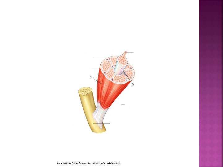

Figure 6. 7
- Slides: 28