MUSCLE TISSUE 1 Skeletal muscle 2 Cardiac muscle

")


")


: - Very long - Cylindrical -")


 tubules & Sarcoplasmic")
: 1 - Terminal")


 striations. - Are")


 in ventricular muscle.")

 Endocardium. • (2) Myocardium. • (3) epicardium.")


 L/M : - Do not show cross")


 Skeletal muscle fibers ( cells): - can not divide. -")
- Slides: 32
MUSCLE TISSUE 1 - Skeletal muscle. 2 - Cardiac muscle. 3 - smooth muscle.
SKELETAL MUSCLE LIGHT MICROSCOPIC STRUCTURE (L. S. )
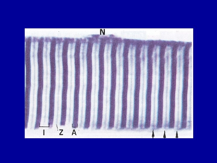
SARCOMERE
T tubules, Triads, Sarcoplasmic reticulum
SARCOMERE (E/M)
ACTIN
MYOSIN
SKELETAL MUSCLE C. T. COMPONENTS
SKELETAL MUSCLE L/M of skeletal muscle fibers (cells): - Very long - Cylindrical - multinucleated - Nuclei are oval & peripheral - Show cross (transverse) striations - Sarcoplasm is acidophilic
ORGANIZATION OF SKELETAL MUSCLE • C. T. Component: 1 - Epimysium : dense C. T. surround the entire muscle 2 - Perimysium: dense C. T. around each bundle (fascicle) of muscle fibers. 3 - Endomysium: surrounds each muscle fiber. delicate C. T. composed mainly of reticular fibers & external lamina. Endomysium contains continuous blood capillaries and lymph vessels. * Muscle fibers are arranged in regular bundles.
E/M of skeletal muscle fibers: • Sarcomere: Definition Structure N. B. M line consists of myomesin & C protein.
E/M of skeletal muscle fibers: • Sarcolemma, Transverse ( T ) tubules & Sarcoplasmic reticulum. T tubules: invaginations of the sarcolemma forming anastomosing network of tubules that encircles the boundaries of the A-I bands of each sarcomere in every myofibril.
E/M of skeletal muscle fibers: Sarcoplasmic reticulum ( S. R. ): 1 - Terminal cisternae: 2 lateral portions of S. R. 2 - Sarcotubules: branching network of S. R around each myofibril. • Triad: components = T. T. + 2 T. C.
E/M of skeletal muscle fibers: * Mitochondria: numerous, elongated with many cristae. • Myoglobin: more in red fibers than in white fibers. • Glycogen granules.
TYPES OF SKELETAL MUSCLE FIBERS 1 - Red Muscle Fibers. 2 - White Muscle Fibers. 3 - Intermediate Muscle Fibers
CARDIAC MUSCLE
CARDIAC MUSCLE L/M of cardiac muscle cells: - Have cross (transverse) striations. - Are usually mononucleated (may be binucleated). - Nuclei are oval & central. - are elongated , branched cells. - are parallel to each other. - Have intercalated disks ( at sites of end-to-end contact of cells in the same fiber). - Mitochondria: about 40%. - Lipofuscin pigments.
E/M of cardiac muscle cells: • Intercalated disks: Types: 1 - Straight. 2 - Steplike (stepwise) pattern. Junctions: 1 - Fascia adherent. 2 - Macula adherent (Desmosomes). 3 - Gap junctions.
INTERCALATED DISK
• T Tubules: - are more numerous and larger (wider) in ventricular muscle. - are found at the level of Z lines. • Sarcoplasmic reticulum: - is not as well developed. • Diads. • Mitochondria: occupy 40 % of the sarcoplasmic volume. • Glycogen. • Lipofuscin pigment granules (aging pigment) • Secretory granules: more in Rt atrium, atrial natriuretic factor.
MODERATOR BAND & PURKINJE MUSCLE FIBERS
WALL OF THE HEART • (1) Endocardium. • (2) Myocardium. • (3) epicardium.
SMOOTH MUSCLE FIBERS
SMOOTH MUSCLE
SMOOTH MUSCLE
SMOOTH MUSCLE
SMOOTH MUSCLE FIBERS ( SMOOTH MUSCLE CELLS) L/M : - Do not show cross striations. - are fusiform. - have a single nucleus located in the center.
E/M of smooth M. F. : • No T tubules , No sarcomeres. • Abundant intermediate filaments: coursing through the sarcoplasm. *Dense bodies ( D. B. ): Types: 1 - membrane-associated. 2 - cytoplasmic. Both contain α-actinin ( are thus similar to Z lines ). Both actin & intermediate filaments insert to D. B. * Abundant gap junctions.
FUNCTIONS OF S. M. F. : 1 - Contractile activity. 2 - Synthesis of extracellular products e. g. collagen, elastin & proteoglycans.
REGENERATION OF MUSCLE (1) Skeletal muscle fibers ( cells): - can not divide. - limited regeneration by satellite cells ( inactive myoblasts ). (2) Cardiac muscle cells: Have almost no regenerative capacity beyond early childhood. (3) Smooth muscle fibers ( cells ): a- Can divide. b- Pericytes. ----→active regenerative response.