Muscle Tissue Types of Muscle Tissue w Skeletal


 w Muscle action potentials arise at the NMJ. w The NMJ")
 w The neuron cell communicates with the second by releasing a")


 fibers. n n Smallest of")
 fibers. n n Intermediate in")
 fibers. n n n Largest")


 smooth muscle.")
- Slides: 48
Muscle Tissue
Types of Muscle Tissue w Skeletal muscle tissue w Cardiac muscle tissue n Autorhythmicity - pacemaker w Smooth muscle tissue
Functions of Muscle Tissue w Producing body movements w Stabilizing body positions w Storing and moving substances within the body n n Sphincters – sustained contractions of ringlike bands prevent outflow of the contents of a hollow organ Cardiac muscle pumps nutrients and wastes through Smooth muscle moves food, bile, gametes, and urine Skeletal muscle contractions promote flow of lymph and return blood to the heart w Generating heat - thermogenesis
Properties of Muscle Tissue w Electrical excitability n Produces electrical signals – action potentials w Contractility n n Isometric contraction – tension without muscle shortening Isotonic contraction – constant tension with muscle shortening
Properties of Muscle Tissue w Extensibility – ability of a muscle to stretch without being damaged w Elasticity n Ability of a muscle to return to its original length
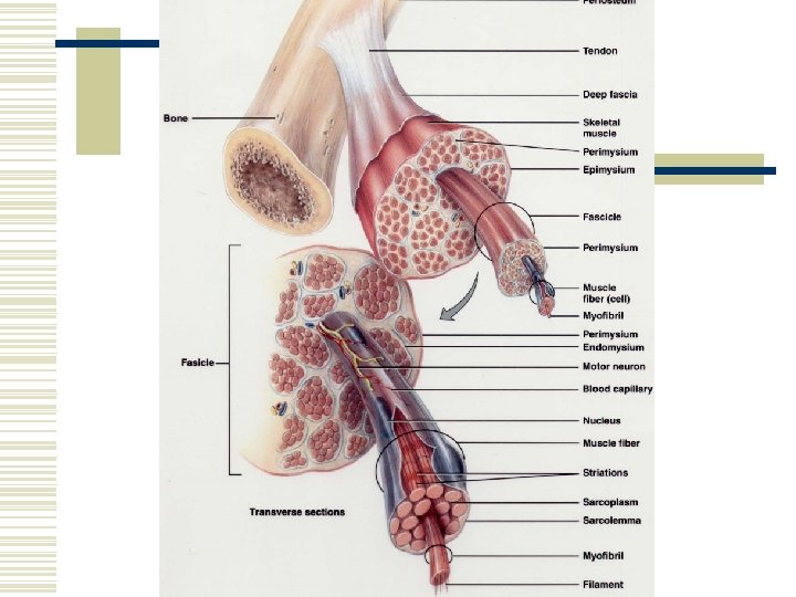
Connective Tissue Components w Fascia – a sheet of fibrous CT that supports or surrounds muscles and other organs n n Superficial fascia (subcutaneous layer) – separates muscle from skin Deep fascia – holds muscles with similar functions together w Epimysium – outermost layer – encircles whole muscles w Perimysium n Surrounds groups of 10 – 100 individual muscle fibers separating them into bundles called fascicles
Connective Tissue Components w Endomysium n Separates individual muscle fibers within the fascicle w Tendon n All 3 CT layers may extend beyond the muscle to form a cord of dense regular CT that attaches muscle to the periosteum of bone w Aponeurosis n A broad, flat layer of CT
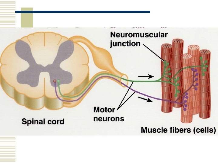
Nerve and Blood Supply w Skeletal muscles are well supplied with nerves and blood vessels w Neuromuscular junction – the structural point of contact and the functional site of communication between a nerve and the muscle fiber w Capillaries are abundant – each muscle fiber comes into contact with 1 or more
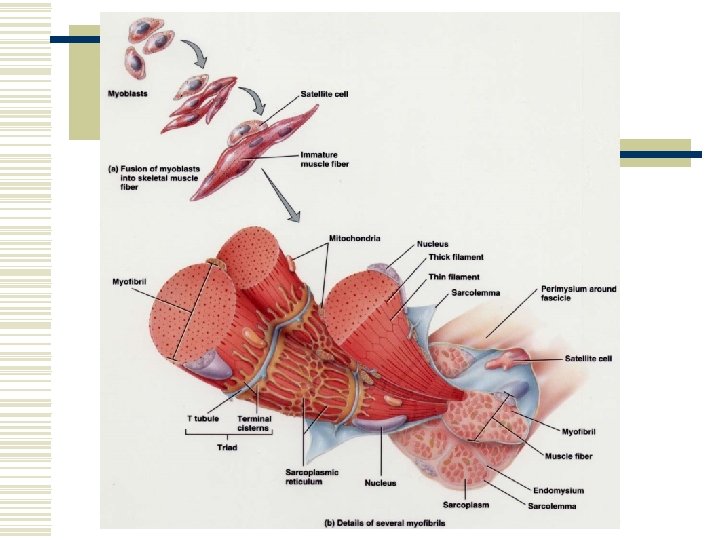
Sarcolemma, T Tubules, and Sarcoplasm w Sarcolemma – the plasma membrane of a muscle cell w T (transverse) tubules – Propogate action potentials – extend to the outside of the muscle fiber w Sarcoplasm – cytoplasm of the muscle fiber n Contains myoglobin – protein that binds with oxygen
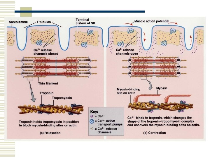
Myofibrils and Sarcoplasmic Reticulum w Myofibril – the contractile elements of skeletal muscle w Sarcoplasmic reticulum (SR) – encircles each myofibril – stores CA 2+ (its release triggers muscle contractions)
Atrophy and Hypertrophy w Muscular atrophy – wasting away of muscles n n Disuse Denervation w Muscular hypertrophy – an excessive increase in the diameter of muscle fibers
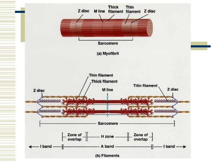
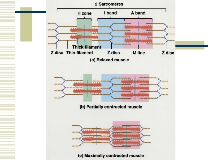
Filaments and the Sarcomere w Filaments – structures within the myofibril n n Thick w Sarcomere – basic functional unit of a myofibril w Z discs – separate one sarcomere from the next
Filaments and the Sarcomere w A band – predominantly thick filaments n n Zone of overlap at the ends of the A bands H zone – contains thick, but no thin filaments w I band – thin filaments w M-line – middle of the sarcomere
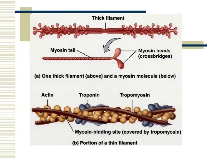
Muscle Proteins w Contractile proteins – generate force n n Myosin Actin w Regulatory proteins – switch contraction on and off w Structural proteins
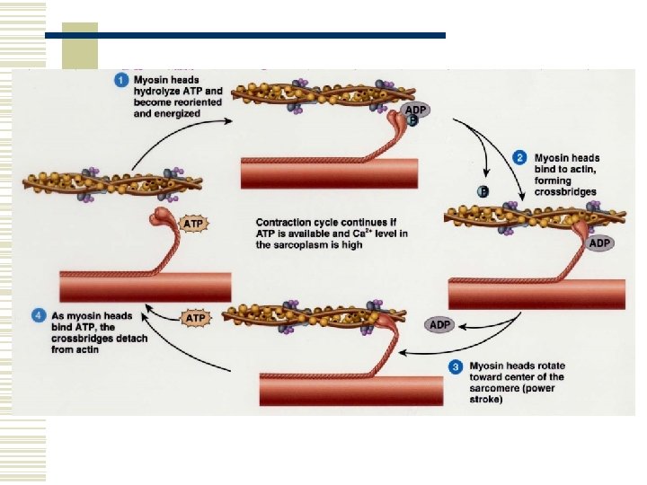
Sliding Filament Mechanism w Muscle contraction occurs because myosin heads attach to the thin filaments at both ends of the sarcomere and pull them toward the M line. w The length of the filaments does not change; However, the sarcomeres shorten, thereby shortening the entire muscle.
Role of Ca 2+ in Contraction w An increase in calcium ion concentration in the cytosol initiates muscle contraction and a decrease in calcium ions stops it.
Rigor Mortis w After death the cellular membranes become leaky. w Calcium ions are released and cause muscular contraction. w The muscles are in a state of rigidity called rigor mortis. w It begins 3 -4 hours after death and lasts about 24 hours, until proteolytic enzymes break down (digest) the cross-bridges.
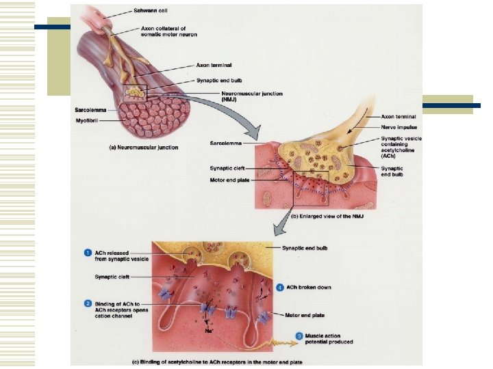
Neuromuscular Junction (NMJ) w Muscle action potentials arise at the NMJ. w The NMJ is the site at which the motor neuron contacts the skeletal muscle fiber. w A synapse is the region where communication occurs.
Neuromuscular Juntcion (NMJ) w The neuron cell communicates with the second by releasing a chemical called a neurotransmitter. w Synaptic vesicles containing the neurotransmitter acetylcholine (ach) are released at the NMJ. w The motor end plate is the muscular part of the NMJ. It contains acetylcholine receptors. w The enzyme acetlycholineesterase (ACh. E) breaks down ACh.
Production of ATP w 1. From creatine phosphate. n When muscle fibers are relaxed they produce more ATP than they need. This excess is used to synthesize creatine phosphate (an energy rich compound).
Production of ATP w 2. Anaerobic cellular respiration. n n Glucose undergoes glycolysis, yielding ATP and 2 molecules of pyruvic acid. Does not require oxygen.
Production of ATP w 3. Aerobic cellular respiration. n n The pyruvic acid enters the mitochondria where it is broken down to form more ATP. Slower than anaerobic respiration, but yields more ATP. Utilizes oxygen. 2 sources of oxygen. l l Diffuses from bloodstream. Oxygen released from myoglobin.
Muscle Fatigue w Muscle fatigue is the inability of a muscle to contract forcefully after prolonged activity. w Central fatigue – a person may develop feelings of tiredness before actual muscle fatigue.
Oxygen Debt or Recovery Oxygen Uptake w Added oxygen, over and above resting oxygen consumption, taken in after exercise. w Used to restore metabolic conditions. n n n 1. To convert lactic acid back into glycogen stores in the liver. 2. To resynthesize creatine phosphate and ATP in muscle fibers. 3. To replace the oxygen removed from hemoglobin.
Motor Units w A motor unit consists of the somatic motor neuron and all the skeletal muscle fibers it stimulates. w A single motor neuron makes contact with an average of 150 muscle fibers. w All muscle fibers in one motor unit contract in unison.
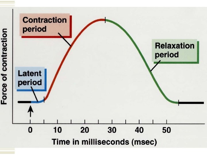
Twitch Contraction w A twitch contraction is the brief contraction of all the muscle fibers in a motor unit in response to a single action potential. w A myogram is a record of a muscle contraction and illustrates the phases of contraction.
Refractory Period w A period of lost excitability during which a muscle fiber cannot respond to stimulation.
Motor Unit Recruitment w The process in which the number of active motor units increases. w The weakest motor units are recruited first, with progressively stronger units being added if the task requires more force.
Muscle Tone w Even at rest a muscle exhibits a small amount of muscle tone – tension or tautness. w Flaccid – when motor units serving a muscle are damaged or cut. w Spastic – when motor units are overstimulated.
Isotonic and Isometric Contractions w Concentric isotonic contraction – a muscle shortens and pulls on another structure. w Eccentric isotonic contraction – the length of a muscle increases during contraction. w Isometric contraction – muscle tension is created; However, the muscle doesn’t shorten or lengthen.
Types of Skeletal Muscle Fibers w Slow oxidative (SO) fibers. n n Smallest of the fibers. Least powerful. Appear dark red – much myoglobin and many capillaries. Resistant to fatigue.
Types of Skeletal Muscle Fibers w Fast oxidative-Glycolytic (FOG) fibers. n n Intermediate in diameter. Appear dark red – much myoglobin and many capillaries. High level of intracellular glycogen. Resistant to fatigue.
Types of Skeletal Muscle Fibers w Fast Glycolitic (FG) fibers. n n n Largest in diameter. Contain the most myofibrils, therefore more powerful contractions. Appear white – low myoglobin and few capillaries. Large amounts of glycogen – anaerobic respiration. Fatigue quickly.
Distribution and Recruitment of Different Types of Fibers w Most skeletal muscles are a mixture of all three types. w The continually active postural muscles have a high concentration of SO fibers.
Distribution and Recruitment of Different Types of Fibers w Muscles of the shoulders and arms are used briefly and for quick actions, therefore they have many FG fibers. w Muscle of the legs support the body and participate in quick activities, therefore they have many SO and FOG fibers.
Cardiac Muscle Tissue w The principle tissue in the heart is cardiac muscle tissue. w Cardiac muscle fibers have intercalated discs, which connect the ends of the cardiac muscle fibers together. w Cardiac muscle tissue remains contracted 10 to 15 times longer than skeletal muscle. w Requires a constant supply of oxygen and contains larger and more numerous mitochondria.
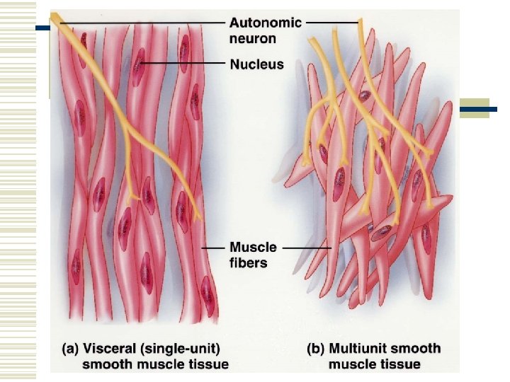
Smooth Muscle Tissue w Activated involuntarily. w Two types. n Visceral (single-unit) smooth muscle. l n Walls of small blood vessels and walls of hollow organs (I. E. Stomach, intestines, uterus, and urinary bladder). Multi-unti smooth muscle. l Walls of large ateries, in the airways of lungs, in arrector pili muscles.