Muscle physiology Structure of various muscles In higher


- Slides: 27

Muscle physiology Structure of various muscles • In higher animals such as vertebrates, most movements are caused by skeletal, cardiac and smooth mucles.

• Striated or striped, muscle constitute a large fraction of the total body weight. • Both the ends of most striated muscles are attached to the skeleton and thus are often called skeletal muscles. • They are attached to the bones by tendons.

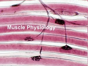
The muscle fibre • Living muscle is composed of many long, cylindrical shaped fibres from 0. 02 to 0. 08 millimetre in diameter. • The fibres in living muscle appear transparent when viewed in an ordinary optical microscope. • Each fibre is surrounded by a rather complex, multilayered structure, the sarcolemma. • Sarcoplasm is the special name for cytoolasm of a muscle fibre. Each striated muscle has a nucleus.

Myofibril • Myofibril is about one or two mm, or microns in diameter and extends the entire length of muscle fibre. • Myofibrils are shaped like cycindrical column.

Cardiac muscle • The heart consists mostly of muscle, the myocardial cells (collectively termed the myocardium), arranged in several ways that set it apart from other types of muscle.

The myocardial cells • The sheets or bands of muscle fibres of the ventrides are arranged in a complex pattern. Some encircle only the cavity of the left ventricle. • Others surround both ventricles in a helical pattern. • Cardiac muscle seems to depend on the influx of Ca 2+ ions from either the extra cellular space or some other area during contraction, to supplement the Ca 2+ ions released from the sacoplasmic reticulum.

Smooth muscle or non striated muscle • The term smooth muscle derive from the uniform appearance of the cytoplasm of stained sections when examined under the low magnification of a light microscope. • Many organs of vertebrates contain smooth muscles; the stomach, intestines, bile ducts, bladder and urinary tracts, uterus and blood vessels.

Visceral or angle unity muscle • The individual smooth muscle cells of the intestine is roughly spindle shaped. • some are ribbon shaped or rod shaped. • The length of smooth muscle cell ranges bet 50 and 500 m.

Multi unit muscle • These types of muscles are found in vasdeferns.

Striated muscle. • The muscle is made up of a large number of parallel fibers, which are between 0. 1 and 0. 01 mm in diameter, but may be several centimeters long. These fibers in turn are made up of thinner fibrils. • The region between two Z-lines is called a sarcomere.

• From the narrow Z-line very thin filaments extend in both direction. • In vertebrates the thin filaments are about 0. 005 µm in diameter and the thick filaments are about twice that size, about 0. 01 µm in diameter. • The length of the thick filaments is about 1. 5 µm. • In striated muscle thick filaments consist of myosin and the thin filaments of actin. • The thick and thin filaments are linked together by a system of molecular cross linkages.

Cardiac muscle • Cardiac muscle is cross-striated, like skeletal muscle, but its functional properties differ in two important respects. • One is that, when a contraction starts in one area of the heart muscle, it rapidly spreads throughout the muscle mass. • Another important property of cardiac muscle is that a contraction is immediately followed by a relaxation period during which the muscle cannot be stimulated to contract again.

Smooth muscle • Smooth muscle lacks the cross striations characteristic of skeletal muscle, but the contraction depends on the same proteins as in striated muscle, actin and myosin, and on a supply of energy from ATP.

Substances for energy storage • The immediate source of energy for a muscle contraction is adenosine triphosphate. • In spite of its great importance, ATP is present in muscle in very small amounts. • Contraction of the muscle causes a force to be exerted on the points of attachment; because no change in length takes place, this us called an isometric contraction.

• The thin filaments are more complex, with three important protein actin, a relatively osmall globular protein, arranged in the filament as a twisted double strand of beads. • Each actin molecule is slightly asymmetrical. • The next important protein in the thin filament is tropomyosin, long and thin molecules attached to each other end to end, forming a very thin threadlike structure.

• A third protein molecule, troponin, is attached to each tropomyosin molecule. • Troponin, a calcium-binding protein, is a key to the contraction process. • The interaction between thick and thin filaments consists of a cyclic attachment and detachment of cross bridges between the two filaments.

Molluscan catch muscle • Clams and mussels protect themselves by closing the shells. • One or more adductor muscles pull the shells together and keep them closed against the springy action of the elastic hinge. • In some speices, but not all, the closing muscles are divided into two portions, one of smooth and one of striated fibers. Functionally, the striated portion contracts quickly and is referred to as the fast or phasic (twitch) portion; the nonstriated is much slower and is known as the slow or tonic portion.

• The most studied muscle of the catch type is the anterior byssus retractor muscle (ABRM) of the blue mussel (Mytilus edulis), which contains only smooth fibers.

Crustacean muscle • Crustacean muscles show in pure form some characteristics that to varying degrees are present in the muscles of a number of other animals. • Nearly everybody who has handled live crabs or lobsters has made contact with the impressive force these animals can exert with their claws. • strength of the muscle is extraordinarily high. • In the claw of the crab the muscle is pinnate.

• Another advantage of the pinnate muscle in the crab claw is related to the confined space in which it works. • The most interesting aspect of crustacean muscle is the multiple innervation: Individual muscle fibers may be innervated by two or more nerve fibers. • The jumping muscle of the hind-leg of a locust is innervated by three axons: one fast excitatory, one slow excitatory, and one inhibitory.

• As stated above, the most striking characteristic of crustacean muscle is that it is supplied by two or more different nerve fibers.

Muscle metabolism and function

Dark muscle • Salmonids and tunas have layer of dark muscle under the shell. • In perch the propulsion is through pectoral fin. • See horses and pipe fish propel themselves primarily with pectoral fin.

Composition and metabolism of dark and white muscle • low Na and Cl- and high K+. • Light muscle is characterized by high levels of glycogen dark muscle has high levels of lipids. • In fish blood lactate is 200 mg/100 ml blood. • Loss of lactate across the gill also possible.

Specialized muscles • Increased innervations of the region around the base of the fin, increased number of nerve endings in the cerebellum to co-ordinate the increased activity.

• A number of fish make humming, grunting or other low pitched sounds tonal fish (Opsanus tall), squirrel fish (Holocertrus rufus), northern midshipman (Porichthys notatus). • Mechanism is a pair of striated muscle located symmetrically on and completely attached to the wall of the swimbladder.

Muscle modified as electric organ • All muscle contraction involve an electrical event called an action potential during which a resting potential of about – 60 mv in resting light muscle flip – flops to about +5 mv.
 Muscle physiology
 Physiology of muscle contraction
 Muscle physiology
 Muscle physiology
 Physiology of muscle contraction
 Nerve muscle physiology
 Muscles and muscle tissue chapter 9
 Refractory period cardiac
 What muscle fibers run in circles around your eye
 Various control structure in vb
 Woody stem parts
 Sentence structure examples higher english
 Higher order structure of proteins
 Advanced higher modern studies understanding standards
 Higher modern studies example essays
 Higher modern studies 12 mark essay examples
 Higher business structures
 English ruae higher
 Higher business structures
 Higher history essay example
 Muscle cell structure
 Starter which muscles do you already know
 Sarcomere structure
 Thin filament
 Skeletal muscle tissue structure
 Transverse tubules
 It is a collection of websites
 Alterations in various aspects of society overtime
