MUSCLE PHYSIOLOGY Specific learning objectives After the end

MUSCLE PHYSIOLOGY

Specific learning objectives: After the end of this topic students must know about: • Types of muscles • Sarcomere • Myofilaments • Sarcotubular System

SKELETAL MUSCLE q Long cylindrical cells q Many nuclei per cell q Striated q Voluntary

CARDIAC MUSCLE q Branching cells q One or two nuclei q Striated q Involuntary

SMOOTH MUSCLE q Fusiform cells q One nucleus per cell q Nonstriated q Involuntary

SKELETAL MUSCLE • Human body contains over 400 skeletal muscles and constitutes 40 – 50% of total body weight • They are attached to the skeleton by tendons • The tendons transmit the muscle force to the bone • Skeletal muscle causes the skeleton to move at joints
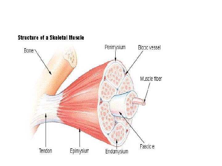

Structure of Skeletal Muscle: Connective Tissue Covering • Epimysium – Surrounds entire muscle • Perimysium – Surrounds bundles of muscle fibers • Fascicles • Endomysium – Surrounds individual muscle fibers

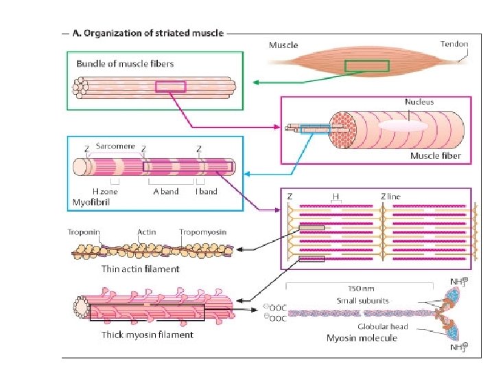
• Muscle fiber 10 to 80μ in diameter each and is composed of 1000 s of myofibrils • Each Myofibril is divided into smaller functional units called, Sarcomere • The Sarcomere contains two sets of Myofilaments • (i) Actin filament (ii) Myosin filament

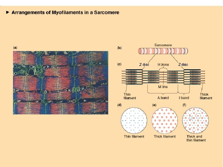
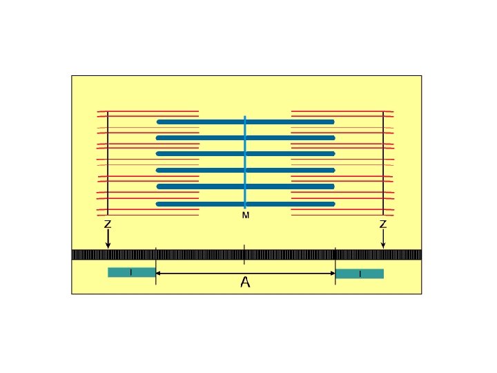
• Sarcomere- between two z-lines. Basic unit of muscle • Cross striations due to alternate dark and light bands • Light band - Isotropic band - I band - Thin filaments • Dark band - Anisotropic band - A band - Thick filaments • H zone - lighter zone in A band • Z line - in the center of I band • M line - in the center of A band
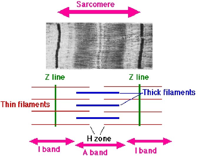

Sarcomere: functional unit of striated muscle
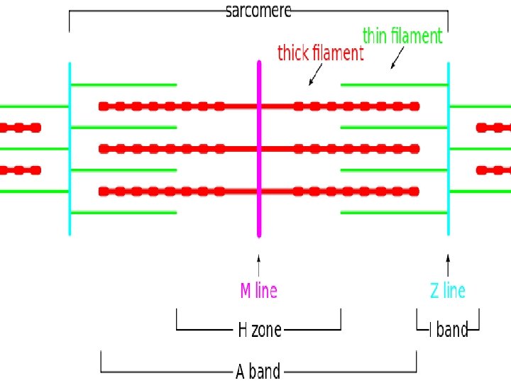

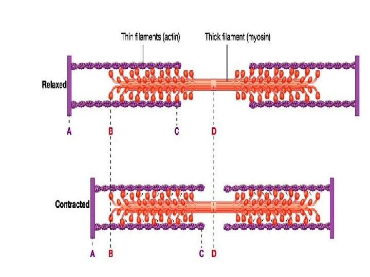
• Sarcomere • Distance between two Z lines. • Whole of A band one half of I band on either side of A band. • At rest the sarcomere length is 2 µm • At contracted state its length is 1. 6 µm • At relaxed state its length is 3. 5 µm
 Myofilament • • Many elongated myosin molecules. Single filament contains roughly 300")
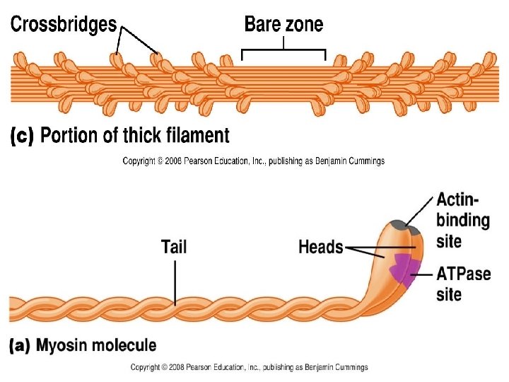
Myosin (Thick) Myofilament • • Many elongated myosin molecules. Single filament contains roughly 300 myosin molecules Molecule consists of two heavy chain and four light chain. Myosin heads 1. Can bind to active sites on the actin molecules to form cross-bridges. (Actin binding site) 2. Attached to the rod portion by a hinge region that can bend and straighten during contraction. 3. Have ATPase activity: activity that breaks down adenosine triphosphate (ATP), releasing energy. Part of the energy is used to bend the hinge region of the myosin molecule during contraction

• Has a diameter of 11 nm and a length of 1. 6 micron • Located in the centre of the A band • Cross bridges project from the lateral sides in a helical fashion
 1. F (fibrous) actin Myofilaments 2. Tropomyosin 3.")
• Thin Filament: Actin (Thin) 1. F (fibrous) actin Myofilaments 2. Tropomyosin 3. Troponin • Two strands of fibrous (F) actin form a double helix – G actin each of which has a myosin-binding site – Actin site can bind myosin during muscle contraction. • Tropomyosin: an elongated protein winds along the groove of the F actin double helix.

TROPONIN: Globular units located at intervals along the tropomyosin molecule. Has 3 components • Troponin C- contain binding site for calcium • Troponin T – binds the troponin component to tropomyosin • Troponin I –inhibit the interaction of myosin with actin.

• • Other muscle proteins Actinin-Z-lines to actin Titin-Z lines-M-line Desmin-Z-lines to cell membrane
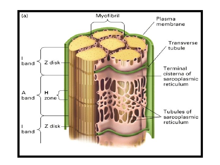

SARCOTUBULAR SYSTEM The myofibrils are surrounded by some important membranous structures which appear in the form of vesicles and tubules. These structures are together called Sarcotubular system • T-tubules (Transverse Tubules) • L-tubules (Sarcoplasmic Reticulum)

T –tubule: They are inwardly directed extensions of the sarcoplasm into the muscle fibers at the junction between A and I bands. Each Sarcomere has 2 tubules. Its function is the rapid transmission of the action potential from the cell membrane to all the fibrils in the muscle. Depolarization of T – tubules activate the L- tubule via dihyropyridine receptors- release of Ca 2+ from L- tubules

Longitudinal Sarcoplasmic Reticulum Longitudinally on the either side of tubular system Vesicles /sacs- Terminal cisternae. Rich in glycogen and Ca 2+ Dihydropyridine receptor Ryanodine receptor Transverse tubule with 2 terminal cisterns - TRIAD L-tubules: • Stores a large quantity of calcium ions. • Triggers the process of muscle contraction

Sarcomere Completely Contracted
- Slides: 29