Muscle muscle tissue tendons Dr Anna L Kiss


- Slides: 33

Muscle, muscle tissue tendons Dr. Anna L. Kiss Department of Anatomy, Histology and Embryology Semmelweis University Budapest 2018

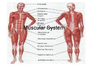
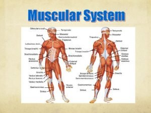
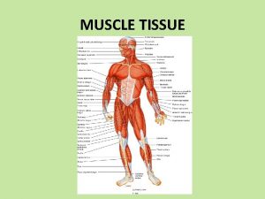
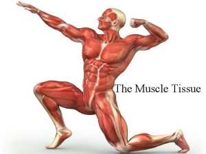
General myology Musculature: actíve component of the movement Structure: striated muscle (skeletal) tendons: dense connective tissue sheath: fascia (epimysium)

Muscle tissue • Smooth muscle • Stiated muscle • Cardiac muscle Function: contraction Origin: mesoderm Structure: cells or „fibers”

Smooth muscle • phylogenetically the most ancient type • spindle shaped cells • slow, non-synchronized, unvoluntary contraction • actin and myosin are NOT arranged in registers • internal organs wall

Smooth muscle Nucleus: in the middle of the cells

Smooth muscle thick filaments: myosin thin fialments: actin dense bodies: to help the contraction

Contraction of smooth muscle

Smooth muscle contraction For the contraction: Ca ion: caveolae Ca-binding protein: calmodulin Contractile protein: actin+myosin ATP

Striated(skeletal) muscle

Striated muscle: a. ) skeletal b. ) visceral c. ) cardiac Structure: muscle fibers: multinucleated giant cells (syncitium: fusion of the embryonic myoblasts) lenght: > 30 cm diameter: 10 -100 µm

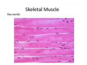
Skeletal muscle

Electron Microscopic picture Sarcomere: functional unit

Structural unit of the striated muscle: sarcomera Muscle fibers myofilaments: contractile proteins: actin and myosin regularly arranged cross striation

Sarcomere during contraction

Skeletal muscle

Contractile proteins Thin filaments Myosin head: actin binding site + ATP binding thick filaments

Sarcomere

mysosine head mysosine binding site


For the contraction of the skeletal muscle: • • contractile proteins: actin and myosin Ca 2+ (stored in s. ER) impulse transfer from the sarcoplasm to s. ER-re (triads) ATP (directly from kreatin phosphate, 20 mmól/kg) aerob (biological oxidation) glikogén anaerob (fermentation) • mitochondria • oxigen (myoglobin + haemoglobin)

Triád: a. ) voltage-gated Ca 2+ channels: T-tubules (transverse) SR ciszterna Ca 2+ outflow b. ) Ca 2+ -ATP-ase Ca 2+ back to the SER

Types of the skeletal muscle size diameter contraction color fat in the cytoplasm glycogen in the cytoplasm resistancy „red” muscle „white” muscle small large fibers slow dark (red) numorou s numorous fewer bigger fast light (white) few fewer larger amount smaller

Cardiac muscle

Cardiac muscle • cells (bifurcation; X or Y shaped branching ce • cross striation: actin and myosin are in register • nucleus is in the middle of the cells • intercalated disc (Eberth’s line – junctions) • lots of capillaries • lipofuscin by aging

Cardiac muscle Intercalated disc: • special junctions between cells • fast impulse cinduction

Intercalated disc (Eberth’s line) • fascia adherens • desmosoma • gap junction (nexus)

Cardiac muscle

Fine structure of the cardiac muscle • diad • large amount of mitochondria

impulse condacting cells: Purkinje cells non-differentiated muscle cells!!

proximal end: origin belly distal end: insertion tendon

Structure of the tendon sheath a. ) outer, fibrous layer b. ) inner, synovial layer mesotendon

Muscle • shape: spindle, triangular, quadrangular, flat • venter (belly), caput (head), tendineous intersection, aponeurosis, • unipennatus, bipennatus,

spindle biceps unipennate bipennate
 Similar pictures
 What type of connective tissue are tendons and ligaments
 Lanz point
 Submucosa
 Anconeus
 Bad horse conformation pictures
 Knee muscle anatomy
 Ligament
 Orbicularis oculi origin and insertion
 Diagram of heart with labelling
 Youtube.com
 Perforation plates
 Types of muscle tissue
 Types of muscle tissue
 Four basic tissues
 Sarcomere
 Muscle tissue
 Properties of heart muscle
 Skeletal muscle tissue structure
 Skeletal muscle tissue description
 Types of muscle tissue
 What makes up muscle tissue
 Muscle tissue
 Tissue that connects muscle to bone
 Smooth muscle gap junctions
 Muscle tissue
 Characteristics of skeletal smooth and cardiac muscle
 Groups of cells with a common structure and function.
 Muscle tissue parts
 Muscles and muscle tissue chapter 9
 Smooth muscle
 Skeletal muscle tissue 40x
 Structural proteins in muscle
 Tropomyosin muscle contraction
