Muscle Mechanics Muscles are named by Location Shape

Muscle Mechanics

Muscles are named by…………. • Location • Shape • • • Eg Tibialis anterior Eg Rhomboids major/minor Rectus abdominis Eg Ex. Carpi ulnaris –extension of the wrist Action Number of heads or Eg Biceps brachii, Triceps brachii divisions Eg Sternocleidomastoid Attachments muscle Eg Internal oblique Eg Pectoralis major – large Fiber direction Pectoralis minor - small • • Size of the muscle

Arrangement of skeletal muscle fibers

Muscle Fiber Arrangement • Parallel – longer, ROM – Strap Eg Sartorius, Rectus abdominis, Sternocleidomastoid – Fusiform Eg Biceps, Brachialis – Rhomboidal Eg Rhomboids, Gluteus maximus – Triangular Eg Pectoralis major 4

• Oblique – shorter, greater strength – Unipennate Semi membrinosus – Bipennate Rectus femoris – Multipennate Deltoid 5

Muscle Contraction • Nerve impulse reaches myoneural junction • Acetylcholine is released from motor neuron • Ach binds with receptors in the muscle membrane to allow sodium to enter • Sodium influx will generate an action potential in the sarcolemma
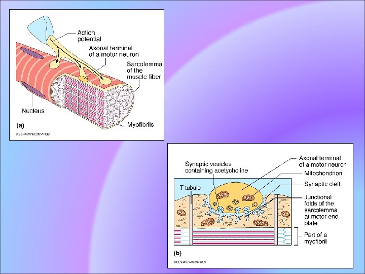
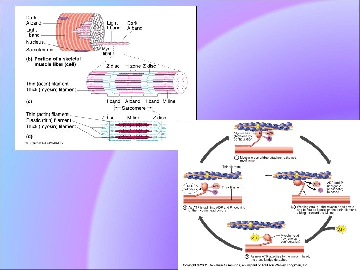

Muscle Contraction Continued • Action potential travels down T tubule • Sarcoplamic reticulum releases calcium • Calcium binds with troponin to move the troponin, tropomyosin complex • Binding sites in the actin filament are exposed

Muscle Contraction Continued • Myosin head attach to binding sites and create a power stroke • ATP detaches myosin heads and energizes them for another contaction • When action potentials cease the muscle stop contracting

Muscular Attachments • Origin – On more stable bone – Closer to the trunk • Insertion – On more mobile bone – Closer to distal end of bone Normal muscle action: Insertion towards origin Eg During elbow flexion 11

• Reversal of muscle action – Origin moves toward insertion – More movable segment is stabilized – Eg; Elbow flexion in a person hanging on a bar, humerus moves towards the elbow 12

Voluntary muscle contraction Main 5 types, • Concentric contraction force generated is sufficient to overcome the resistance • muscle shortens as it contracts. • Eccentric contraction force generated is insufficient to overcome the external load on the muscle • muscle fibers lengthen as they contract. • Use in the means of decelerating a body part or object, or lowering a load gently rather than letting it drop.

• Isometric contraction muscle remains the same length. • force precisely matches the load, and no movement results.

• Isotonic contractions the tension in the muscle remains constant despite a change in muscle length. • This can occur only when a muscle's maximal force of contraction exceeds the total load on the muscle.
, the muscle contraction velocity remains constant,")
• In isovelocity contraction (sometimes called "isokinetic"), the muscle contraction velocity remains constant, while force is allowed to vary. • True isovelocity contractions are rare in the body

Motor Unit All the muscle cells controlled by one nerve cell

Motor Unit Ratios • Back muscles – 1: 100 • Finger muscles – 1: 10 • Eye muscles – 1: 1

Muscle spindle • One type of proprioceptors • Sensory receptors within the belly of a muscle • Primarily detect changes in the length of this muscle • They convey length information to the central nervous system via sensory neurons • Responses of muscle spindles to changes in length also play an important role in regulating the contraction of muscles

Anatomy • Found within the belly of muscles embedded in extrafusal muscle fibers • Muscle spindles are composed of 312 intrafusal fibers • Encapsulated by connective tissue, and are aligned parallel to extrafusal muscle fibers
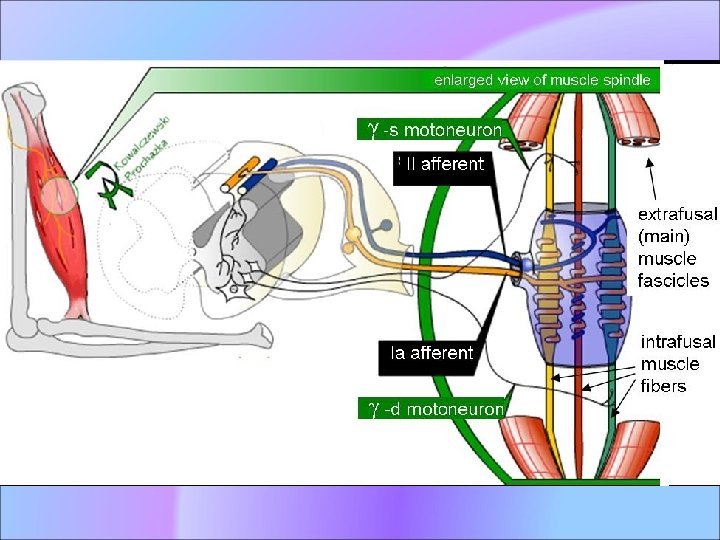

• Muscle spindle has both sensory and motor components. • Primary and secondary sensory nerve fibers spiral around and terminate on the central portions of the intrafusal muscle fibers • Gamma and beta motoneurons are called fusimotor neurons, because they activate the intrafusal muscle fibers. Gamma motoneurons only innervate intrafusal muscle fibers • Alpha motoneurons innervate both extrafusal and intrafusal muscle fibers and so are referred to as skeletofusimotor neurons

Stretch reflex • When a muscle is stretched, primary sensory fibers of the muscle spindle respond • transmit this activity to the spinal cord in the form of changes in the rate of action potentials • many alpha motor neurons of the receptorbearing muscle • The reflexly-evoked activity in the alpha motoneurons is then transmitted via their efferent axons to the extrafusal fibers of the muscle, which generate force and thereby resist the stretch.

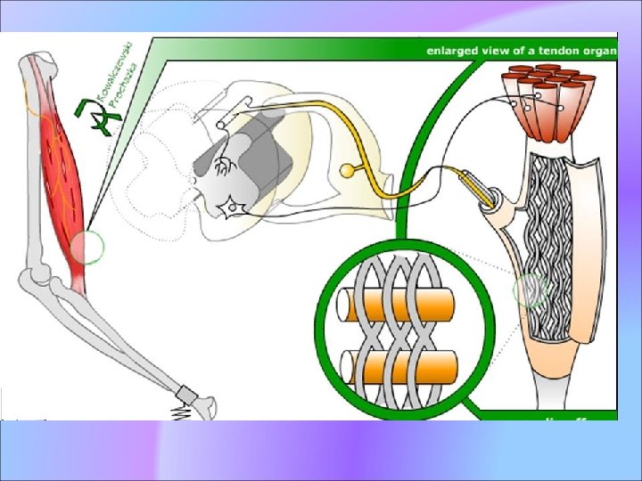
Golgi tendon organ • Another type of proprioceptors which is located at the insertion of tendon into the skeletal muscle. • It provides the sensory component of the Golgi tendon reflex • Provide information about the changes in muscle tension • made up of strands of collagen that are connected at one end to the muscle fibers and at the other merge into the tendon proper.

• innervated by a single afferent type 1 b sensory fiber that branches and terminates as spiral endings around the collagen strands

Function • When the muscle generates force, the sensory terminals are compressed. • This deforms the terminals of the Ib afferent axon • Opening stretch-sensitive cation channels depolarizing and fires nerve impulses that are propagated to the spinal cord • The Ib sensory feed back generates spinal reflex and supraspinal responses which control muscle contraction. • Main spinal reflexes associated with Ib afferent activity is the autogenic inhibition reflex which helps regulate the force of muscle contractions

Skeletal muscle fiber types • 3 types, – Type II A – Type II B Type I slow oxidative or red fibers, generate most of their ATP from glucose have a high oxidative capacity

Type II B fast glycolytic or white fibers • Have the ability to take up glucose and lack myoglobin • Low oxidative capacity, and a high glycolytic capacity Type II A • Very high oxidative capacity and a high glycolytic capacity • Not very much found in humans

Energy sources for muscle contractions Aerobic Anaerobic • Oxygen is used • Break down of fat, protein & carbohydrates • Can provide energy for a long period • Oxygen is not used • Break down of phosphocreatine • Can provide energy for muscle contraction for a period of 20 - 30 sec

Muscle Fatique • Lack of oxygen causes ATP deficit • Lactic acid builds up from anaerobic respiration

Muscle Atrophy • Weakening and shrinking of a muscle • May be caused – Immobilization – Loss of neural stimulation

Muscle Hypertrophy • Enlargement of a muscle • More capillaries • More mitochondria • Caused by – Strenuous exercise – Steroid hormones

Properties of muscle Muscles has following properties • • Excitability/Irritability Contractility Extensibility Elasticity

• Excitability/Irritability: – ability to receive and respond to stimuli inside or outside the body • Contractility: – ability to shorten to ½ its resting length when receive adequate stimulus 35

• Extensibility: – Ability to be stretched 2 times as far as it is able to be shortened • Elasticity: – Ability to resume resting length after stretch or shortening 36

Length tension diagram of a muscle

Strength of muscles • Depends upon the number of fibers in the physiological cross-section • Physiological cross-section - which passes through practically all of the fibers • Anatomical cross section – • unipinnate and bipinnate muscles the physiological cross-section may be nearly at right angles to the anatomical cross-section


- Slides: 40