
Muscle contraction hox genes respiration translocation Hox Genes
- Slides: 32
Muscle contraction, hox genes, respiration & translocation
Hox Genes
Hox Genes Definitions • Homeobox gene sequences • About 180 bp sequence found WITHIN genes • Highly conserved regions • Hox genes • Group of genes that control body plan • Determine type of segment structure in embryo development e. g. leg, antennae
Muscle Contraction
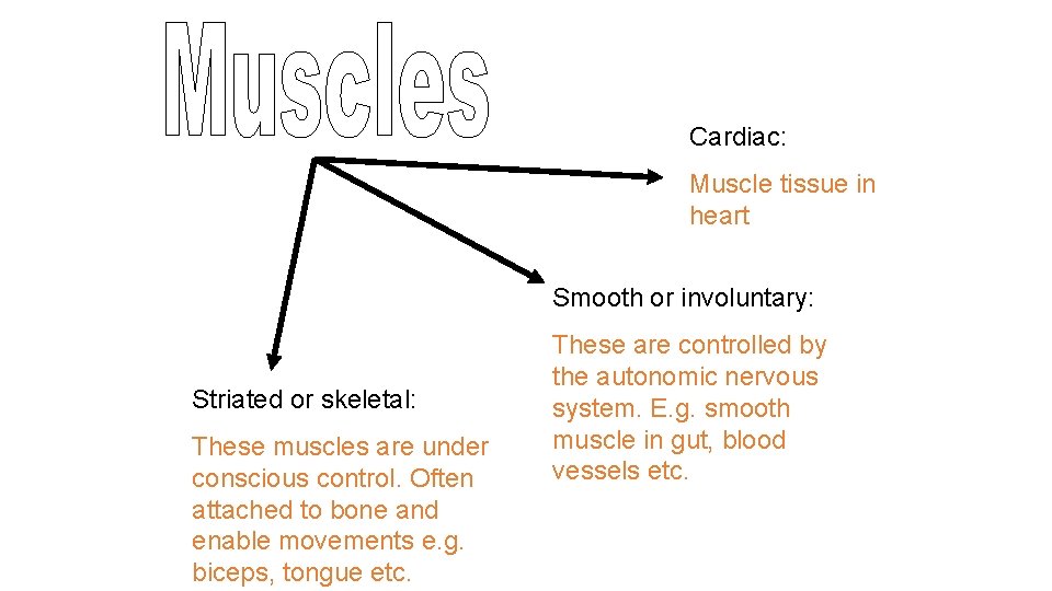
Cardiac: Muscle tissue in heart Smooth or involuntary: Striated or skeletal: These muscles are under conscious control. Often attached to bone and enable movements e. g. biceps, tongue etc. These are controlled by the autonomic nervous system. E. g. smooth muscle in gut, blood vessels etc.
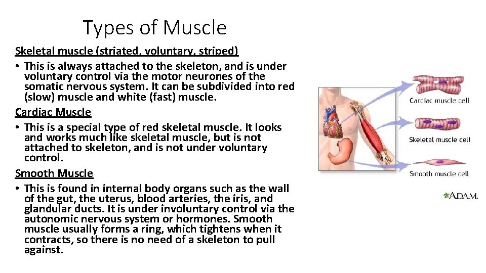
Types of Muscle Skeletal muscle (striated, voluntary, striped) • This is always attached to the skeleton, and is under voluntary control via the motor neurones of the somatic nervous system. It can be subdivided into red (slow) muscle and white (fast) muscle. Cardiac Muscle • This is a special type of red skeletal muscle. It looks and works much like skeletal muscle, but is not attached to skeleton, and is not under voluntary control. Smooth Muscle • This is found in internal body organs such as the wall of the gut, the uterus, blood arteries, the iris, and glandular ducts. It is under involuntary control via the autonomic nervous system or hormones. Smooth muscle usually forms a ring, which tightens when it contracts, so there is no need of a skeleton to pull against.
Neuromuscular Junction
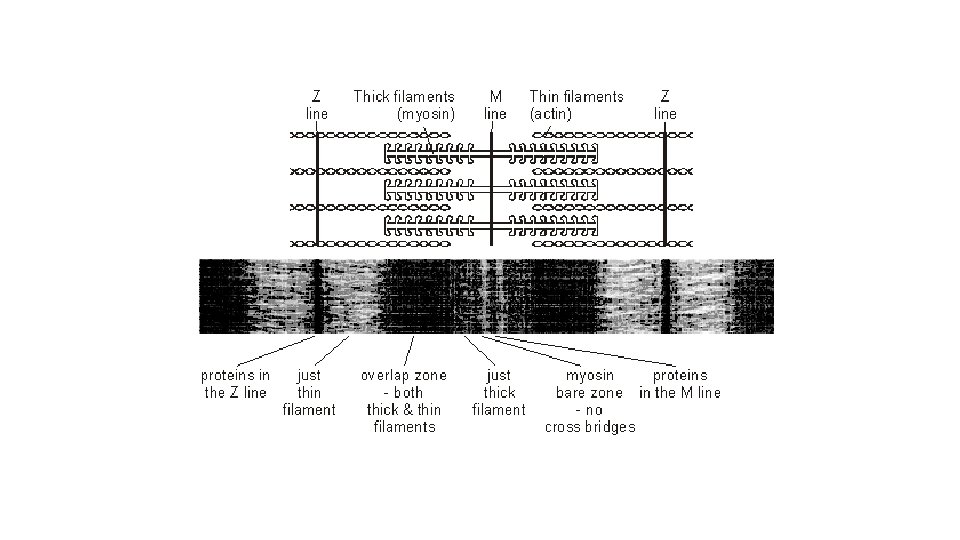
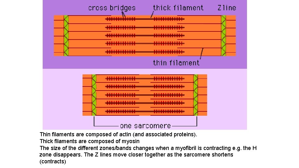
Thin filaments are composed of actin (and associated proteins). Thick filaments are composed of myosin The size of the different zones/bands changes when a myofibril is contracting e. g. the H zone disappears. The Z lines move closer together as the sarcomere shortens (contracts)
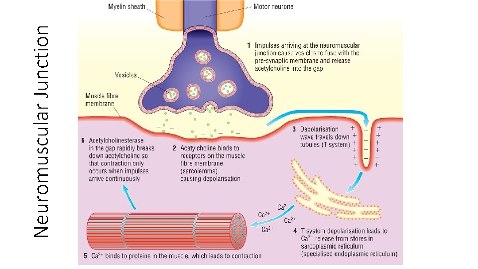
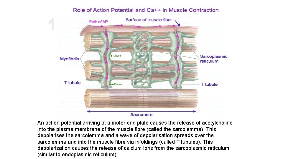
1 An action potential arriving at a motor end plate causes the release of acetylcholine into the plasma membrane of the muscle fibre (called the sarcolemma). This depolarises the sarcolemma and a wave of depolarisation spreads over the sarcolemma and into the muscle fibre via infoldings (called T tubules). This depolarisation causes the release of calcium ions from the sarcoplasmic reticulum (similar to endoplasmic reticulum).
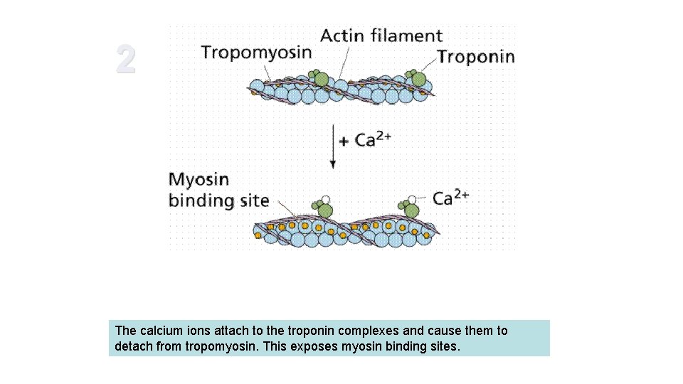
2 The calcium ions attach to the troponin complexes and cause them to detach from tropomyosin. This exposes myosin binding sites.
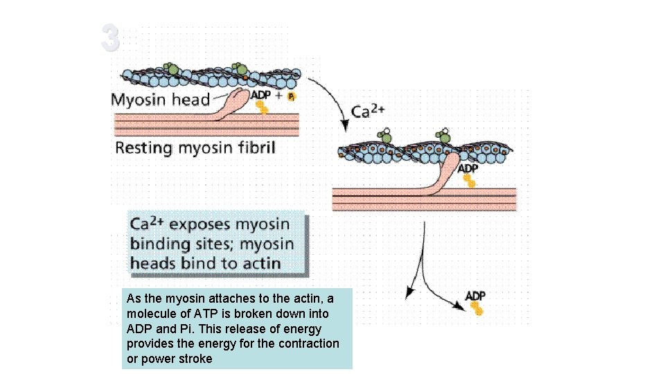
3 As the myosin attaches to the actin, a molecule of ATP is broken down into ADP and Pi. This release of energy provides the energy for the contraction or power stroke
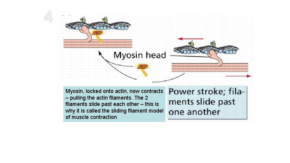
4 Myosin, locked onto actin, now contracts – pulling the actin filaments. The 2 filaments slide past each other – this is why it is called the sliding filament model of muscle contraction
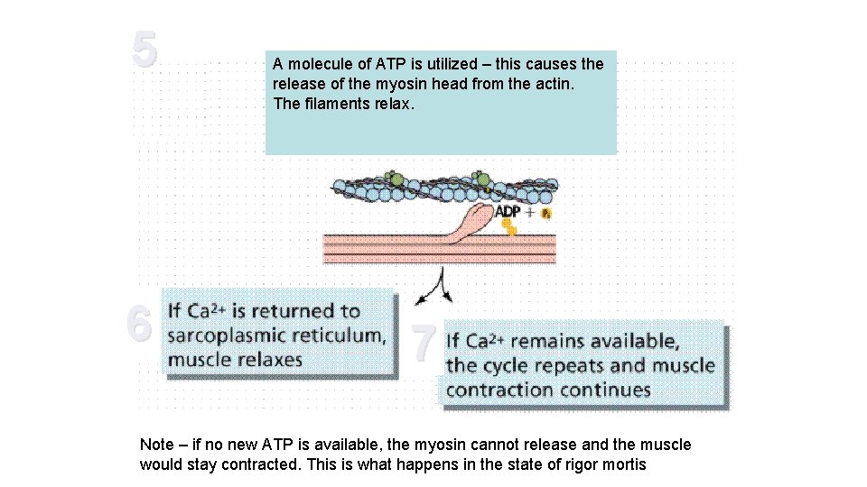
5 6 A molecule of ATP is utilized – this causes the release of the myosin head from the actin. The filaments relax. 7 Note – if no new ATP is available, the myosin cannot release and the muscle would stay contracted. This is what happens in the state of rigor mortis
Maintaining ATP supply 1. Aerobic respiration in muscle cells. Needs a supply of respiratory substrate and oxygen 2. Anaerobic respiration. Produces lactate and can lead to fatigue/cramp. 3. Creatinine phosphate – another chemical present in muscle cells can donate its phosphate to recharge ADP back to ATP
Translocation
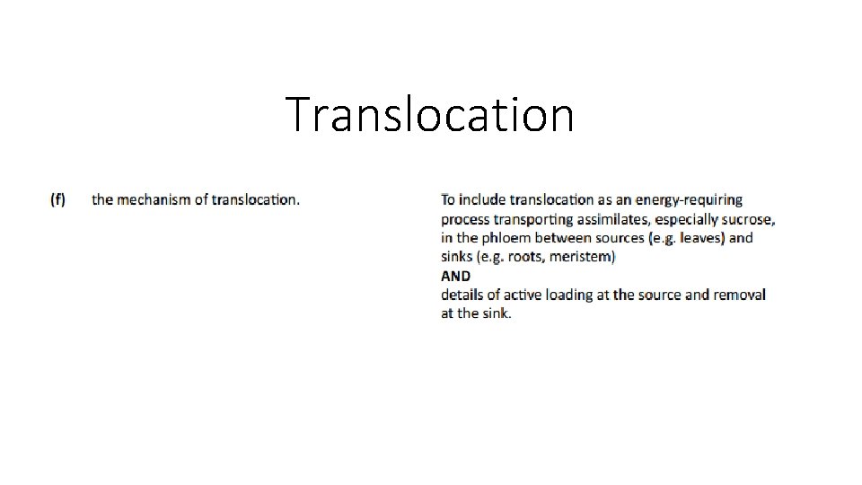
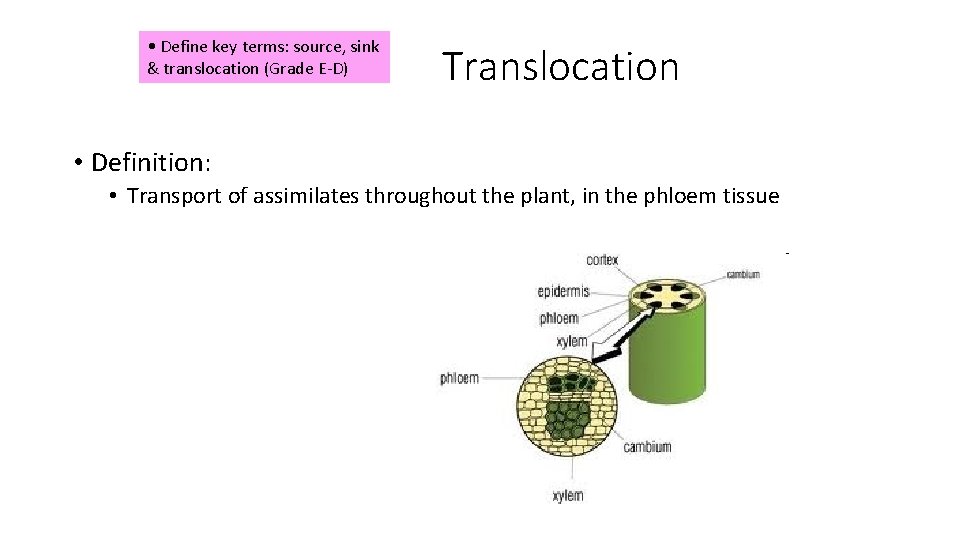
• Define key terms: source, sink & translocation (Grade E-D) Translocation • Definition: • Transport of assimilates throughout the plant, in the phloem tissue
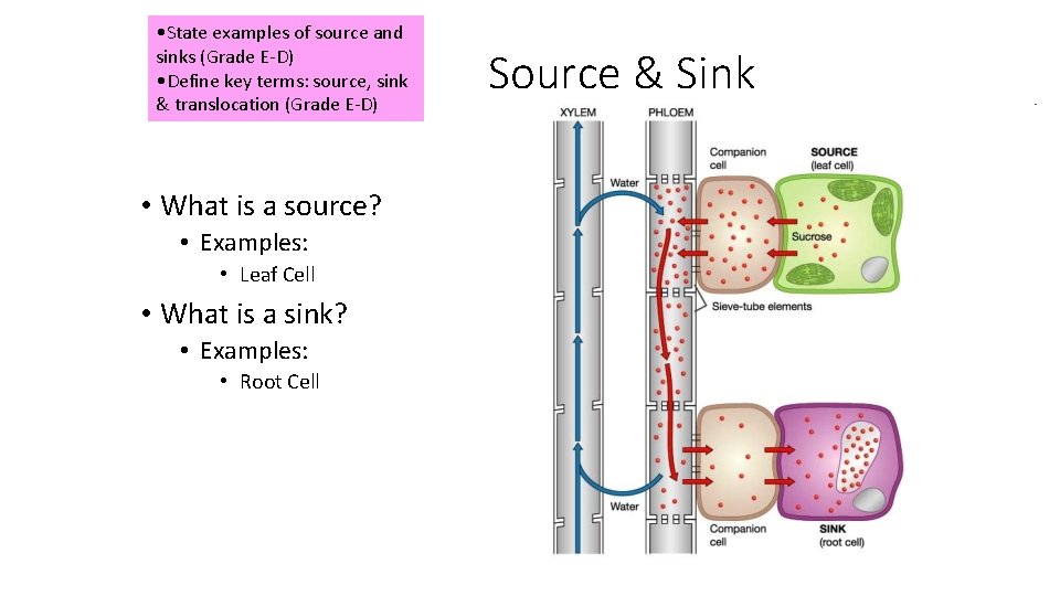
• State examples of source and sinks (Grade E-D) • Define key terms: source, sink & translocation (Grade E-D) • What is a source? • Examples: • Leaf Cell • What is a sink? • Examples: • Root Cell Source & Sink
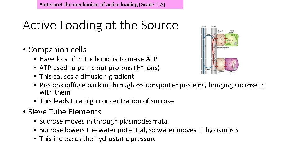
• Interpret the mechanism of active loading (Grade C-A) Active Loading at the Source • Companion cells Have lots of mitochondria to make ATP used to pump out protons (H+ ions) This causes a diffusion gradient Protons diffuse back in through cotransporter proteins, bringing sucrose in with them • This leads to a high concentration of sucrose • • • Sieve Tube Elements • Sucrose moves in through plasmodesmata • Sucrose lowers the water potential, so water moves in by osmosis • This increases the hydrostatic pressure
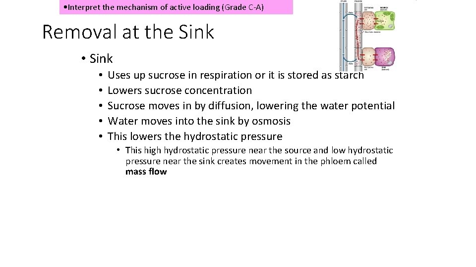
• Interpret the mechanism of active loading (Grade C-A) Removal at the Sink • • • Uses up sucrose in respiration or it is stored as starch Lowers sucrose concentration Sucrose moves in by diffusion, lowering the water potential Water moves into the sink by osmosis This lowers the hydrostatic pressure • This high hydrostatic pressure near the source and low hydrostatic pressure near the sink creates movement in the phloem called mass flow
• Evaluate evidence for this mechanism (Grade C-A) OLD SPEC: Evidence • Active loading and movement of sucrose in the phloem is a theory • Evidence supports this theory
Radioactive labelling of carbon dioxide (for photosynthesis) • Labelled carbon observed in the phloem • Movement of labelled sucrose is faster than it would be by diffusion • Sucrose is moved to all parts of the plant at the same rate, rather than going quicker to areas of low concentration
Tree Ringing • Removal of bark in a ring all around the tree trunk, which removes the phloem but leaves the xylem intact • Tree swells above the ring, where sugar collect Mitochondria • Companion cells have large amounts of mitochondria
Aphids • Feed by inserting mouth parts into phloem • Analysis of fluids collected from aphids contain lots of sugars p. Hthan surrounding cells • Companion Cells p. H have a higher
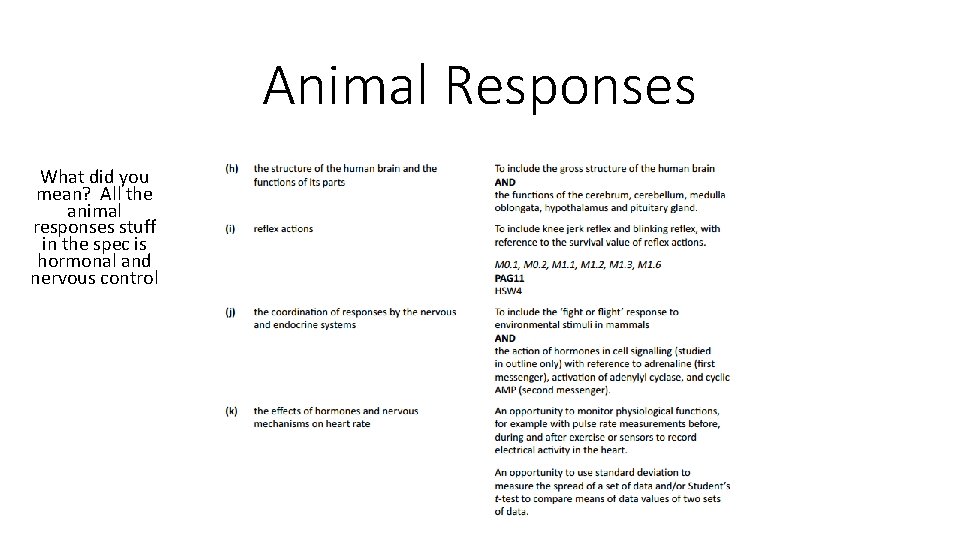
Animal Responses What did you mean? All the animal responses stuff in the spec is hormonal and nervous control
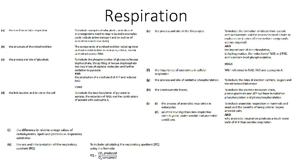
Respiration
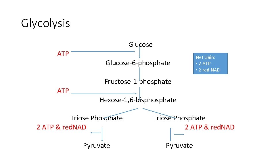
Glycolysis Glucose ATP Glucose-6 -phosphate Net Gain: • 2 ATP • 2 red NAD Fructose-1 -phosphate ATP Hexose-1, 6 -bisphosphate Triose Phosphate 2 ATP & red. NAD Pyruvate
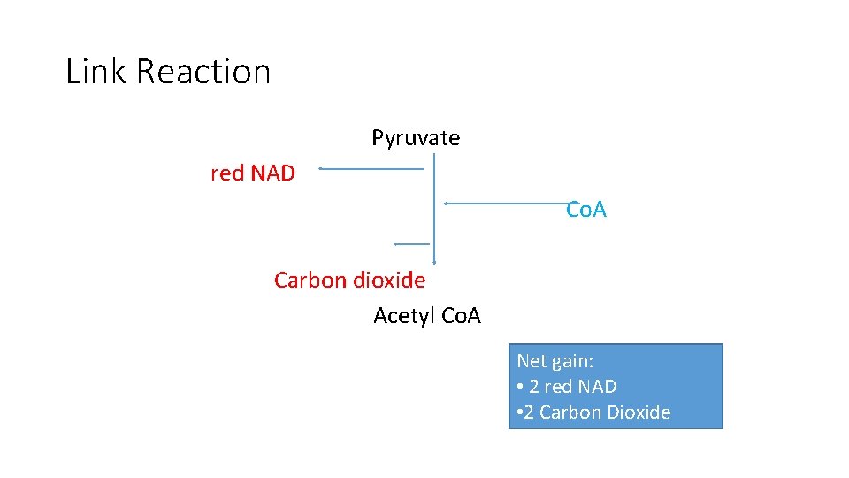
Link Reaction Pyruvate red NAD Co. A Carbon dioxide Acetyl Co. A Net gain: • 2 red NAD • 2 Carbon Dioxide
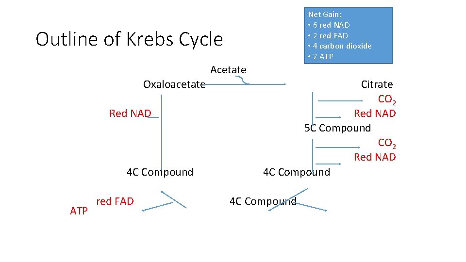
Net Gain: • 6 red NAD • 2 red FAD • 4 carbon dioxide • 2 ATP Outline of Krebs Cycle Acetate Oxaloacetate Red NAD 4 C Compound ATP red FAD Citrate CO 2 Red NAD 5 C Compound CO 2 Red NAD 4 C Compound
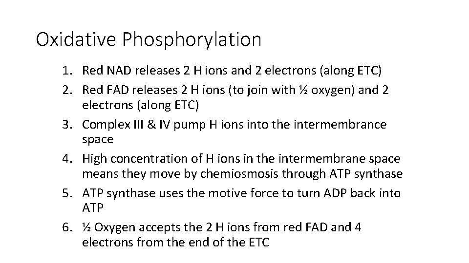
Oxidative Phosphorylation 1. Red NAD releases 2 H ions and 2 electrons (along ETC) 2. Red FAD releases 2 H ions (to join with ½ oxygen) and 2 electrons (along ETC) 3. Complex III & IV pump H ions into the intermembrance space 4. High concentration of H ions in the intermembrane space means they move by chemiosmosis through ATP synthase 5. ATP synthase uses the motive force to turn ADP back into ATP 6. ½ Oxygen accepts the 2 H ions from red FAD and 4 electrons from the end of the ETC
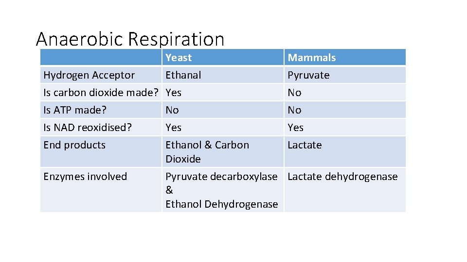
Anaerobic Respiration Yeast Hydrogen Acceptor Ethanal Is carbon dioxide made? Yes Is ATP made? No Mammals Pyruvate No No Is NAD reoxidised? End products Yes Ethanol & Carbon Dioxide Yes Lactate Enzymes involved Pyruvate decarboxylase Lactate dehydrogenase & Ethanol Dehydrogenase
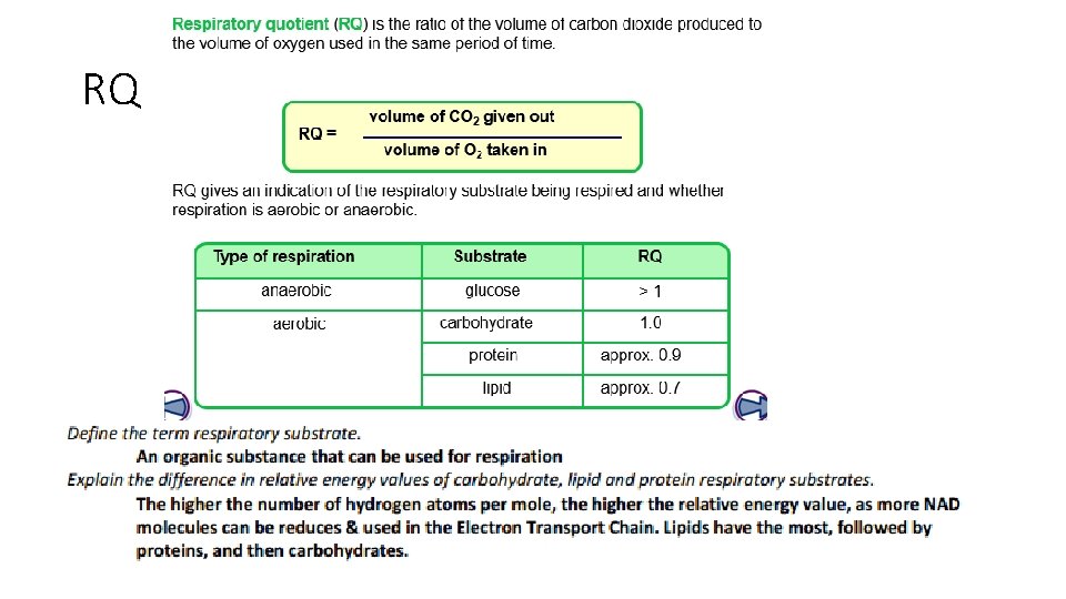
RQ