Musckuloskeletal MCQs Aneurysmal Bone Cyst ABC expansile lesion

Musckuloskeletal MCQs
 • expansile lesion of bone containing thin-walled blood- filled cystic")
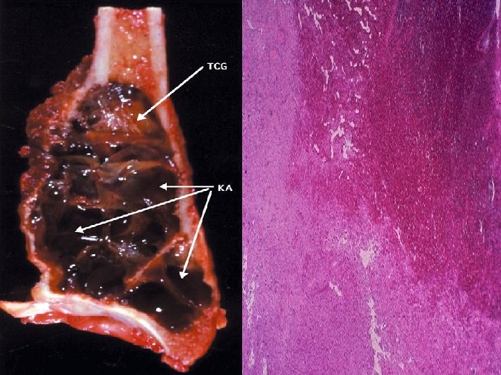
Aneurysmal Bone Cyst (ABC) • expansile lesion of bone containing thin-walled blood- filled cystic cavities Definition Etiology • (a) primary nonneoplastic lesion (2/3) • (b) arising in preexisting bone tumor (1/3): giant cell tumor (39%), osteoblastoma, chondroblastoma, solitary bone cyst, fibrous dysplasia, nonossifying fibroma, metastatic carcinoma • intraosseous arteriovenous malformation" with honeycombed spaces filled with blood + lined by granulation tissue / osteoid; areas of free hemorrhage Histology
 • INTRAOSSEOUS ABC : originating in bone Types: Age: marrow")
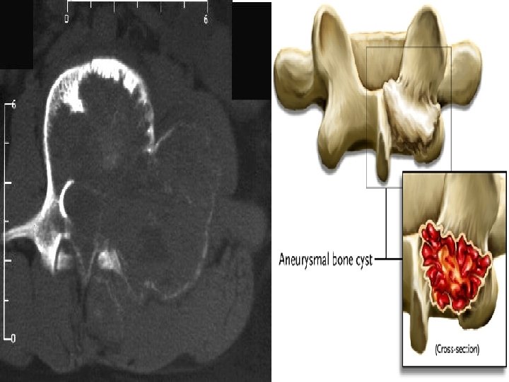
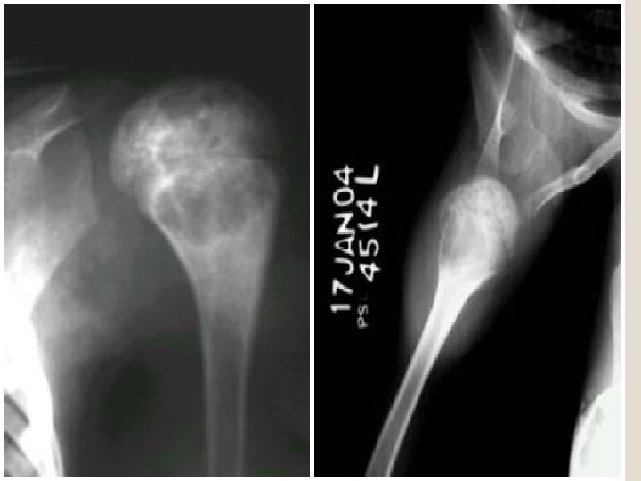
Aneurysmal Bone Cyst (ABC) • INTRAOSSEOUS ABC : originating in bone Types: Age: marrow cavity. • EXTRAOSSEOUS ABC : posttraumatic hemorrhagic : cyst; originating on surface of bones • peak age 16 years (range 10 - 30 years) • Spine: with slight predilection for posterior elements Location: • long bones: eccentric in metaphysis of femur, tibia, humerus, fibula; pelvis

Respect epiphyseal plate soap-bubble Thin internal trabeculations Almost invesible cortex Expansile lesion No periosteal reaction except in case of fracture


Doughnut sign in scintigraphy

Aneurysmal Bone cyst : a. b. c. d. e. Occur secondary to fibrous dysplasia (√ ) Can present with scoliosis (√ ) May contain fluid-fluid level at MRI (√ ) Contain calcified matrix (X ) Doughnut sign at scintigraphy is pathognomonic (X )

Multiple Myeloma

Mulyiple osteolytic Punched-out lesion

Mulyiple osteolytic Punched-out lesion Absence of perilesional sclerosis

Complicated by pathological fracture Associated with osteopenia

PET PET-CT coronal view

Regarding Multiple Myeloma : a. It is the commonest 1 ry neoplasm of bone (√ ) b. Generalised osteopenia is a recognised appearance (√ ) in 15 % c. Scintigraphy over-estimates disaese d. (X ) under estimates Lesions becomes scleotic following extent chemotherapy e. ) also following Vertebral pedicle(√ destruction is an radiotherapy early event (X ) (Ref: Grainger and Allison pp 1913 -1915, Daenhartppp 121 -122, Chapman 2003 pp 575 -576)

Rheumatoid arthritis :

Joint deformity Narrowing of the joint spaces Periarticular osteopenia Soft tissue swelling Joint instability due to ligament rupture Articular erosion

High ridding of the humerus Lateral erosion of the clavicle Marginal erosion Narrowing of the glenohumoral joint

Lateral deviation of toes , Hammer toes Erosion of the MTP

Erosion of Odontoid process ligamentous destruction can result in atlantoaxial impaction Pannus proliferation directly compresses the spinal cord. Erosion of facet joints

Erosion of the lateral 3 rd of the clavicle is seen in the following: a. Rheumatoid arthritis b. Ankylosing spondylitis c. Langerhans cell histiocytosis d. Hypoparathyroidism (X ) Hyperparathyroidism e. Multiple myeloma (√ ) (X )

Concerning Malignant Bone Lesions

Concerning Malignant bone lesion: a. Chordoma is most common in the thoracolumbar spine b. (X ) 90% in basisphenoid and sacrum Fibrosarcoma is the commonest tumour 2 ry to paget’s disease c. Adamantinoma occursin the tibia in over 90% of cases d. Angiosarcoma has a soap bubble appearance at radiography e. Chondroblastoma are typically found in the diaphysis

Sacral Chordoma

Basisphenoid Chordoma

Concerning Malignant bone lesion: a. Chordoma os most common in the thoracolumbar spine b. (X ) 90% in basisphenoid and sacrum Fibrosarcoma is the commonest tumour 2 ry to paget’s disease c. Adamantinoma occurs in the tibia in over 90% of cases d. (√ ) Angiosarcoma has a soap bubble appearance at radiography e. Chondroblastoma are typically found in the diaphysis

Adamantinoma

Concerning Malignant bone lesion: a. Chordoma os most common in the thoracolumbar spine b. (X ) 90% in basisphenoid and sacrum Fibrosarcoma is the commonest tumour 2 ry to paget’s disease c. (√ ) Adamantinoma occursin the tibia in over 90% of cases (√ ) d. Angiosarcoma has a soap bubble appearance at radiography e. (√) Chondroblastoma are typically found in the diaphysis (X ) they are epiphyseal lesions

Chondroblastoma

Thank you
- Slides: 35