Mullerian Anomalies infertility Bahia Namavar Jahromi 12 11


Mullerian Anomalies & infertility Bahia Namavar Jahromi 12. 11. 98

DENITOURINARY TRACT DEVELOPMENT �In females, the external genitalia, gonads, and müllerian ducts each derive from different primordia and in close association with the urinary tract and hindgut. � Abnormal embryogenesis during this process is thought to be multifactorial and can create sporadic anomalies. �Several of these can lead to infertility, subfertility, miscarriage, or preterm delivery. Thus, knowledge of genitourinary system development is essential.

Embryology of the Urinary System �Between the 3 rd and 5 th GA, an elevation of intermediate mesoderm on each side of the fetus—the urogenital ridge—begins development into the urogenital tract. �The urogenital ridge divides into the genital ridge, destined to become the ovary, and into the nephrogenic ridge. The nephrogenic ridges develop into the mesonephros (mesonephric kidney) and paired mesonephric ducts, also termed wolffian ducts, which connect to the cloaca.

Copyrights apply
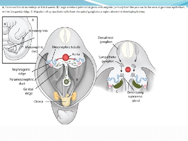

�The early urinary tract develops from the mesonephros and its mesonephric ducts (Fig. 3 -2 A). Recall that evolution of the renal system passes sequentially through the pronephric and mesonephric stages to reach the permanent metanephric system. Between the 4 th and 5 th weeks, each mesonephric duct gives rise to a ureteric bud, which grows cephalad toward its respective mesonephros (Fig. 3 -2 B). � As each bud lengthens, it induces differentiation of the metanephros, which will become the final kidney (Fig. 32 C). Each mesonephros degenerates near the end of the first trimester, and without testosterone, the mesonephric ducts regress as well.
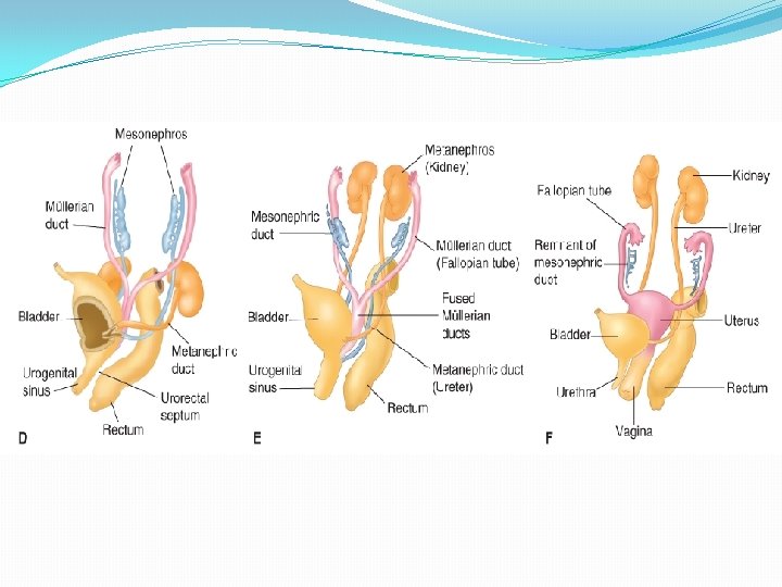

�The cloaca begins as a common opening for the embryonic urinary, genital, and alimentary tracts. By the 7 th week it becomes divided by the urorectal septum to create the rectum and the urogenital sinus. � The urogenital sinus is considered in three parts: (1) the cephalad or vesicle portion, which forms the urinary bladder; (2) the middle or pelvic portion, which creates the female urethra; and (3) the caudal or phallic part, which gives rise to the distal vagina and to the greater vestibular (Bartholin) and paraurethral glands.

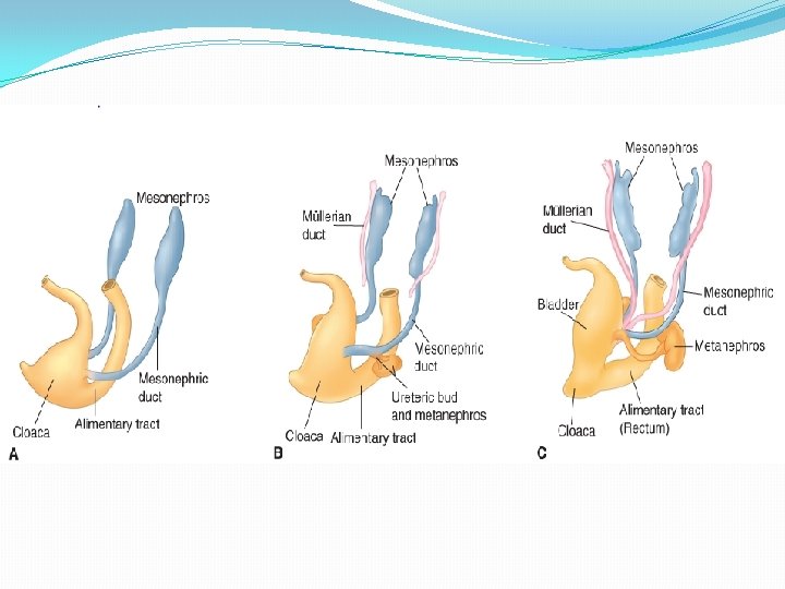
Embryology of the Genital Tract � The fallopian tubes, uterus, and upper vagina derive from the müllerian ducts, also termed paramesonephric ducts, which form adjacent to each mesonephros. � These ducts extend downward and then turn medially to meet and fuse together in the midline. The uterus is formed by this union of the two müllerian ducts at approximately the 10 th week. Fusion to create the uterus begins in the middle and then extends both caudally and cephalad. With cellular proliferation at the upper portion, a thick wedge of tissue creates the characteristic piriform uterine shape. At the same time, dissolution of cells at the lower pole forms the first uterine cavity. � As the upper wedge-shaped septum is slowly reabsorbed, the final uterine cavity is usually formed by the 20 th week. � If the two müllerian ducts fail to fuse, then two separate uterine horns remain. In contrast, resorption failure of the common tissue between them results in various degrees of persistent uterine septum.

�As the distal end of the fused müllerian ducts contacts the urogenital sinus, this induces endodermal outgrowths from the sinus termed the sinovaginal bulbs. These bulbs proliferate and fuse to form the vaginal plate, which later resorbs to form the vaginal lumen. � This vaginal canalization is generally completed by the 20 th week. However, the lumen remains separated from the urogenital sinus by the hymeneal membrane. This membrane further degenerates to leave only the hymeneal ring.

Copyrights apply
 and paramesonephric (müllerian) ducts explains the simultaneous")
�The close association of the mesonephric (wolffian) and paramesonephric (müllerian) ducts explains the simultaneous abnormalities in their end organs. �Kenney and colleagues (1984) showed that up to half of females with uterovaginal malformations have associated urinary tract defects. Anomalies most frequently associated with renal defects are unicornuate uterus, uterine didelphys, and agenesis syndromes, whereas arcuate and bicornuate are less commonly linked. � When müllerian anomalies are identified, the urinary system can be evaluated with magnetic resonance (MR) imaging, sonography, or intravenous pyelography. � With müllerian anomalies, ovaries are functionally normal but have a higher incidence of anatomical maldescent into the pelvis.

�As discussed, the mesonephric ducts usually degenerate, however, persistent remnants may become clinically apparent. Mesonephric or wolffian vestiges can persist as Gartner duct cysts. These are typically located in the proximal anterolateral vaginal wall but may be found at other sites along the vaginal length. � They can be further characterized by MR imaging, which provides excellent image resolution at soft tissue interfaces. Most cysts are asymptomatic and benign and usually do not require surgical excision. �Intra abdominal wolffian remnants in the female include a few blind tubules in the mesovarium—the epoöphoron—and similar ones adjacent to the uterus—paroöphoron (see Fig. 32 F) (Moore, 2013). The epoöphoron or paroöphoron may develop into clinically identifiable cysts in the adult.
 agenesis of both ducts, either focally or along the entire duct")
MÜLLERIAN ABNORMALITIES �(1) agenesis of both ducts, either focally or along the entire duct length; �(2) unilateral maturation of one müllerian duct with incomplete or absent development of the opposite side; �(3) absent or faulty midline fusion of the ducts; �(4) defective canalization. American Fertility Society (1988) classification: �groups with similar clinical characteristics, prognosis for pregnancy, and treatment. It also includes one for abnormalities associated with fetal exposure to diethylstilbestrol (DES).

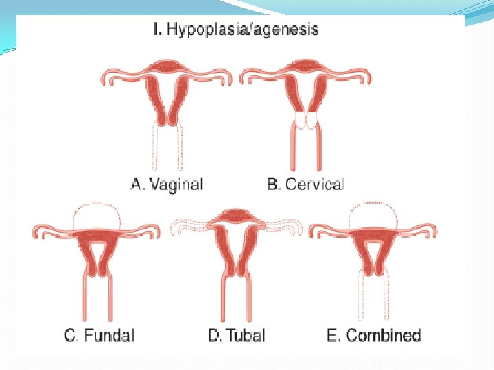
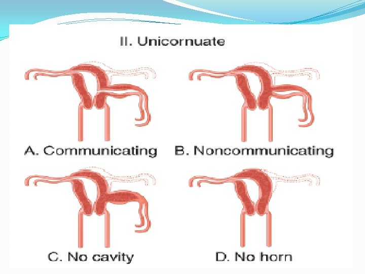
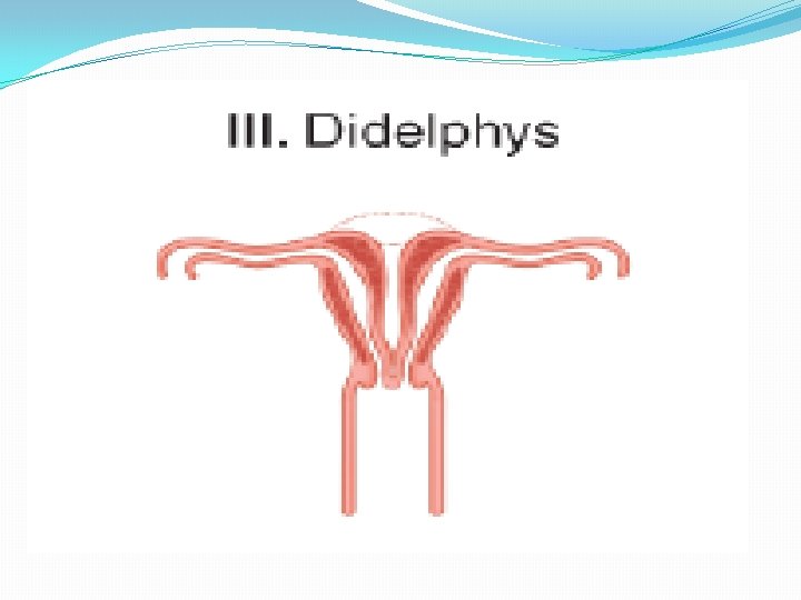
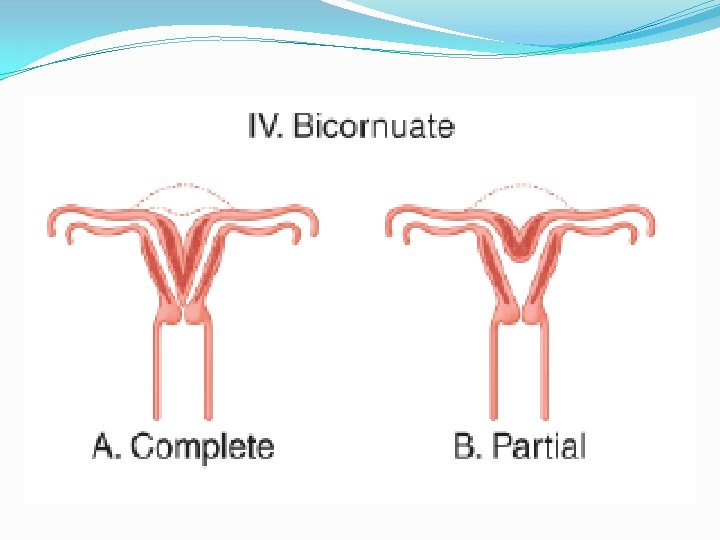
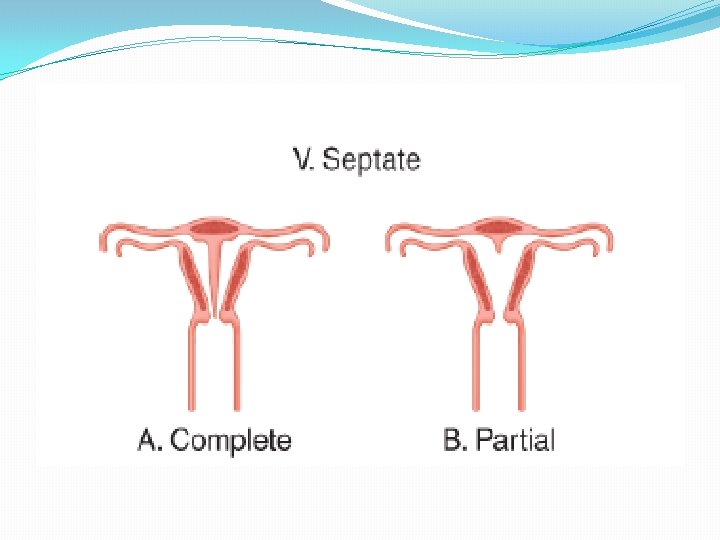
Classification of Müllerian Anomalies � � � I. Segmental müllerian hypoplasia or agenesis a. Vaginal b. Cervical c. Uterine fundal d. Tubal e. Combined anomalies � � � II. Unicornuate uterus a. Communicating rudimentary horn b. Noncommunicating horn c. No endometrial cavity d. No rudimentary horn � III. Uterine didelphys � IV. Bicornuate uterus � a. Complete—division to internal os � b. Partial � V. Septate uterus � a. Complete—septum to internal os � b. Partial � VI. Arcuate � VII. Diethylstilbestrol related

Copyrights apply

Copyrights apply

Copyrights apply

Copyrights apply

Copyrights apply

Copyrights apply

Copyrights apply

PREVALENCE �the prevalence of CUAs was 5. 5% in an unselected population, � 8% in infertile women, � 12. 3% in women with a history of miscarriage, � and 24. 5% in women with miscarriage and infertility [9]. �The prevalence of CUAs in women with primary infertility is not increased; it is approximately the same as that in fertile women with normal reproductive outcomes [12].
, � bicornuate (26 percent),")
Frequency of specific CUAs in affected women �septate (35 percent), � bicornuate (26 percent), �arcuate (18 percent), �unicornuate (10 percent), �didelphys (8 percent), �agenesis (3 percent)

ASSOCIATED ANOMALIES IN OTHER ORGAN SYSTEMS �Women with CUAs are at increased risk of having renal, skeletal, or abdominal wall abnormalities, or a history of inguinal hernia, and vice versa. �Renal anomalies are found in 20 to 30 percent of women with Müllerian defects [7, 8]. Duplex collecting system, horseshoe kidney, pelvic kidney, and unilateral renal agenesis have been associated with an obstructed hemiuterus, obstructed hemivagina, and transverse vaginal septa.

Congenital anomalies of the hymen and vagina �Development of the female genital tract is a complex process that is dependent upon a series of events involving cellular differentiation, migration, fusion, and canalization. Failure of any one of these processes results in a congenital anomaly. �Anomalies of the hymen and vagina may interfere with menstruation, sexual activity, fertility, or childbirth. These typically present after puberty, but are discovered during the neonatal period in some infants

Imperforated hymen
 syndrome, in which upper vaginal agenesis")
Vaginal Abnormalities �vaginal agenesis : Mayer- Rokitansky-Küster-Hauser (MRKH) syndrome, in which upper vaginal agenesis is typically associated with uterine hypoplasia or agenesis. Less often, this syndrome also displays abnormalities of the renal, skeletal, and auditory systems. This triad is known by the acronym MURCS, which reflects müllerian duct aplasia, renal aplasia, and cervicothoracic somite dysplasia �Uterine transplantation is currently experimental but holds future promise for these women.

�congenital septa may form longitudinally or transversely, and each can arise from a fusion or resorption defect. Longitudinal septa divide the vagina into right and left portions.

�A transverse septum poses an obstruction of variable thickness. It may develop at any depth within the vagina, but most are in the lower third. �obstruction or infertility is variably present.

Cervical Abnormalities �Developmental anomalies: �partial or complete agenesis �duplication �longitudinal septa.

Copyrights apply

Copyrights apply

Copyrights apply

�Uncorrected complete agenesis is incompatible with pregnancy, and IVF & gestational surrogacy is an option. � Surgical correction by uterovaginal anastomosis has resulted in successful pregnancy (Kriplani, 2012). � Significant complications for this reason, they recommend hysterectomy for complete cervical agenesis and reserve reconstruction attempts for carefully selected patients with cervical dysgenesis

Uterine Abnormalities �the prevalence found with imaging ranges from 0. 4 to 10 percent. �In a general population, the most common finding is arcuate uterus, followed in descending order by septate, bicornuate, didelphic, and unicornuate classes. �Müllerian anomalies may be discovered during pelvic examination, cesarean delivery, tubal sterilization, or infertility evaluation. � Depending on clinical presentation, diagnostic tools may include sonography, HSG, MRI, laparoscopy, and hysteroscopy.

�In most clinical settings 2 -D TVS is initially performed. �the pooled accuracy for TVS is 90 to 92% �Saline infusion sonography (SIS) improves delineation of the endometrium for a patent endometrial cavity. � 3 -D sonography is more accurate than 2 -D sonography because it provides uterine images from virtually angle.

�MRI: preferred for complex anatomy, especially cases for which corrective surgery is planned. �accuracy of up to 100% for müllerian anomaly �renal or skeletal anomalies can be concurrently evaluated.
 �Look for the underdeveloped or rudimentary horn �If present, it")
Unicornuate Uterus (Class II) �Look for the underdeveloped or rudimentary horn �If present, it may or may not communicate with the dominant horn and may or may not contain an endometrium-lined cavity � General population estimates cite an incidence of 1 in 4000 women � 40% of affected women will have renal anomalies �significant obstetrical risks, including first- and secondtrimester miscarriage, malpresentation, IUGR, fetal demise, PROM, PTB �Rudimentary horns also increase the risk for an ectopic pregnancy within the remnant. prophylactic excision of a horn that has a cavity. ( in pregnancy: MTX or surgically before rupture ) �subsequent pregnancy after excision are scarce. In one series of eight women, all had a preterm cesarean delivery
 was patent*")
NONCOMMUNICATING RUDIMENTARY UTERINE HORN * attached fallopian tube (arrow) was patent*
")
Uterine Didelphys (Class III)

�complete lack of fusion that results in two entirely separate hemiuteri, cervices, and usually two vaginas �It is common among marsupials, for example, the American possum—Didelphys virginiana. Most women have a double vagina or a longitudinal vaginal septum. Uterine didelphys may be isolated. Or, it may compose a triad with an obstructed hemivagina and with ipsilateral renal agenesis (OHVIRA), also known as Herlyn- Werner. Wunderlich syndrome (Tong, 2013). �PE: longitudinal vaginal septum and two cervices. �HSG: two separate endocervical canals. These open into separate noncommunicating fusiform endometrial cavities that each ends with a solitary fallopian tube.

� 2 - or 3 -D TVS: separate divergent uterine horns with a large intervening fundal cleft are seen. Endometrial cavities are uniformly separate. � MRI: valuable in cases without classic findings. �Adverse obstetrical outcomes : similar but less frequent than those seen with unicornuate uterus. Increased risks include miscarriage, preterm birth, and malpresentation. �Metroplasty for either uterine didelphys or bicornuate uterus involves resection of intervening myometrium and fundal recombination (Alborzi, 2015). �for highly selected patients with otherwise unexplained miscarriages. Moreover, no evidence-based data confirm the efficacy of such surgical repair.

IMAGING MODALITIES IN DIDELPHYS UTERUS HSG 3 DUSG MRI

Copyrights apply
 � fusion anomaly : two hemiuteri. �a single cervix (bicornuate")
Bicornuate Uterus (Class IV) � fusion anomaly : two hemiuteri. �a single cervix (bicornuate unicollis) or reach the external os (bicornuate bicollis). a coexistent longitudinal vaginal septum is not uncommon. �Radiological discrimination of a bicornuate uterus from a septate uterus can be challenging. �important because septate uterus can be treated with hysteroscopic septal resection. �With these, an intercornual angle greater than 105 degrees typifies a bicornuate uterus, whereas one less than 75 degrees indicates a septate uterus. �an intrafundal downward cleft measuring ≥ 1 cm or more is indicative of bicornuate uterus. A septate uterus shows a cleft depth <1 cm, or it may have a normal fundal contour. �adverse obstetrical outcomes: miscarriage, PTB, and malpresentation. �metroplasty is reserved for highly selected patients.

Copyrights apply

Copyrights apply

Copyrights apply

IMAGING MODALITIES IN BICORNUATE UTERUS HSG 3 D USG MRI

SURGICAL MANAGEMENT �Metroplasty is reserved only in recurrent aborters �Strassmann procedure either by Laparoscopy or Laparotomy
 �resorption defect: complete or partial rarely a complete vaginocervicouterine septum")
Septate Uterus (Class V) �resorption defect: complete or partial rarely a complete vaginocervicouterine septum is found. �infertility or RPL. �adverse pregnancy outcomes: miscarriage, preterm delivery, and malpresentation �Hysteroscopic septal resection: improve pregnancy rates and outcomes (63% Pregnancy rate and 50% live birth rate following resection).

IMAGING MODALITIES IN SEPTATE UTERUS HSG USG 3 DUSG MRI

Copyrights apply

Copyrights apply

SEPTATE AND BICORNUATE UTERUS Anna Lev-Toaff, MD , Thomas Jefferson University, PA
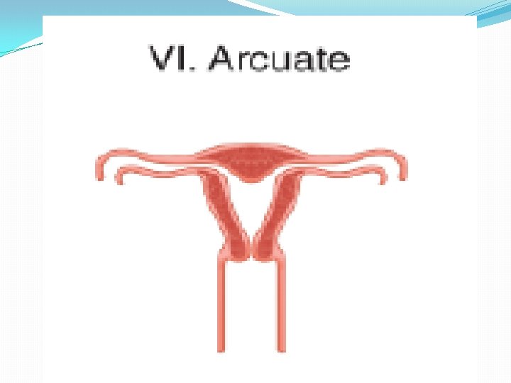
 �mild deviation from the normally developed uterus.")
Arcuate Uterus (Class VI) �mild deviation from the normally developed uterus.

Copyrights apply

Copyrights apply

�Clinical significance — Although previously believed to have clinical significance, an arcuate uterus is now considered to be a normal variant. Patients are asymptomatic, have no compromise of fertility, and similar pregnancy outcomes as those in the general obstetric population.

Treatment with Cerclage �Some women with uterine anomalies and repetitive pregnancy losses may benefit from transvaginal or transabdominal cervical cerclage �Others with partial cervical atresia or hypoplasia may also benefit.
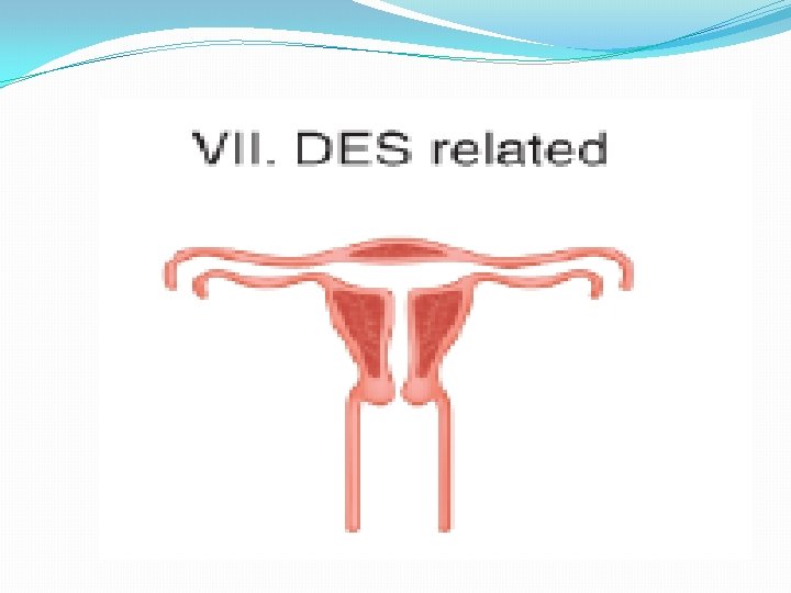
 � During the 1960 s, a synthetic nonsteroidal")
Diethylstilbestrol Reproductive Tract Abnormalities (Class VII) � During the 1960 s, a synthetic nonsteroidal estrogen—diethylstilbestrol (DES)—was used to treat pregnant women for threatened abortion, PTB, preeclampsia, and diabetes. � The treatment was remarkably ineffective. � reproductive-tract abnormalities: transverse septa, circumferential ridges, and cervical collars. Uteri potentially had smaller cavities, shortened upper uterine segments, or T-shaped and other irregular cavities. � vaginal clear cell adenocarcinoma � CIN � small-cell cervical carcinoma � vaginal adenosis � Infertility, miscarriage, ectopic pregnancy, PTB � Now, more than 50 years after DES use was proscribed, most affected women are past childbearing age, but higher rates of earlier menopause, cervical intraepithelial neoplasia, and breast cancer are reported in exposed women.

Thanks for Your Attention. . 74
- Slides: 74