
Mucosal Immune System Dr Andrew Exley Immunology Lab

Mucosal Immune System Dr Andrew Exley Immunology Lab & Lung Defence Unit Papworth Hospital AR Exley Immunology, Papworth Hospital, Cambridge
Introduction u Mucosal Defence u Mucosal Immune system – Antigen - induced expansion of lymphoid tissue – Lymphocyte homing – Functional and phenotypic diversity – Regulatory T cells – Immunoglobulin Ig. A – Immunity thru’ Vaccination AR Exley Immunology, Papworth Hospital, Cambridge
Mucosal Defence u. Direct interface with exterior u. First line defences u. Innate immunity u. Antigen load - food, inhaled antigen, microbes – Germ-free animals AR Exley Immunology, Papworth Hospital, Cambridge
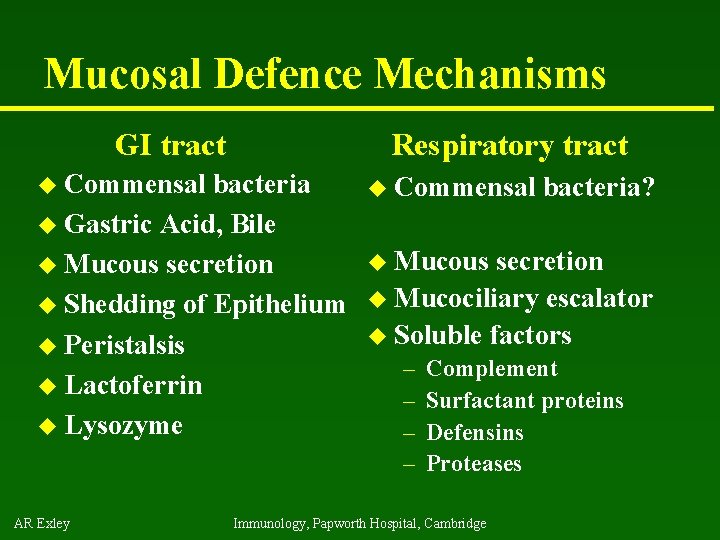
Mucosal Defence Mechanisms GI tract u Commensal Respiratory tract bacteria u Gastric Acid, Bile u Mucous secretion u Shedding of Epithelium u Peristalsis u Lactoferrin u Lysozyme AR Exley u Commensal bacteria? u Mucous secretion u Mucociliary escalator u Soluble factors – – Complement Surfactant proteins Defensins Proteases Immunology, Papworth Hospital, Cambridge
Mucosal Immune System u Immune response – Induction & Expression within same system u Mucosal Lymphocytes – Remain within the mucosal immune system – Lymphocyte migration / homing / retention u Special T cells – CD 8 a/a, gd , and regulatory T cells AR Exley Immunology, Papworth Hospital, Cambridge
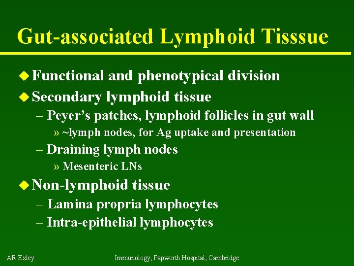
Gut-associated Lymphoid Tisssue u Functional and phenotypical division u Secondary lymphoid tissue – Peyer’s patches, lymphoid follicles in gut wall » ~lymph nodes, for Ag uptake and presentation – Draining lymph nodes » Mesenteric LNs u Non-lymphoid tissue – Lamina propria lymphocytes – Intra-epithelial lymphocytes AR Exley Immunology, Papworth Hospital, Cambridge
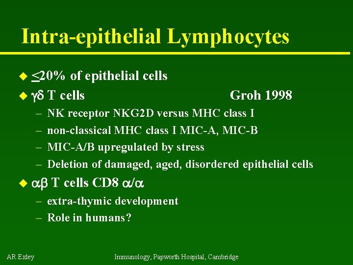
Intra-epithelial Lymphocytes u <20% of epithelial cells u gd T cells – – Groh 1998 NK receptor NKG 2 D versus MHC class I non-classical MHC class I MIC-A, MIC-B MIC-A/B upregulated by stress Deletion of damaged, disordered epithelial cells u ab T cells CD 8 a/a – extra-thymic development – Role in humans? AR Exley Immunology, Papworth Hospital, Cambridge
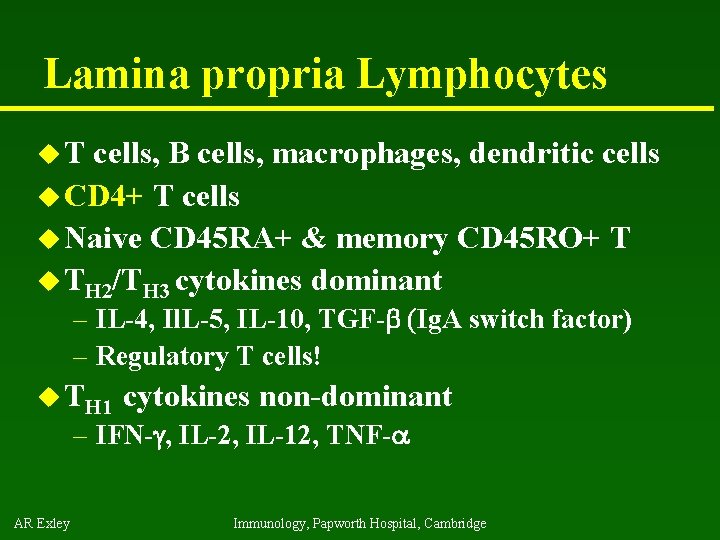
Lamina propria Lymphocytes u. T cells, B cells, macrophages, dendritic cells u CD 4+ T cells u Naive CD 45 RA+ & memory CD 45 RO+ T u TH 2/TH 3 cytokines dominant – IL-4, Il. L-5, IL-10, TGF-b (Ig. A switch factor) – Regulatory T cells! u TH 1 cytokines non-dominant – IFN-g, IL-2, IL-12, TNF-a AR Exley Immunology, Papworth Hospital, Cambridge
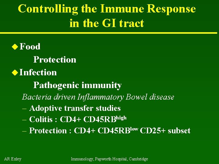
Controlling the Immune Response in the GI tract u Food Protection u Infection Pathogenic immunity Bacteria driven Inflammatory Bowel disease – Adoptive transfer studies – Colitis : CD 4+ CD 45 RBhigh – Protection : CD 4+ CD 45 RBlow CD 25+ subset AR Exley Immunology, Papworth Hospital, Cambridge
CD 4+ low CD 45 RB CD 25+ T cells u Naturally activated u ~10% of CD 4+ T cells u Unresponsive in vitro u Inhibit wide range of immune responses – To self – To pathogens u Transcription Maloy 2003 factor Foxp 3 u Effector function present in thymus AR Exley Immunology, Papworth Hospital, Cambridge
Regulatory T cells - natural u. T cells from thymus / peripheral blood u Classical transfer experiments u CD 4+ CD 25+ T cells in mice inhibit – autoimmune diabetes – inflammatory bowel disease – anti-tumour immunity – expansion of other T cells in vitro – Promote tolerance to skin allografts AR Exley Immunology, Papworth Hospital, Cambridge
Regulatory T cells - induced u Foxp 3 transcription factor positive – Physiological inducers? – Trl IL 10 producers – Tr 3 TGFb producers » Inducible with TGFb Chen 2003 – Inhibit Th 1, Th 2 responses in vivo u Classical – Ag delivered at mucosal sites induces peripheral + mucosal unresponsiveness Ostroukhova 2004 – Soluble Peptide induced tolerance » oral, nasal, peritoneal, subcutaneous, intradermal route » Peptides must be ~ naturally occurring epitopes AR Exley Immunology, Papworth Hospital, Cambridge
Oral Tolerance – Clinical Use? u Good data in autoimmune disease models – Prophylaxis prevents / attenuates disease – Treatment suppresses disease – Dose dependent effects u Poor – – AR Exley results to date from studies of Clinical diseases Monitor state & mode of oral tolerance induced Phase III: Oral MBP analogue in Multiple sclerosis NIH study: oral insulin in juvenile diabetes mellitus Myasthenia gravis : anti-ACHreceptor responses Immunology, Papworth Hospital, Cambridge
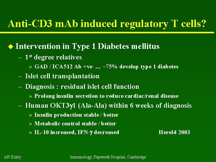
Anti-CD 3 m. Ab induced regulatory T cells? u Intervention in Type 1 Diabetes mellitus – 1 st degree relatives » GAD / ICA 512 Ab +ve … ~75% develop type 1 diabetes – Islet cell transplantation – Diagnosis : residual islet cell function » Prolong insulin secretion to reduce cardiac/renal disease – Human OKT 3 g 1 (Ala-Ala) within 6 weeks of diagnosis » Insulin production stable / better » Metabolic control stable / better » IL-10 increased, IFN-g decreased AR Exley Immunology, Papworth Hospital, Cambridge Herold 2003
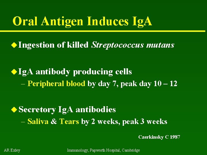
Oral Antigen Induces Ig. A u Ingestion u Ig. A of killed Streptococcus mutans antibody producing cells – Peripheral blood by day 7, peak day 10 – 12 u Secretory Ig. A antibodies – Saliva & Tears by 2 weeks, peak 3 weeks Czerkinsky C 1987 AR Exley Immunology, Papworth Hospital, Cambridge
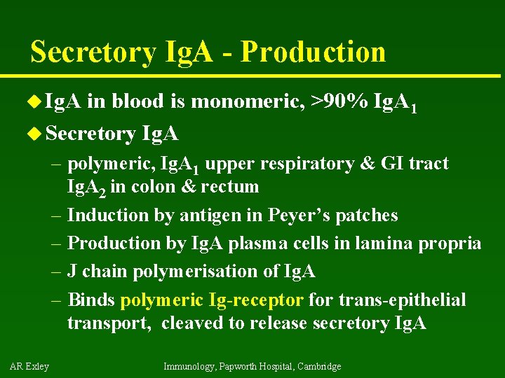
Secretory Ig. A - Production u Ig. A in blood is monomeric, >90% Ig. A 1 u Secretory Ig. A – polymeric, Ig. A 1 upper respiratory & GI tract Ig. A 2 in colon & rectum – Induction by antigen in Peyer’s patches – Production by Ig. A plasma cells in lamina propria – J chain polymerisation of Ig. A – Binds polymeric Ig-receptor for trans-epithelial transport, cleaved to release secretory Ig. A AR Exley Immunology, Papworth Hospital, Cambridge
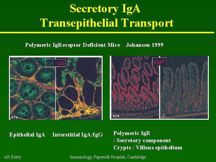
Secretory Ig. A Transepithelial Transport Polymeric Ig. Receptor Deficient Mice Epithelial Ig. A AR Exley Interstitial Ig. A/Ig. G Johansen 1999 Polymeric Ig. R / Secretory component Crypts / Villous epithelium Immunology, Papworth Hospital, Cambridge
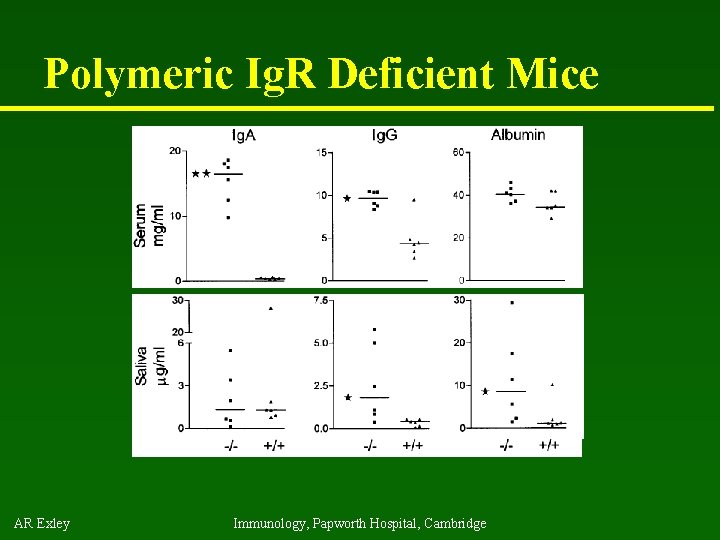
Polymeric Ig. R Deficient Mice AR Exley Immunology, Papworth Hospital, Cambridge
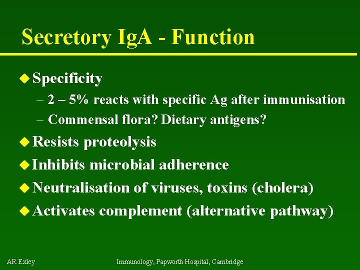
Secretory Ig. A - Function u Specificity – 2 – 5% reacts with specific Ag after immunisation – Commensal flora? Dietary antigens? u Resists proteolysis u Inhibits microbial adherence u Neutralisation of viruses, toxins (cholera) u Activates complement (alternative pathway) AR Exley Immunology, Papworth Hospital, Cambridge
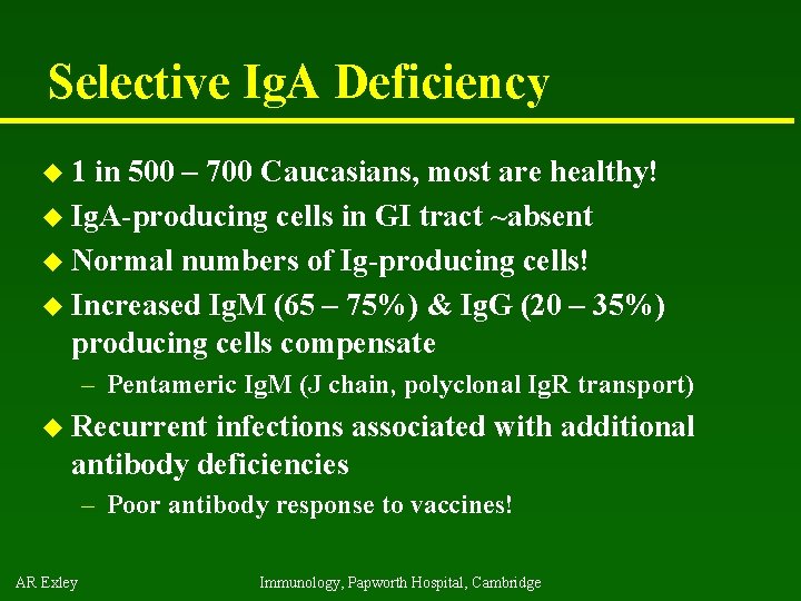
Selective Ig. A Deficiency u 1 in 500 – 700 Caucasians, most are healthy! u Ig. A-producing cells in GI tract ~absent u Normal numbers of Ig-producing cells! u Increased Ig. M (65 – 75%) & Ig. G (20 – 35%) producing cells compensate – Pentameric Ig. M (J chain, polyclonal Ig. R transport) u Recurrent infections associated with additional antibody deficiencies – Poor antibody response to vaccines! AR Exley Immunology, Papworth Hospital, Cambridge
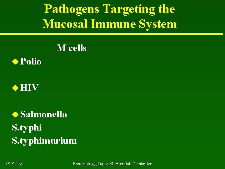
Pathogens Targeting the Mucosal Immune System M cells u Polio u HIV u Salmonella S. typhimurium AR Exley Immunology, Papworth Hospital, Cambridge
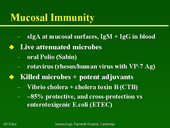
Mucosal Immunity – s. Ig. A at mucosal surfaces, Ig. M + Ig. G in blood u Live attenuated microbes – oral Polio (Sabin) – rotavirus (rhesus/human virus with VP-7 Ag) u Killed microbes + potent adjuvants – Vibrio cholera + cholera toxin B (CTB) – ~85% protective, and cross-protection vs enterotoxigenic E. coli (ETEC) AR Exley Immunology, Papworth Hospital, Cambridge
Conclusion u Mucosal Immune system – Antigen - induced expansion of lymphoid tissue – Lymphocyte homing – Functional and phenotypic diversity – Regulatory T cells – Immunoglobulin Ig. A – Immunity thru’ Vaccination AR Exley Immunology, Papworth Hospital, Cambridge
References u u u AR Exley Chen W et al. Conversion of Peripheral CD 4+CD 25 - Naive T Cells to CD 4+CD 25+ Regulatory T Cells by TGF-beta. Induction of Transcription Factor Foxp 3. J Exp Med 2003; 198 (12): 18751886. Groh V et al. Recognition of stress-induced MHC molecules by intestinal epithelial gammadelta T cells. Science 1998; 279 (5357): 1737 -1740. Herold et al. Activation of human T cells by Fc. R nonbinding anti -CD 3 m. Ab, h. OKT 3 g 1(Ala-Ala). J. Clin. Invest. 2003; 111 (3): 409418. Maloy et al. CD 4+CD 25+ TR Cells Suppress Innate Immune Pathology Through Cytokine-dependent Mechanisms. J Exp Med 2003; 197 (1): 111 -119. Ostroukhova O et al. Tolerance induced by inhaled antigen involves CD 4+ T cells expressing membrane-bound TGF-b and FOXP 3. J. Clin. Invest. 2004; 114 (1): 28 -38. Immunology, Papworth Hospital, Cambridge
- Slides: 24