mucles Anterior Muscles Origin on pelvis or vertebral

mucles

Anterior Muscles • Origin on pelvis or vertebral column – Iliacus – Psoas major

Illiopsoas Muscle • Three muscles: – Illiacus – Psoas major – Psoas minor • Action – Hip flexion

Iliacus • Origin – illiac fossa • Insertion – Lesser trochanter of the femur

Psoas Major and Minor • Origin – Transverse processes of L 1 -5 • Insertion – Minor: pectineal line – Major: lesser trochanter

Pectineus Muscle • Origin – Superior ramus of pubis • Insertion – pectineal line of femur • Action – Hip flexion – adduction

Tensor Fasciae Latae Muscle • Origin – Anterior iliac crest and surface of the ilium • Insertion – Ilio-tibial band • Action – Abduction about the hip – Hip flexion

Posterior Muscles Gluteal muscles • Origin on pelvis or sacrum – Gluteus maximus – Gluteus medius – Gluteus minimus • Lateral rotators – Piriformis – Obturator externus – Obturator internus – Superior and inferior gemellus – Quadratus femoris

Posterior Muscles

MUSCLES THIGH

Muscles of the Anterior Compartment of the Thigh • Quadriceps femoris – Has four separate heads – Has a common insertion at the quadriceps tendon – Powerful knee extensors • • Rectus femoris Vastus lateralis Vastus medialis Vastus intermedius – Tensor fasciae latae

Muscles of the Anterior Compartment of the Thigh

SARTORIUS Flexes Thigh, & Rotates Thigh Laterally O: I: Anterior Superior Iliac. Spine Medial Side of Tibia

VASTUS LATERALIS Extends Lower Leg

RECTUS FEMORIS Flexes Thigh, Extends Lower Leg O: Ilium I: Patella & Tibial Tuberosity

VASTUS MEDIALIS Extends Lower Leg

Muscles of the Posterior Compartment of the Thigh • • Hamstrings Biceps femoris Semitendinosus Semimembranosus Figure 11. 21 a

Muscles of the medial compartment – Adductor longus – Adductor brevis – Adductor magnus – Pectineus – Gracilis

ADDUCTOR LONGUS Adduct, Rotate & Flex Thigh Laterally

Anterior and Medial Muscles

This is formed by the inguinal ligament, the sartorius laterally and the adductor longus on the medial side.

MUSCLES of the LEG
")
Muscles of the Anterior Compartment • • Tibialis anterior Extensor digitorum longus Fibularis (peroneus) tertius Extensor hallucis longus Figure 11. 22 a

Muscles of the Posterior Compartment • Superficial muscles – Triceps surae • Gastrocnemius • Soleus – Plantaris

Muscles of the Posterior Compartment • Deep muscles – Popliteus – Flexor digitorum longus – Flexor hallucis longus – Tibialis posterior
 longus • Fibularis (peroneus) brevis •")
Muscles of the Lateral Compartment • Fibularis (peroneus) longus • Fibularis (peroneus) brevis • Fibularis tertius Figure 11. 23 a

Muscles of the Lateral Compartment

Intrinsic Muscles of the Foot • Muscle on the dorsum of the foot – Extensor digitorum brevis • Muscles on the sole of the foot – First layer • Flexor digitorum brevis • Abductor hallucis • Abductor digiti minimi

Intrinsic Muscles of the Foot • Second layer – Flexor accessorius – Lumbricals

Intrinsic Muscles of the Foot • Third layer – Flexor hallucis brevis – Adductor hallucis – Flexor digiti minimi brevis • Fourth layer – Plantar and dorsal interossei


Arteries

Branches of the Ascending Aorta • Coronary arteries – Supply the heart’s cardiac muscle with oxygen and nutrients
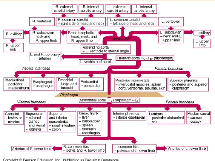

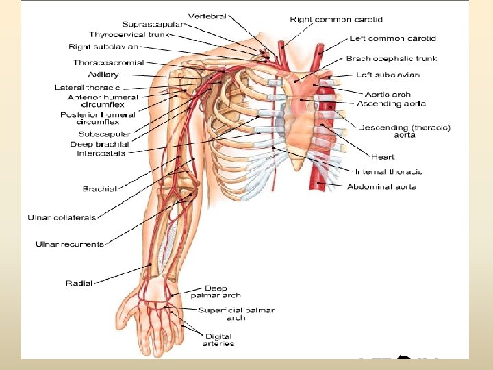
– Aortic Arch 1. Brachiocephalic Trunk 1. Right Common Carotid Artery 2. Right Subclavian Artery 2. Left Common Carotid 1. Brain 2. Neck and head 3. Left Subclavian

Branches of the Aortic Arch Right common carotid artery • First branch – Brachiocephalic trunk – Right common carotid and right subclavian • Second branch – Left common carotid • Third branch – Left subclavian Vertebral artery Right subclavian artery Brachiocephalic trunk Left common carotid artery Left subclavian artery Aortic arch Descending thoracic aorta

Blood Vessels entering or leaving the heart
 Left subclavian")
Left common carotid artery Brachiocephalic trunk Ascending aorta (gives off coronary arteries) Left subclavian artery Aortic arch Thoracic (descending) aorta Abdominal aort Common iliac arteries

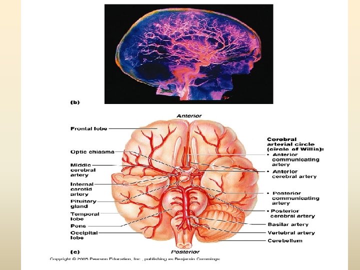
The Carotid Arteries and Brain Blood Supply • External carotid artery neck, pharynx, esophagus, larynx, mandible, & face • Internal carotid artery brain (IC branches): – Ophthalmic artery -eyes; – anterior cerebral artery -frontal/parietal; – middle cerebral -midbrain, lat. cerebrum • Vertebral> basilar>posterior cerebral>posterior communicating arteries>middle cerebral> anterior communicating>anterior cerebral

External carotid artery Vertebral artery Subclavian artery Brachiocephalic trunk
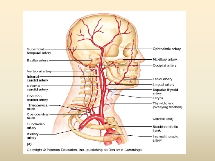

• Thyrocervical trunk-neck, shoulder & upper back • Internal thoracic -pericardium/ant. thoracic wall – Vertebral artery -brain/spinal cord • Axillary artery -pectoral region/axilla – Brachial artery -upper limb – Radial/ulnar arteries -antebrachium – Superficial/deep palmar arch -palm – Digital artery -thumb/fingers

Subclavian artery Vertebral artery Axillary artery Brachial artery Radial artery Ulnar artery

Subclavian artery Vertebral artery Axillary artery • Left and Right Subclavian Arteries Brachial artery – Subclavian becomes Axillary Radial artery Ulnar artery

• Axillary Artery – Axillary becomes Brachial

Subclavian artery Vertebral artery Axillary artery Brachial Artery • Brachial artery Radial Artery – Ulnar artery

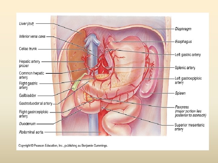
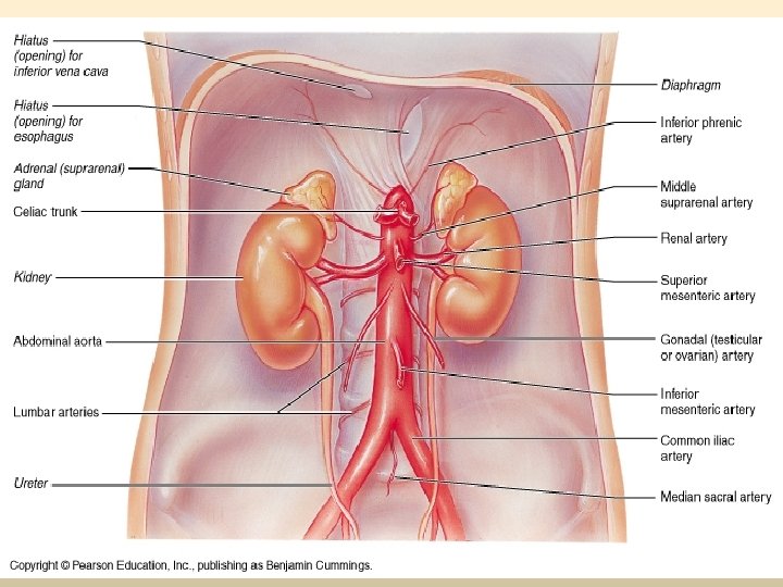
Branches of the Descending Aorta: Arteries of the Abdominal Aorta Celiac trunk Right renal artery Descending abdominal aorta Inferior mesenteric artery Left renal artery Superior mesenteric artery Common iliac artery Left internal iliac artery Left external iliac artery Left femoral artery • Three major branches (in order from superior to inferior along abdominal aorta) – Celiac trunk – Superior mesenteric artery – Inferior mesenteric artery

The Descending Aorta Thoracic Aorta & Branches • Visceral branches -Bronchial, pericardial, mediastinal, esophageal arteries. • Parietal branches Intercostal, superior phrenic.

The Descending Aorta Abdominal Aorta & Branches Unpaired arteries : Ø Celiac trunk liver, stomach, spleen; Branches: Ø left gastric Ø Splenic Ø common hepatic arteries. Ø Superior mesenteric pancreas, small intestine, most of large intestine. Ø Inferior mesenteric terminal colon & rectum

 Paired arteries: Inferior phrenic Ø Suprarenal Ø Renal Ø")
Abdominal Aorta & Branches (cont’d) Paired arteries: Inferior phrenic Ø Suprarenal Ø Renal Ø Gonadal Ø Lumbar Ø

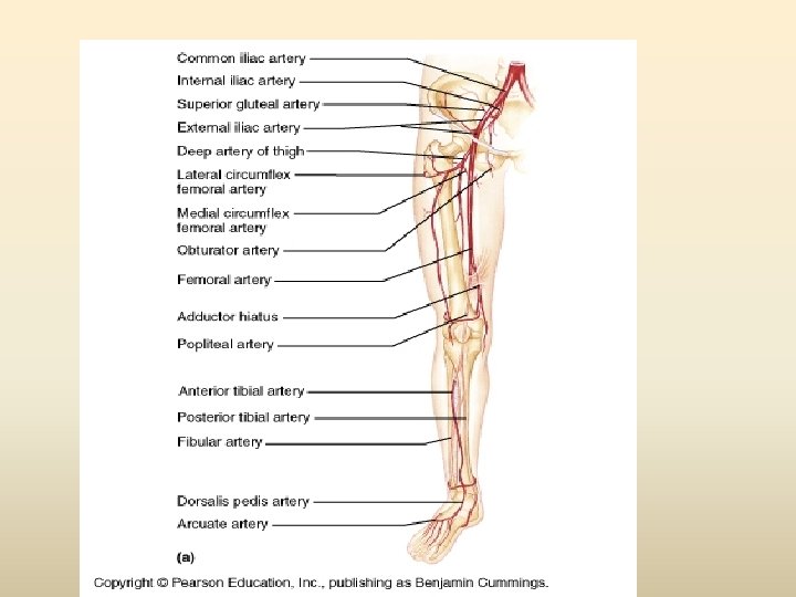
Arteries of the Pelvis & Lower Limbs • Right/Left Common Iliacs – Internal Iliac -urinary bladder, int. , ext. walls of pelvis, genitalia – External Iliac -lower limbs
 The internal iliac (hypogastric) artery: • • •")
Blood supply of the pelvis a) The internal iliac (hypogastric) artery: • • • Arises from the common iliac artery opposite the sacroiliac joint Descends under cover of peritoneum into the true pelvis for about 4 cm before dividing into anterior and posterior divisions The posterior division: – – • Has three parietal branches: Iliolumbar lateral sacral superior gluteal The anterior division has three parietal branches – obturator artery – inferior gluteal artery – internal pudendal artery) which supply the pariets of the anterolateral quadrant of the pelvic wall, the buttock and perineum – It has four visceral branches which are: 1) Umbilical artery 3) Vaginal artery 2) Uterine artery 4) Middle rectal artery

View of iliac and femoral arteries

Arteries of Thigh & Leg • Femoral – Deep femoral – Popliteal • Post. Tibial – Peroneal • Ant. tibial

Major Systemic Arteries • Ant. Tibial Artery – Dorsalis pedis • Posterior Tibial Artery – Fibular (peroneal) Artery • Medial, Lateral plantar

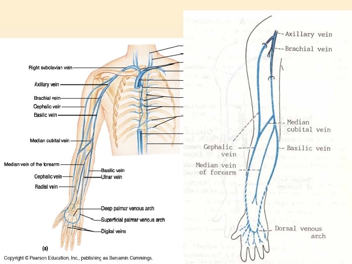
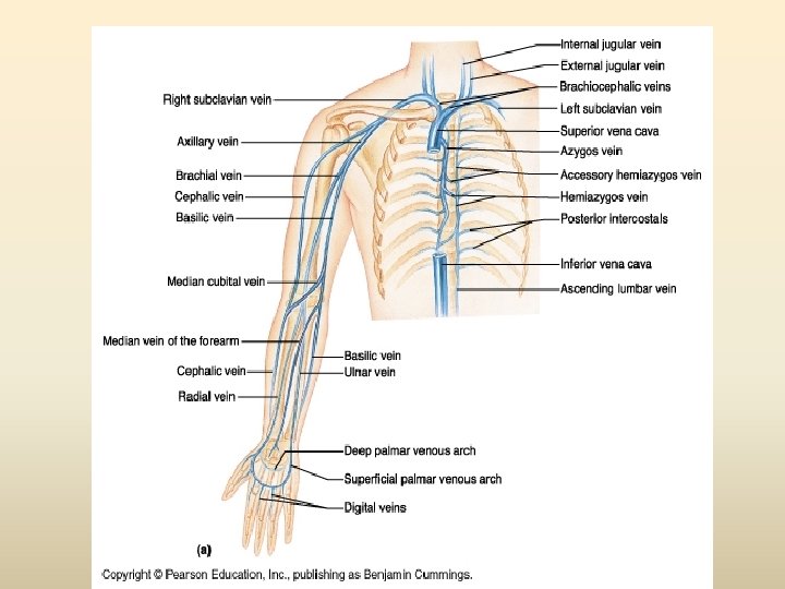
Systemic Veins Brachium venous return • • Digital veins Superficial/deep palmar Palmar venous arches Cephalic Median antebrachial Basilic Median cubital (cephalic, basilic) Axillary (basilic, brachial)
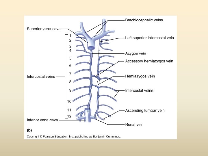
 • Azygos(hemiazygos)chief blood")
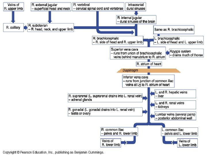
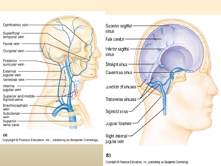
Systemic Veins SVC formation • Subclavians • Brachiocephalics(vert ebrals, ext/int jugulars) • Azygos(hemiazygos)chief blood collectors of thorax


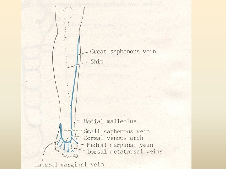
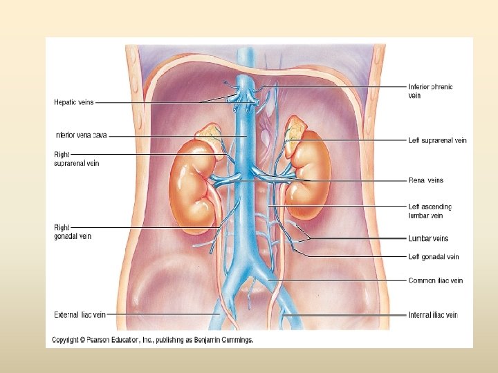
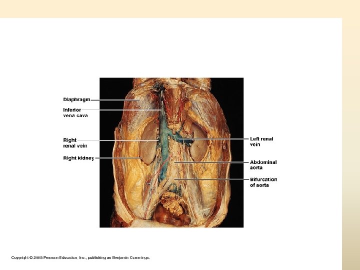
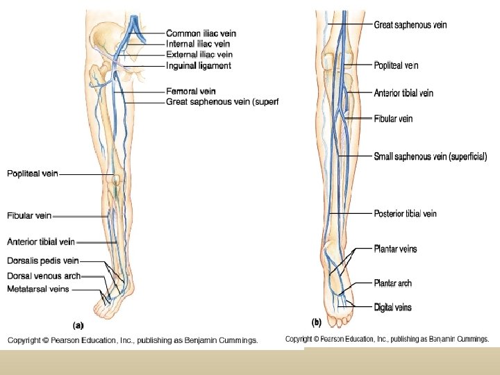
Systemic Veins Tributaries of the IVC Pelvic limb venous drainage • Plantar/dorsal venous arch • Anterior/ posterior tibial • Peroneal • Popliteal • Femoral • Great/small saphenous • External iliac

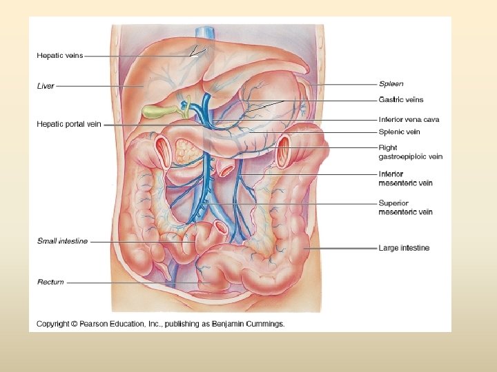
Hepatic Portal System Tributaries • Inferior mesenteric • Splenic • Superior Mesenteric * Hepatic portal vein formed by fusion of superior mesenteric and splenic

Vascular system within the liver

Systemic Venous System

Venous System of the Trunk and Upper Limb
- Slides: 80