MRI x 40 x 400 x 400 x


 who developed lymph node")

- Slides: 33
MRI
x 40
x 400
x 400
x 400
x 400
x 400
x 1000
x 1000
x 1000
x 1000
x 1000
x 1000
x 1000
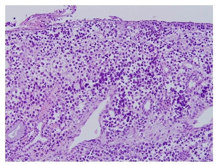
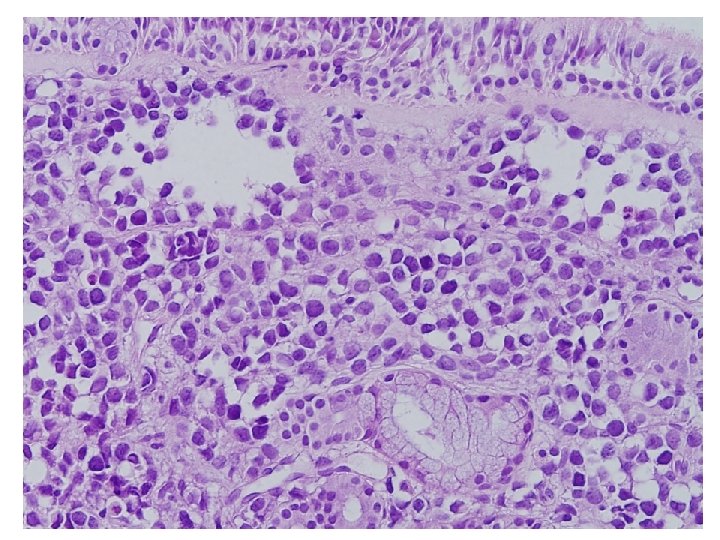
Cytologic features • Cellularity : High • Background : Clear, but necrotic cytoplasmic materials • Dispersed isolated round cells • Cytoplasm : dense, well defined border • Nucleus : small, eccenteric, round or curved, hyperchromatic • Mulinucleated, and binucleated cells • Nucleoli : indistinct, occasionally small nucleoli • Mitosis
Small round cell tumor • Rhabdomyosarcoma • Lymphoma – Lymphoblastic lymphoma – Burkitt’s lymphoma – DLBCL • • • Ewing’s sarcoma Neuroblastoma Desmoplastic small round cell tumor Round cell liposarcoma Undifferentiated carcinoma
Neuroblastoma • • • Small round cell Monotonous Granular chromatin Homer Wright rosettes Fibrillary cytoplasm • • Pleomorphism Vesicular and coarse Pseudorosettes Rhabdoid feature
Ewing’s sarcoma • • • Small round cell Monotonous Hyperchomasia Pseudorosettes Scanty cytoplasm Background necrosis • Pleomorphism • Rhabdoid feature
Burkitt's Lymphoma • Round medium-sized cells with multiple nucleoli • Moderate amount of cytoplasm • Mitoses, apoptoses • Binucleated, multinucleated cells • Rhabdoid feature
Lymphoblastic lymphoma • Small to medium-sized cells • Round blastic nucleus • Monomorphic • Scanty cytoplasm • Multinucleated cell • Round hyperchromatic nucleus • Rhabdoid feature
Rhabdomyosarcoma • • • High cellularity Small round cell Rhabdoid feature Eccentric hyperchromatic nucleus Occasionally small nucleoli
Desmin
Subtype of rhabdomyosarcoma Embryonal Alveolar Pleomorphic Age Less than 5 year Young child Adult Site of involvement Head and neck GU system Extremity Paraspinal, perianal, and paranasal Deep soft tissue of the lower extremities Cell size Small Large Cell Spindle, tadpole Round cell -shaped Nucleus Centric Eccentric Binucleated and multinucleated cell + +++ Pleomorphic cell
Diagnosis FNA from left level Ⅲ lymph node : Metastatic rhabdomyosarcoma, alveolar type
Desmin CD 99 LCA S-100
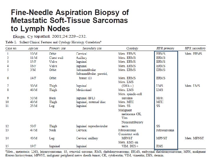
LN metastasis of sarcoma • Less than 4% of cases having nodal metastases at presentation (Enzinger FM, Weiss SW, editors. Soft tissue tumors, 3 rd ed. Mosby, 1995. p 17– 38. ) • In a review of over 2, 500 patients in the world literature (Weingrad DW, Rosenberg SA. Early lymphatic spread of osteogenic and soft tissue sarcoma. Surgery 1978; 84: 231– 240. ) – Only 5% of patients developed nodal metastases
• Fong et al. found 46 patients (2. 6%) who developed lymph node metastases. Ann Surg 1993; 217(1): 72– 7. ) – Epitheloid sarcoma – Embryonal rhabdomyosarcoma – Angiosarcoma