MRI of acute abdominal pain in pregnancy Tips

MRI of acute abdominal pain in pregnancy: Tips for success Rex A. Parker, MD Kaiser Los Angeles Medical Center

I have no disclosures

Goals • Briefly review the safety of MRI in imaging the pregnant patient with abdominal pain. • Review a basic protocol for MR imaging of the pregnant patient with abdominal pain. • Highlight a series of TIPS which will aid in performance and interpretation of MRI in pregnant patients.

MRI in pregnancy: Is it safe? • Probably • No known deleterious effects to the human fetus documented. • ACR approves of MR imaging of the pregnant patient in any trimester. • Although there is no ionizing radiation, radiofrequency energy used in MR does deposit energy in the patient as heat. The fetal temperature rise is felt to be less than those required for teratogenesis. • Other theoretic risks include noise exposure. • Potential risks versus benefits should be assessed on a case by case basis. • Gadolinium based contrast agents are known to cross the placenta and be taken up by fetus. They are classified as Category C in pregnancy and should be avoided.

MRI for RLQ Pain in Pregnancy: How I do it • T 2 Single Shot Fast Spin Echo • Axial, Sagittal and Coronal • Balanced Steady State Free Precession (b. SSFP, FIESTA, Tru. FISP, etc) • Axial and Coronal • Axial t 1 gradient echo, dual echo, in and out of phase • Axial T 2 SSFSE with fat suppression • Axial T 1 fast spoiled gradient echo with fat suppression

Aortocaval compression and respiratory motion • Pregnant women, especially later in 2 nd and 3 rd trimesters can have limited breath hold capacity. • Compression of IVC can contribute to hypotension, tachycardia, even fainting. • Prone positioning or 10 -20 degrees of left lateral tilt by placing a foam pillow under the patients right side can be helpful.

Use T 2 Single Shot Fast Spin Echo Sequences • Single shot: entire matrix for an individual image obtained with one 90 degree excitation. • Half acquisition makes acquisition rapid, with each image typically obtained in under 1 second. • Images are relatively insensitive to fetal and maternal motion. • Acquire in transverse, coronal and sagittal planes. • Downside: The multiple 180 degree pulses mean that these sequences deposit more energy (higher SAR) than gradient echo or conventional spin echo/fast spin echo sequences.

Fat stranding on T 2 SSFSE can appear as hazy HYPOINTENSITY in the fat

Fat suppressed T 2 SSFSE or STIR aid in identifying fluid and inflammation

Can’t find the appendix? Don’t worry • Nikolaidis, et al. and colleagues at PENN published a study in AJR in 2004 looking at the likelihood of acute appendicitis if the appendix was not seen and there were no secondary inflammatory findings in the RLQ. • Retrospective study. 366 CTs were reviewed by 2 experienced radiologists. The appendix could not be seen in 46 patients. • Only 1 of the 46 ended up with pathologically proven acute appendicitis, and that patient had a paucity of intra-abdominal fat. • Although a similar study has not been published on MRI, the overall sensitivity and specificity of MR and CT are similar and this experience could potentially be extrapolated to MR as well. • Take home: If there is a fair amount of fat in the belly, the appendix is not seen, and there are no right lower quadrant inflammatory findings, then the patient does not have appendicitis. Nikolaidis P, Hwang CM, Miller FH, et al. The nonvisualized appendix: incidence of acute appendicitis when secondary inflammatory changes are absent. AJR Am J Roentgenol. 2004; 183(4): 889– 92.
 images • Normal appendix is")
Identifying the normal appendix: Blooming on gradient echo (GRE) images • Normal appendix is frequently airfilled. • GRE images demonstrate a pronounced blooming effect due to magnetic susceptibility due to the lack of 180 refocusing pulses present on SSFSE. • Longer TE on gradient echo images results in more blooming effect. If dual echo T 1 GRE (in and opposed phase) is used, blooming will be more evident on the in phase image.
 can")
SSFSE b. SSFP Balanced Steady State Free Precession (b. SSFP, Fiesta, Tru. FISP) can aid in differentiating a normal appendix from dilated peri-uterine and peri-ovarian veins. • Both will be hypointense on SSFSE • The properties of b. SSFP lead to hyperintense, or bright blood in veins. • b. SSFP is a gradient echo sequence, meaning a normal gas filled appendix will demonstrate blooming related to magnetic susceptibility.

Blooming can aid in identifying appendicoliths and renal calculi T 2 SSFSE Out of Phase GRE In Phase GRE

Hydronephrosis can be physiologic during pregnancy • Due to extrinsic compression of ureter by gravid uterus and hormonally induced ureteral smooth muscle relaxation. • Common, seen in up to 90% of pregnancies. • Usually right side. • Look for tapering of the ureter to the level of the sacral promontory.

Fat Suppressed T 1 weighted gradient echo sequences can make identifying blood products easier.

Hemorrhage Free Fluid Subchorionic Hemorrhage

Don’t assume the pregnancy is intrauterine • Findings of Ectopic Pregnancy on MRI • • • Lack of intrauterine gestational sac Isolated hemoperitoneum Tubal mass, frequently cystic Cornual or Interstitial mass Hematosalpinx

Don’t assume the pregnancy is intrauterine • Findings of Ectopic Pregnancy on MRI • • • Lack of intrauterine gestational sac Isolated hemoperitoneum Tubal mass, frequently cystic Cornual or Interstitial mass Hematosalpinx

Don’t assume the pregnancy is intrauterine • Findings of Ectopic Pregnancy on MRI • • • Lack of intrauterine gestational sac Isolated hemoperitoneum Tubal mass, frequently cystic Cornual or Interstitial mass Hematosalpinx

Don’t assume the pregnancy is intrauterine • Findings of Ectopic Pregnancy on MRI • • • Lack of intrauterine gestational sac Isolated hemoperitoneum Tubal mass, frequently cystic Cornual or Interstitial mass Hematosalpinx

Cornual vs Interstitial vs Angular Pregnancy? • Some clinicians use the term cornual to describe interstitial and angular pregnancy • Others reserve cornual for implantation in one horn of a bicornuate uterus

Interstitial pregnancy • Implantation in the most proximal portion of the fallopian tube that traverses the myometrium • Important clinically because interstitial pregnancies have higher mortality than other ectopics • Surrounding myometrium can cause confusion for intrauterine pregnancy on US • Presence of surrounding myometrium has been postulated to allow for the pregnancies to be carried longer prior to presentation/rupture, although there is no good evidence to support this • Implantation is LATERAL to the uterine angle

Angular Pregnancy • Implantation in the lateral angle of the uterus, medial to the uterotubal junction • Completely surrounded by myometrium and can potentially be carried to term • However, high rates of rupture, placenta accreta and spontaneous loss • May present or rupture later in pregnancy

Don’t forget to look at the placenta Normal Placenta • Placenta should have homogenous signal intensity • Uterus should have thin inner and outer bands of T 2 hypointensity with middle band of relative T 2 hyperintensity

Normal Placenta • Tortuous subplacental T 2 hypointense flow voids which represent subplacental vascularity • Can have thin T 2 hypointense lines running through the placenta, likely corresponding with septae Radio. Graphics, http: //pubs. rsna. org/doi/abs/10. 1148/rg. 287085060 Published in: W. Christopher Baughman; Jane E. Corteville; Rajiv R. Shah; Radio. Graphics 2008, 28, 1905 -1916. DOI: 10. 1148/rg. 287085060 © RSNA, 2008

Don’t forget about the placenta • Placenta Previa • Placental implantation in lower uterine segment. • Can be partial, marginal or complete, based on degree to which placenta covers the internal os of the cervix. • Majority of marginal and partial previas resolve. • Complete previa less likely to resolve. Most are treated with Caesarean section. • May present with painless bleeding.

Don’t forget the placenta: Placenta Accreta • Term describing abnormal placentation wherein chorionic villi invade myometrium. • Accreta: Villi attached to myometrium but no muscular invasion. • Increta: Villi partially invade myometrium. • Percreta: Villi invade up to or beyond uterine serosa.

Placenta Accreta • MR Findings Include • Loss of normal T 2 hypointense inner myometrial layer • Focal uterine bulge • Heterogeneous placental signal intensity • Dark, thick, intraplacental bands • Frank extension of placental tissue through the uterine wall

Placenta Accreta • MR Findings Include • Loss of normal T 2 hypointense inner myometrial layer • Focal uterine bulge • Heterogeneous placental signal intensity • Dark, thick, intraplacental bands • Frank extension of placental tissue through the uterine wall

Placenta Percreta • MR Findings Include • Loss of normal T 2 hypointense inner myometrial layer • Focal uterine bulge • Heterogeneous placental signal intensity • Dark, thick, intraplacental bands • Frank extension of placental tissue through the uterine wall

Think outside of the appendix • Like CT for appendicitis in the non-pregnant patient, part of the value of the MRI is in identifying mimics of appendicitis • Pathologic processes that affect reproductive age women occur at similar (or sometimes greater) rates during pregnancy • Look for adnexal, urinary tract, biliary and bowel pathology

Think outside of the appendix: SBO

Think outside of the appendix: Cholecystitis • Findings similar to other imaging modalities • • Distended gallbladder Sludge or stones Gallbladder wall thickening Pericholecystic inflammation

Think outside of the appendix: Choledocholithiasis

Think outside of the appendix: Colitis

Think outside of the appendix: Diverticulitis
")
Think outside of the appendix: Degenerating Fibroids (and subchorionic hemorrhage)

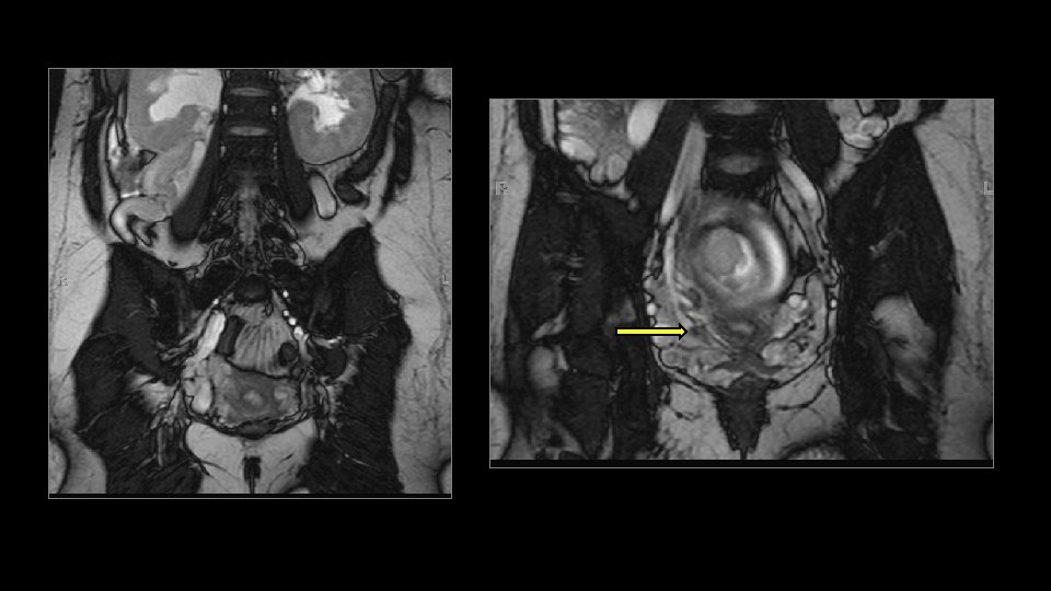
Think outside of the appendix: Ovarian Torsion

Conclusion • MRI can be performed safely and quickly in the pregnant patient with abdominal pain • SSFSE sequences are the mainstay of the protocol, and are relatively insensitive to maternal and fetal motion • Blooming on gradient echo sequences can aid in identifying a normal, gas-filled appendix • Blooming can help to diagnose appendicoliths as well as urinary tract calculi • b. SSFP images can aid in differentiating a gas-filled appendix from adjacent dilated veins • Adding fat suppression to T 1 weighted spoiled gradient echo images can make identifying hemorrhage easier • Don’t assume the pregnancy is intrauterine • Don’t forget to look at the placenta • Think about other common pathologic processes besides appendicitis
- Slides: 40