MRI methods of cerebral vasculature reconstruction on the

MRI methods of cerebral vasculature reconstruction on the example of small laboratory animals G. Jankova 1, 2, A. Akulov 3, S. Maltseva 2, 4, D. Parshin 2, A. Khe 2, A. Cherevko 2, 1 Novosibirsk State University 2 Lavrentyev Institute of Hydrodynamics SB RAS 3 Institute of Cytology and Genetics SB RAS 4 Sobolev Institute of Mathematics SB RAS

Purposes Study of the relationship of phenotype, genotype and hemodynamics of the brain for various lines of laboratory animals according to high field MRI scanning
 - Radon transformation MRI (magnetic field) - Fourier transformation Important stages in")
CT (X-rays) - Radon transformation MRI (magnetic field) - Fourier transformation Important stages in the MRI development 1946 1952 1973 1975 1977 1991 2003 the Discovery of NMR - Bloch & Purcell Nobel prize - Bloch & Purcell Radon transform based MRI- Lauterbur Modern technique of MRI (Fourier transform) – Ernst Echo-planar MRI ("MRI animation") - Mansfield Nobel prize - Ernst Nobel prize - Lauterbur & Mansfield

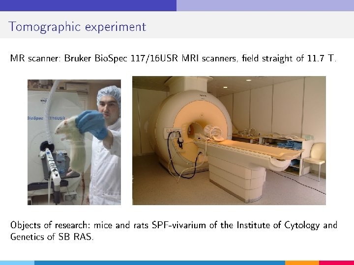
Tomographic equipment and its operation

MRI scanner Philips The Philips Intera Achieva MRI scanner with a field strength of 1. 5 T, belongs to a class of modern high field scanners. The device is intended for conducting diagnostic studies: brain and spinal cord, spine, heart, vessels of the pelvis, of the thorax. MRI scanner Brucker MRI scanner Bruker Bio. Spec 117/16 USR field strength of 11. 7 T, designed for molecular and preclinical magnetic resonance imaging. High-field magnets deliver high spatial resolution. Used on small animals in biomedical science, biomedical and preclinical research.

MRI is based on the resonance equation for the spins of atomic nuclei: n = g Bo n – the resonant frequency, Bo – the applied magnetic field, g – gyromagnetic ratio, different for each chemical element, determined by the characteristics of the atomic nucleus. For hydrogen n =63. 855 МHz for Bo=1. 5 T MRI signal is obtained from the difference between the energy absorbed by the spins that have undergone a transition from lower energy level to higher and energy emitted by the spins which simultaneously moved from a higher energy level to lower. A signal proportional to the difference in population levels.
 The spin-spin relaxation")
90 о RF The spin-lattice relaxation time T 1 (~500 ms) The spin-spin relaxation time T 2 (~20 ms) MXY =MXY 0 exp(-t/T 2) MZ= M 0 ( 1 – exp(-t/T 1) ) T 2 is always less than T 1. The net magnetization in the XY plane goes to zero and then the longitudinal magnetization increases until M 0 along Z.

The superconducting magnet superconducting ring Vacuum layer liquid helium thermal insulating layers magnetic shield Typically, a few kilometers of niobium-titanium wire compacted in a copper matrix

RF-coils:

Gradient coils: Z gradient X gradient Y gradient Conventional MRI coordinate system: external magnetic field directed along the axis Z.

Frequency encoding Signal amplitude is proportional to the number of spins in the plane perpendicular to the gradient.

The general scheme of scanner device

The choice of slice Slice selection is achieved by using a one-dimensional linear magnetic field gradient during the RF pulse. 90 о pulse applied together with a gradient magnetic field will rotate only the spins located in the slice.

Frequency encoding When frequency coding, the examined object is excited in the absence of x and y gradients, after which the signal is recorded in the presence of

Phase encoding is performed before the signal is recorded, but in the presence of the gradient. Disadvantage – time expensive method: For each phase direction should be one phase encoding gradient step.

Full encoding Slice selection gradient - selects the cutting plane. The phase-encoding gradient - along one side of the cut plane. The frequency encoding gradient - along the other side of the cut plane.

Slice selection Phase encoding Frequency encoding Each of the nine vectors of the magnetization is characterized by a unique phase angle and frequency of the precession.

Data extraction X Fourier data Frequency grad. phase Phase grad. First, the signals are subjected to Fourier transformation in the X direction for extracting the frequency component information. Then in the phase encoding direction Y to extract information about the phase position. time frequency фаза phase Y Fourier frequency

Chemical shift The resonant frequency of the protons depends on the environment. At 1. 5 Tesla the difference in the frequencies of fat and water is approximately 220 Hz. Benefits - different brightness of different tissues in the images. Disadvantage - a geometric shift of various tissues relative to their actual position.

Spin-echo sequence Even the smallest magnetic field inhomogeneity causes dephasing of the magnetization vectors. This dephasing is amplified by gradients of the field. As a result, significantly decreases the value of the magnetic resonance signal. A superposition 180º pulse at time τ after applying 90 ° pulse causes a phase shift, whereby after a time 2τ the effects of magnetic field inhomogeneity are canceled mutually, and gives a strong echo signal.

The flow-rate measurement Time-of-flight angiography Spin-echo sequence in which a slice-selective 90 o and 180 o pulses have different frequencies. 90 o pulse excites spins in the one plane. A 180 opulse in the other. If there is no flow, there is no signal, as there is no spin that undergoing 90 o-, and 180 o-pulse. At a flow availability and correct echo time (TE), blood from 90 o-plane flow into 180 o-plane and produces an echo.

The flow-rate measurement Phase-contrast angiography A bipolar gradient pulse: or A bipolar gradient pulse has no cumulative effect on stationary spins. It will cause a phase shift only spins with the speed component in the direction of the gradient. The phase shift is proportional to the velocity component. No flow For optimal signal it is necessary to match the maximum speed of 90 ° phase shift of each of the bipolar pulse. Spins with lower current velocities get smaller phase shifts.

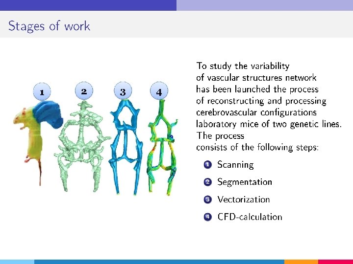
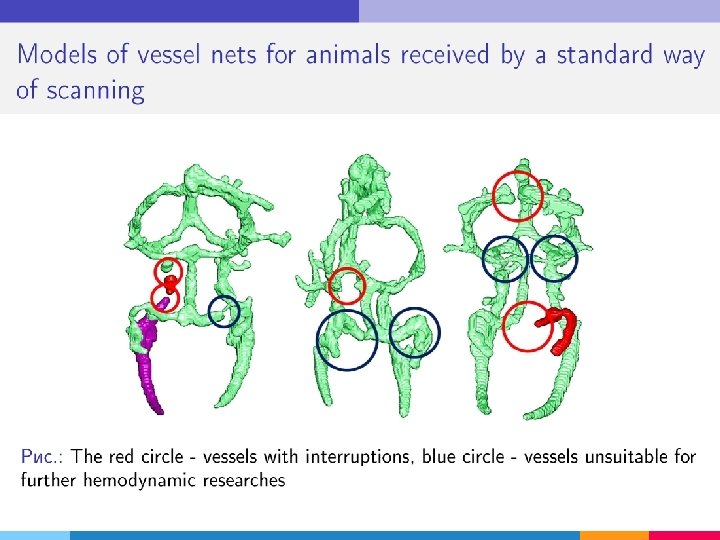
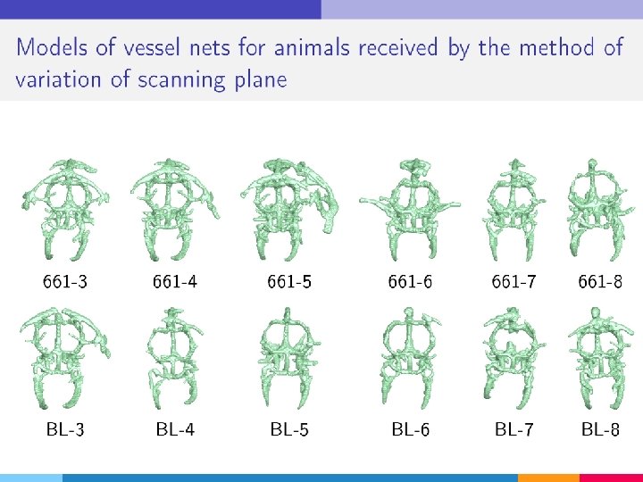
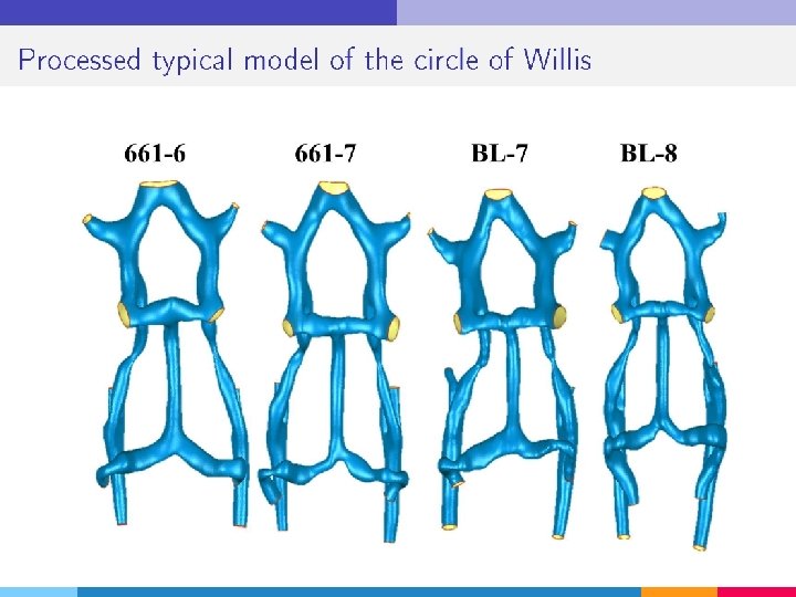
Reconstruction of the cerebral vascular network of the laboratory mouse

Purposes Study of the relationship of phenotype, genotype and hemodynamics of the brain for various lines of laboratory animals according to high field MRI scanning


The standard scanning scheme


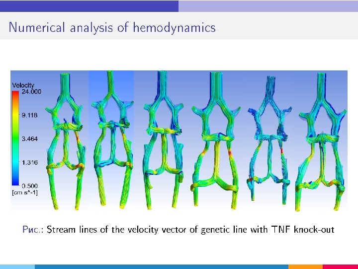
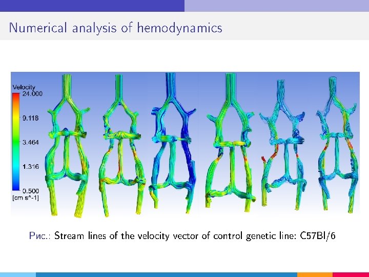

and other hemodynamic parameters


- Slides: 43