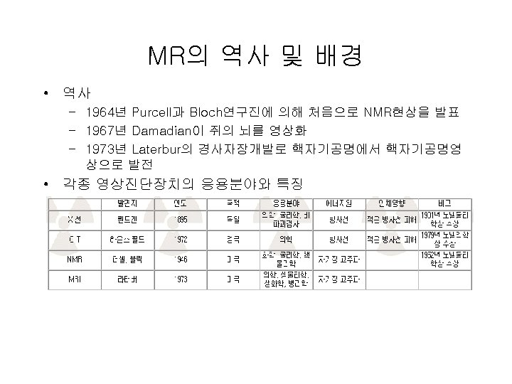
MRI Magnetic resonance imaging IntroductionMRI soft tissue anatomy
 Introduction-MRI 출처 : 고려병설대학")


, Nuclear Magnetic Resonance Computed Tomography, Spin")
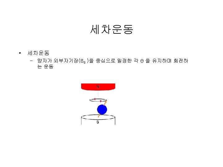
 – 라모어 방정식 • F=γB 0 , F(Larmor): 세차주파수(MHz), B")


 구별 • Gradient in the Z-direction – Resonant frequency varying as a")

 : TE↓, TR↓=>T")


- Slides: 52
MRI (Magnetic resonance imaging) Introduction-MRI 출처 : 고려병설대학
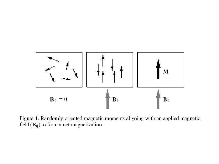
소개 • soft tissue anatomy • MRI – 큰 자장이 걸려있는 검사기에서 고주파와 인체 속 에 있는 수소핵(proton)과의 상호작용에 의한 에너 지 방출을 검출하여 영상 구현 – Nuclei having odd number of neutrons, and odd number of protons, or both will have a net magnetic moment – H or “Proton” NMR • High concentration and high sensitivity
용어 해설 • 자기공명영상 : Magnetic Resonance Imaging(MRI), Nuclear Magnetic Resonance Computed Tomography, Spin Mapping, Hydrogen Mapping, Zeugmatography, Magnetic Resonance Imaging • MRS : Magnetic Resonance Spectroscopy • MRA : Magnetic Resonance Agiography • f. MRI : functional MRI
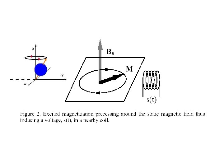
세차주파수 • 세차주파수(그림 B) – 라모어 방정식 • F=γB 0 , F(Larmor): 세차주파수(MHz), B 0 : 외부자기장(tesla), γ: 자기회전비 • γ: 자기회전비 는 nuclear 종류에 따라 결정 – 수소의 자기 회전비 : 42. 57 MHz – 라모어 방정식에서 얻어진 주파수를 걸면 Oscillating magnetic (RF) field • the spins will absorb energy and become excited: excitation – Tipping Magnetization, Induce a voltage in a nearby coil – Figure 2.
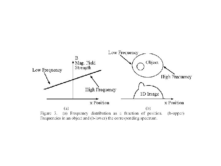
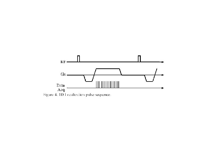
Pulse 순서 • RF pulse • Frequency encoding • Fourier transform – Magnitude of a frequency represents magnetization(induced voltage) at a specific location • Measure T 2(signal decay) and T 1(magnetization recovery)
Phase encoding • Same frequency • Different phase => phase encoding
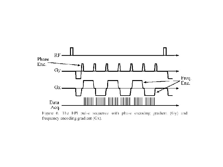
2차원 위치 구별 • Two gradient is not working => 45 degree gradient • X direction: frequency encoding • Y direction: phase encoding • Frequency encoding -> Phase encoding -> frequency encoding • Echo Planar Imaging(EPI) for f. MRI
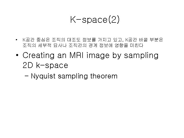
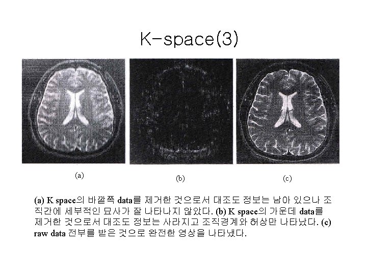
K-space • Data acquired form a continuous pathway through k-space
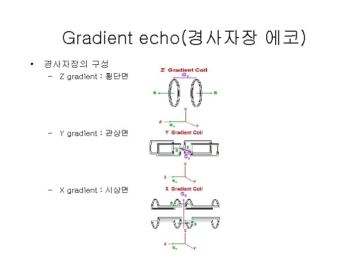
3차원 위치(slice) 구별 • Gradient in the Z-direction – Resonant frequency varying as a function of z – Resonant frequency at specific location • Moving slice location – Shifting the RF frequency up or down – Shifting the overall magnetic field up or down • Any oblique oriented slices by rotating x, y. z
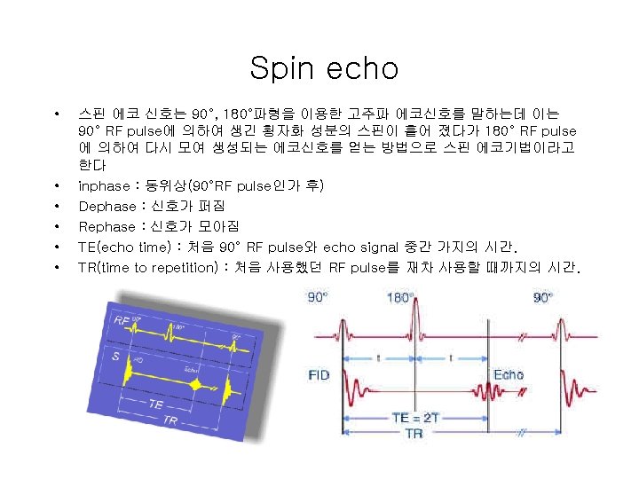
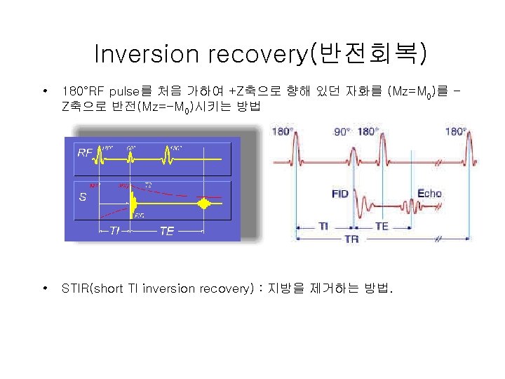
Pulse sequence • MR 영상에서 기본이 되는 4가지 pulse sequence – Partial saturation recovery(부분 포화 회복) • 신호가 미약하여 peak치 신호를 얻을 수 없어 현재는 사 용하지 않음 – Spin echo – Gradient echo – Inversion recovery
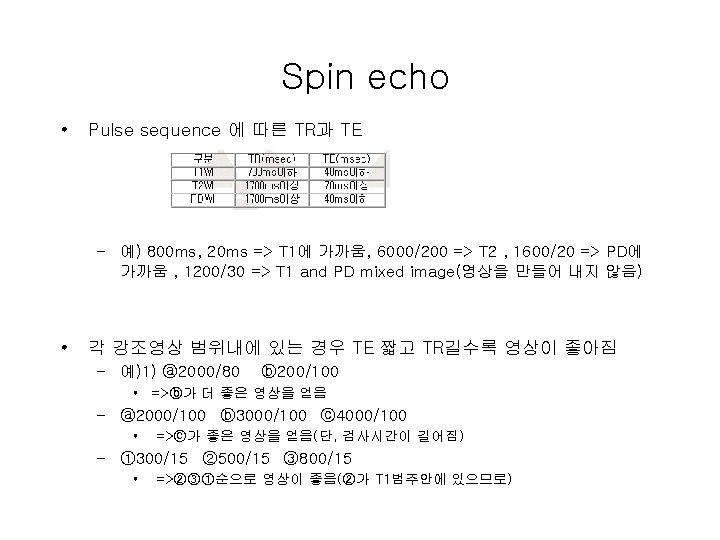
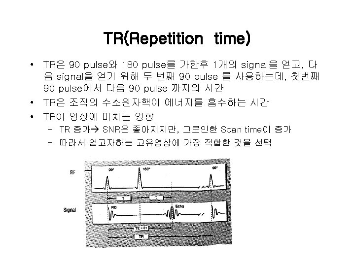
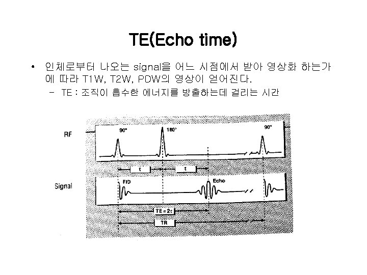
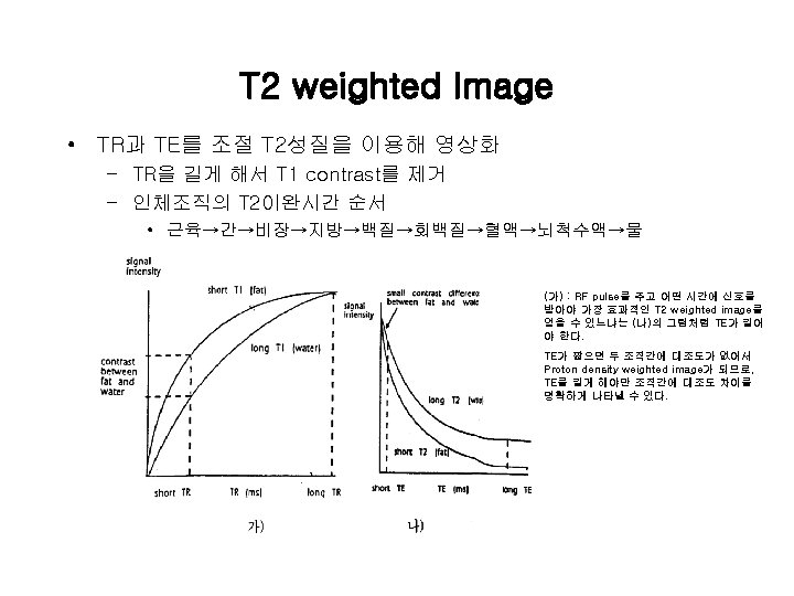
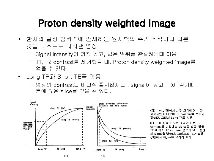
Spin echo • Spin echo영상 – T 1 weighted(T 1 강조) : TE↓, TR↓=>T 1 WI : 해부학적 영상, 검사시간 단축 – T 2 weighted(T 2강조) : TE↑, TR↑=>T 2 WI : 병리학적 영상. – Spin density weighed(Proton Density, 스핀밀도 강조, 수소밀도 강조) : TE↓, TR↑=>PDWI(=SDWI), 대조도↓ A : TR=2500, TE=15(PDW 1) B : TR=500, TE=15(T 1 W 1) C : TR=2500, TE=90(T 2 W 1)
MR Parameter
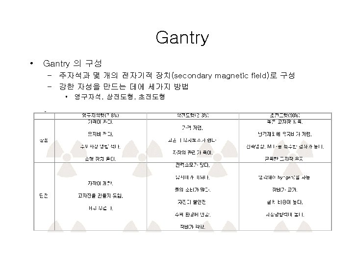
MRI의 구성 • Gantry • Operating Console • Computer
Operation Console & Computer • Operation – MR영상을 보여주는 Monitor와 Keyboard 그리고 Scan 조건과 Scan 상황 을 보여주는 Monitor와 Keyboard로 구성 – 대부분 CT의 Console을 응용하여 제작 • Computer – 용량이 크고 처리속도가 빠른 Minicomputer • 128*128 Pixel에 50 Image 1. 6 MB • 256*256 Pixel에 31 Slice, 4 Echo의 Spin Echo Image에 3 D를 구성하는 요소포 함 8. 1 MB