
MRI Magnetic Resonance Imaging Definition A nonionizing technique



- Slides: 15
MRI Magnetic Resonance Imaging
Definition • A non-ionizing technique with full three dimensional capabilities, excellent soft-tissue contrast, and high spatial resolution.
MRI Facts • Whole body imager = $1. 5 million • Slower then x-ray and CT – Takes between 3 and 10 minutes PER scan
MRI Basics • The person is placed inside a strong magnet. – This produces a static magnetic field typically more than 10, 000 times stronger than the earth’s magnetic field. • Protons inside the body is considered a magnet. – Remember angular momentum? – MRI signal is received by these protons in the body (water and lipids)9
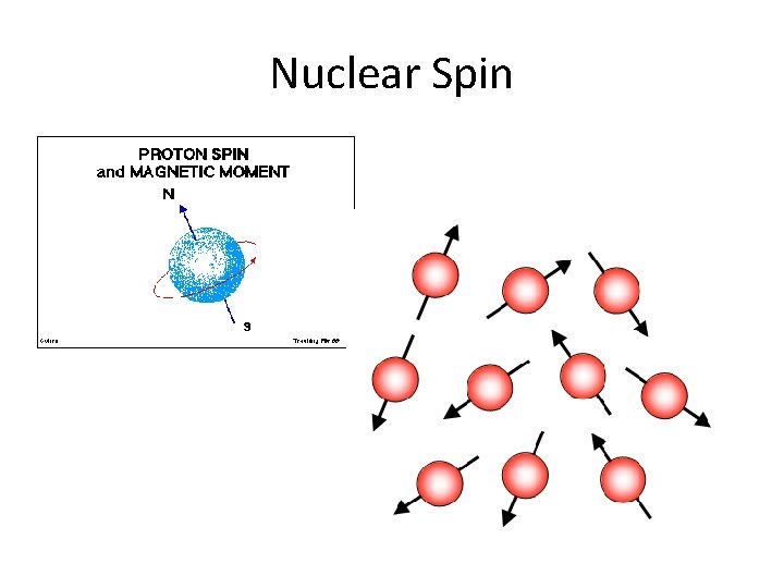
Nuclear Spin
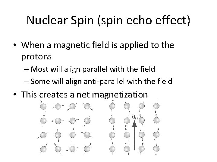
Nuclear Spin (spin echo effect) • When a magnetic field is applied to the protons – Most will align parallel with the field – Some will align anti-parallel with the field • This creates a net magnetization
So here is how it all works… • The magnet is turned on, creating the net magnetization • This magnetization needs to be disturbed, so… – An RF pulse is created by the coil – A 90 degree pulse will throw the protons 90 degrees • A signal is collected as the protons realign with the magnetic field • The material they are contained in will determine how quickly they are able to realign – This is why there are different shades of grey
MRI Instrumentation
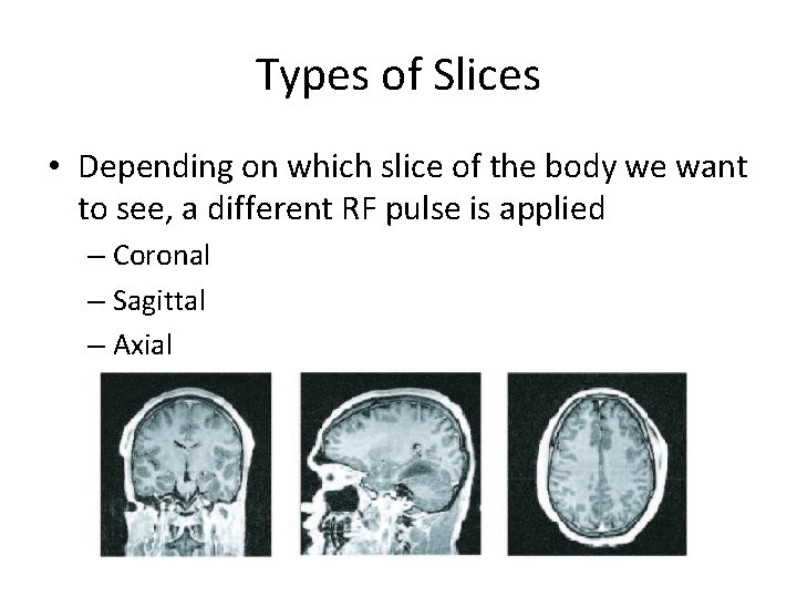
Types of Slices • Depending on which slice of the body we want to see, a different RF pulse is applied – Coronal – Sagittal – Axial
T 1 and T 2 • T 1 = How long for protons to realign or “relax” • T 2 = How long for protons to fall out of phase • Fat: – T 1 = 280 ms, T 2 = 80 ms • Muscle – T 1 = 870 ms, T 2 = 45 ms • Cerebrospinal fluid – T 1 = 2400 ms, T 2 = 160 ms
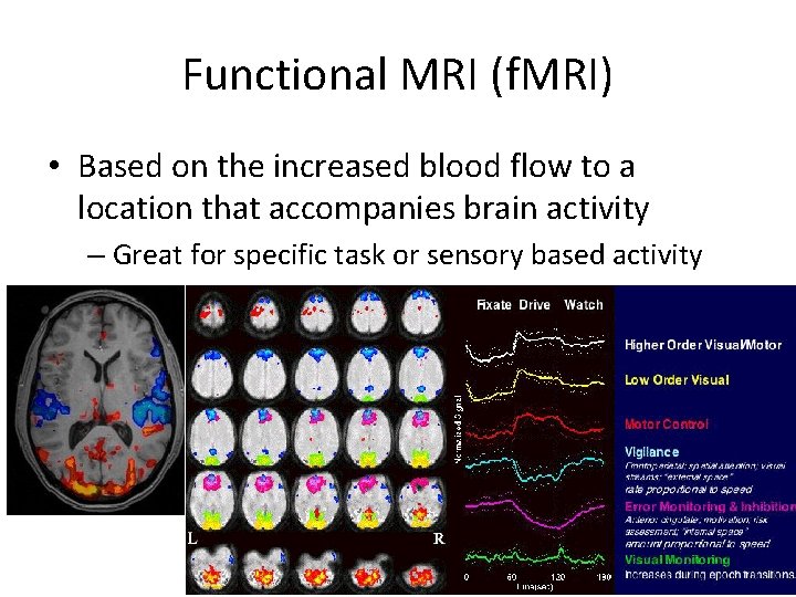
Functional MRI (f. MRI) • Based on the increased blood flow to a location that accompanies brain activity – Great for specific task or sensory based activity