
MRI in epilepsy E Khalili Pouya The specific

- Slides: 104
MRI in epilepsy E. Khalili Pouya
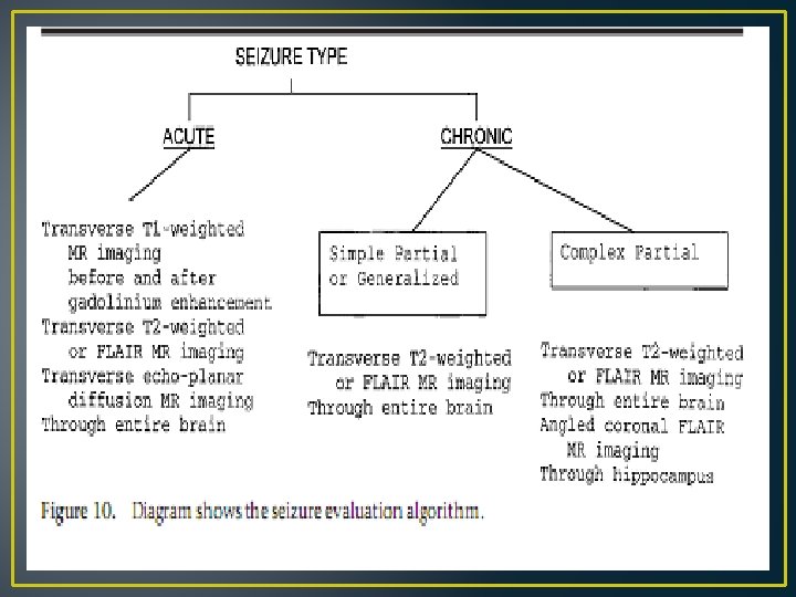
• The specific MR imaging teqnique used depends on the specific type of seizure the patient has. • Recent onset/choronic • Generalized/partial(simple: motor or sensory / complex: altered cognition) • New onset in adult : T 1, T 2, WC • Chronic in adult : T 1, T 2, no WC unless changing in frequency, intensity or nature of seizure(cost effectiveness not harm pts) • Children , young adult(13 -30 yrs)with complex partial seizure objective of MRI is to depict mesial temporal sclerosis(MTS) or temporal or frontal lesions
New onset seizure in an adult • >50% acute(DWI) /chronic cerebrovascular dis • Metastatic dis (parenchymal mets : FLAIR , WC • leptomeningial hematogenous mets (leptomeningeal carcinomatosis)which irritate the cortex : T 1+_WC(recent study : FLAIR wc more sensitive than FLAIR /T 1 wc for cortical process that couse seizure ) • Primary brain tumores(below 60 yrs) : FLAIR/T 2 , T 1+_wc • Higher grade of tumor greater degree of enhancement(exp grade I pillocytic astrocytoma enhances despite its low grade) • FLAIR/T 2 : necrotic portion os tumor in GBM and vasogenic edeme • T 1 -c : subacute hemorrhage(meth Hb), high signal
• Hemorrhagic mets , higer grade(geade III, IV) • MRI is guide prior to BX : avoid necrotic portion and focusing on solid portions(enhances in wc images), more vascular portion likely to have highest grade • DWI : cytotoxic edema (infarction)/high ratio of nuclear cytoplasmic ratio (lymphoma)
Chronic seizure • Surgically treatable/not (not distinguish on basis of clinical presentation alone, imaging is performed) • Not surgically treatable : small cortical scars coused by prior trauma, infarctions, inflammation • Scars: Depend on the size of scar may or may not seen az regions of cortical gliosis (high signal T 2/FLAIR), focal encephalomalacia, sulcal widening on T 1, T 2 • Surgically treatable : benign tomurs(grade I, II) , vascular malformations(AVMs) cavernous angiomas, demylinating lesions, cortical dysplasia
• Most studies in chronic seizure are negative(65% are idiopathic)
Hyppocampal sclerosis (MTS/Ammon’s horn sclerosis) • Hippocampal sclerosis is by far the most common cause of temporal lobe epilepsy. • The familiar reader detects it on MRI in more than 95% of cases but should be aware of typical ‘‘pitfalls’’, namely bilateral hippocampal sclerosis‘‘dual pathology’’ and insufficient image Quality
• Pathology: • half of the patients undergoing surgery have experienced a precipitating injury before the age of 4 years (complex fever seizures, 70%; birth trauma, meningitis, head injury, 30% • Mean age at the onset of complex partial seizures is between 9 and 11 years • mean age at the time of epilepsy surgery around the age of 30 • The long latency between a possible initial precipitating injury, the onset of epileptic seizures, and epilepsy surgery renders assessment of the pathogenesis of hippocampal sclerosis difficult
• Clinical presentation: starts with an epigastric aura (definition of aura = initial part of a partial seizure, that is remembered after the seizure has terminated). The aura is followed by objective phenomena like staring, restlessness, oroalimentary automatism, and (ipsilateral) head deviation, which last from around 30 seconds to several minutes. In the postictal phase, gradual reorientation occurs which may be accompanied by dysphasia and other symptoms
• Pathology: Hippocampal sclerosis is characterized by neuronal loss and gliosis, most prominent in the CA 1 field of the hippocampus, followed by the hilus, CA 3 field, and dentate granule layer, while the CA 2 field is relatively spared
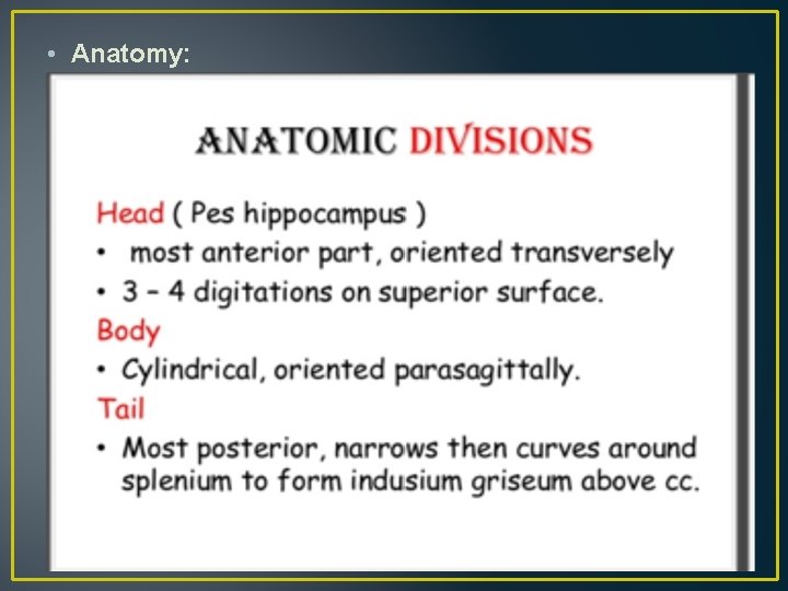
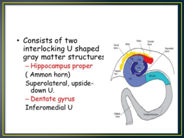
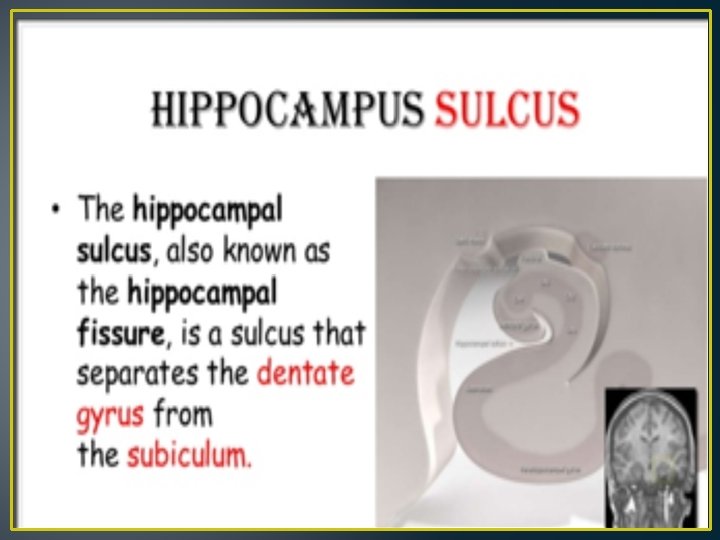
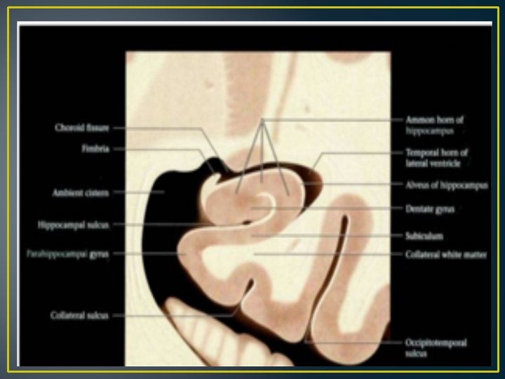
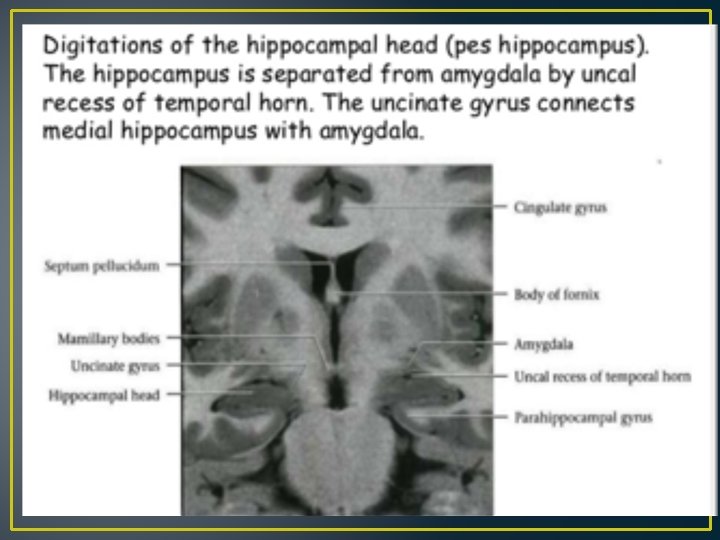
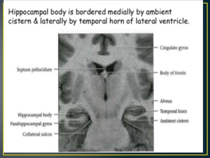
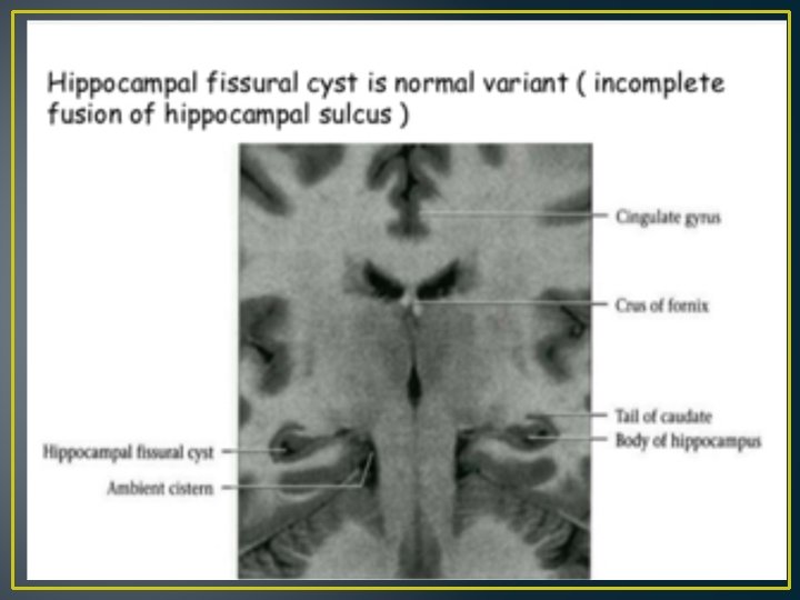
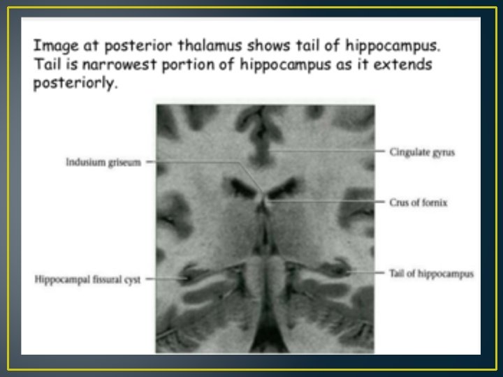
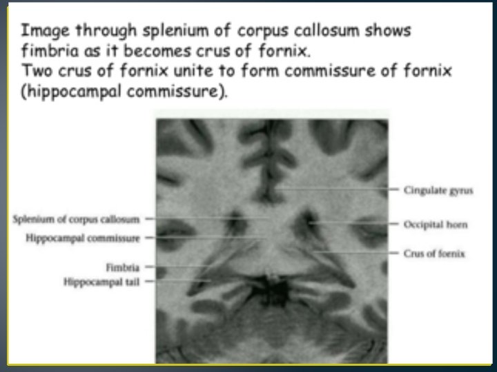
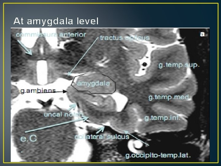
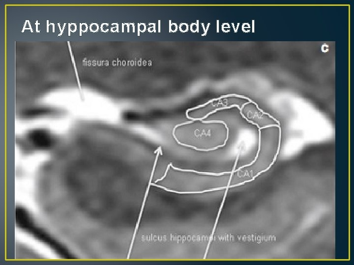
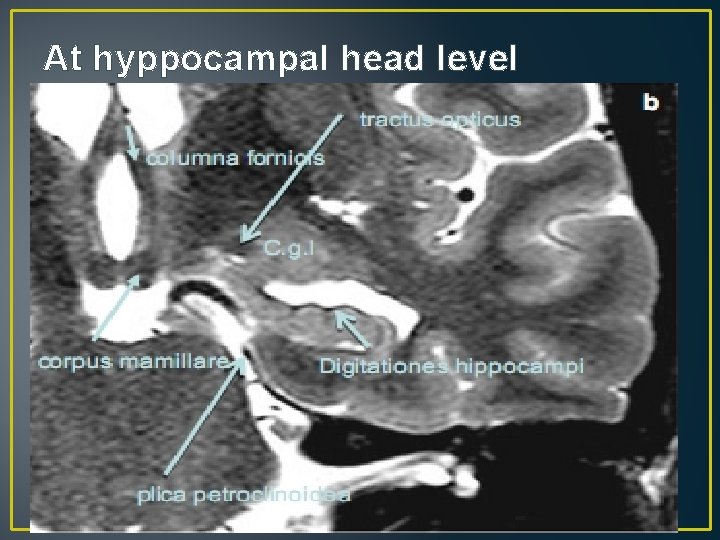
• Anatomy:
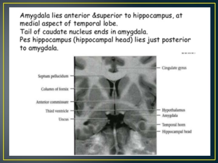
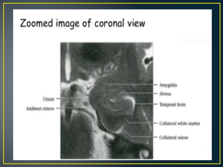
At amygdala level
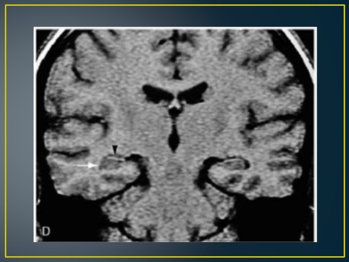
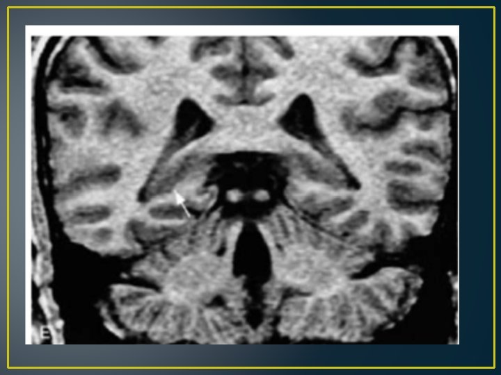
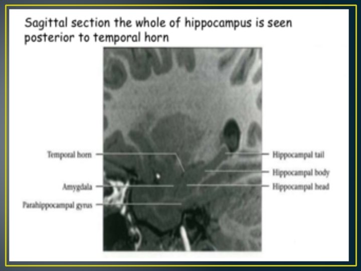
At hyppocampal body level
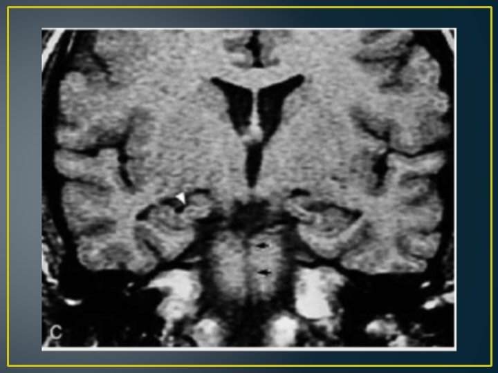
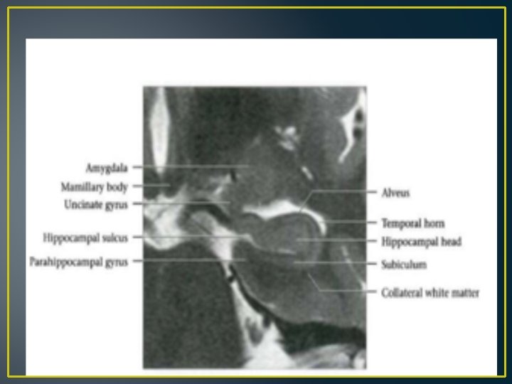
At hyppocampal head level
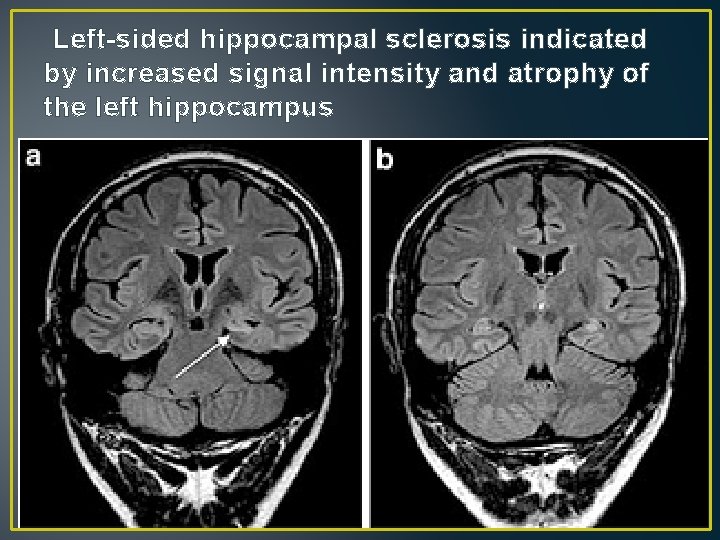
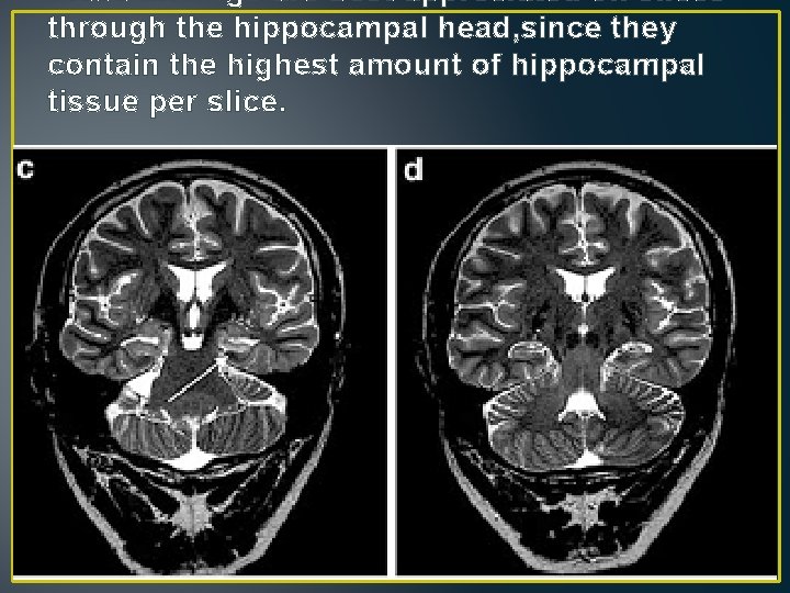
• Imaging: atrophy and increased signal intensity best visualized on coronal FLAIR and T 2 -weighted fast spin echo images angulated perpendicularly to the hippocampal long axis Increased signal intensity T 2 -signal abnormalities appears to correlate with gliosis and may not be directly related to the degree of neuronal loss On FLAIR sequences, contrast to noise ratio (C/N) is higher than on T 2 -weighted sequences, however, one has to be aware that normal limbic structures already have a higher FLAIR signal than the remaining cortex
Left-sided hippocampal sclerosis indicated by increased signal intensity and atrophy of the left hippocampus
these findings are best appreciated on slices through the hippocampal head, since they contain the highest amount of hippocampal tissue per slice.
• T 2 -weighted sequences display the hippocampal substructures in more detail • In order to assess atrophy and signal intensity, side comparisons are helpful • However, 10– 20% of patients have bilateral hippocampal sclerosis which can be overlooked when side comparison is the only criterion and no ‘‘engramm’’ of a normally sized hippocampus exists. T 2 volumetry or T 2 relaxometry can be helpful in these cases. • Hippocampal sclerosis is usually diagnosed on coronal slices through the hippocampal head
• displays the highest relative volume of hippocampal tissue on a slice • More subtle hippocampal sclerosis signs are a loss of the internal structure and loss of hippocampal head digitations which are both best appreciated on high resolution T 2 -weighted image • Dilatation of the temporal horn is common, but occurs also in healthy • Hippocampal sclerosis with atrophy but without increased signal intensity has been described in 5% of pts, it is likely due to poor image quality not suited to visualize increased signal intensity.
• Hippocampal sclerosis as incidental finding is extremly rare. There may be signal increase in healthy patients, however, signal increase and atrophy together almost never occur • MRI scans of older patients, however, often show some degree of atrophy including loss of digitations of the hippocampal head and increased signal intensity (on FLAIR) images. The histopathological substrate typically remains unclear, it may be related to normal ageing or Alzheimer’s disease or so-called pure hippocampal sclerosis which occurs in around 10% of individuals older than 85 years and
• Secondary findings: • Apart from hippocampal sclerosis the following structures of the limbic system can be atrophic: • amygdala, • ipsilateral mamillary body, • ipsilateral fornix, • posterior thalamus (with increased signal), • cingulate gyrus, • contralateral cerebellum
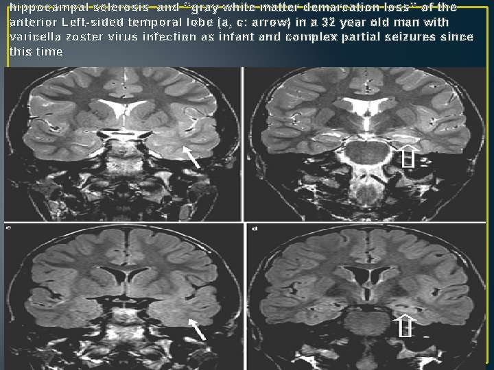
• There is more often a temporal lobe or even hemispheric atrophy with atrophy pronounced in the anterior temporal lobe. • The anterior temporal lobe shows reduced white matter volume and white matter signal is increased as compared to the opposite side or remaining white matter. • Since white matter has a higher signal, contrast to gray matter is reduced and the term ‘‘gray white matter demarcation loss’’ has been designated to describe this condition early seizure onset (often before the age of two) of patients with a ‘‘gray white matter demarcation loss’’ as compared to those who do not have these change
hippocampal sclerosis and ‘‘gray white matter demarcation loss’’ of the anterior Left-sided temporal lobe (a, c: arrow) in a 32 year old man with varicella zoster virus infection as infant and complex partial seizures since this time
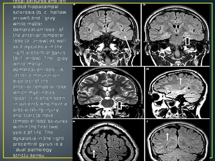
• 10% of patients hippocampal sclerosis is associated with another extrahippocampal epileptogenic lesion (Fig. 7). This is called dual pathology …… poorer px post surgery • Most common dual lesions are cortical dysplasias and gliotic lesions acquired in early childhood. • Dual Pathology refers only to patients with hippocampal sclerosis, who have a second principal lesion affecting the brain (which may be located also outside the ipsilateral temporal lobe), that is, tumor, vascular malformation, glial scar, limbic/Rasmussen encephalitis, or MCD (including FCD Type IIa/IIb). Ipsilateral temporopolar atrophy with increased T 2 signal changes on MRI is not included as its histopathologic correlate has yet to be specified
focal seizures and left sided hippocampal sclerosis (b, c: hollow arrowl) and ‘‘gray white matter demarcation loss’’ of the anterior temporal lobe (a: arrow) as well as a dysplasia in the right precentral gyrus (d–f: arrow). The ‘‘gray white matter demarcation loss’’ is rather a maturation disorder of the anterior temporal lobe which myelinates latest. It is often seen in patients who have a precipitating injury and start to have temporal lobe seizures within the first two years of life. The dysplasia in the right precentral gyrus is a ‘‘dual pathology’’ strictu sensu
• PET: • temporal lobe is hypometabolic for uptake of glucose on the side of the seizure focus during the interictal period. • The region of hypometabolism can be both medial and lateral, • commonly exceeds the size of tissue that needs to be removed for cure of seizures
Vascular Malformations • • • cavernomas, arteriovenous malformations, dural arteriovenous fistulae, developmental venous anomalies capillary telangiectasias
Cavernomas‘cavernous malformation. ’’ • Around 10 % of cavernomas are familial cases with autosomal dominant inheritance • Up to 20 % of cavernomas are accompanied by developmental venous anomalies (DVAs) • Up to 40 % present with epileptic seizures • seizures are most prevalent in supratentorial cavernomas and progress to epilepsy in 40 % of these cases • Around 75 % of patients become seizure-free following microsurgical resection
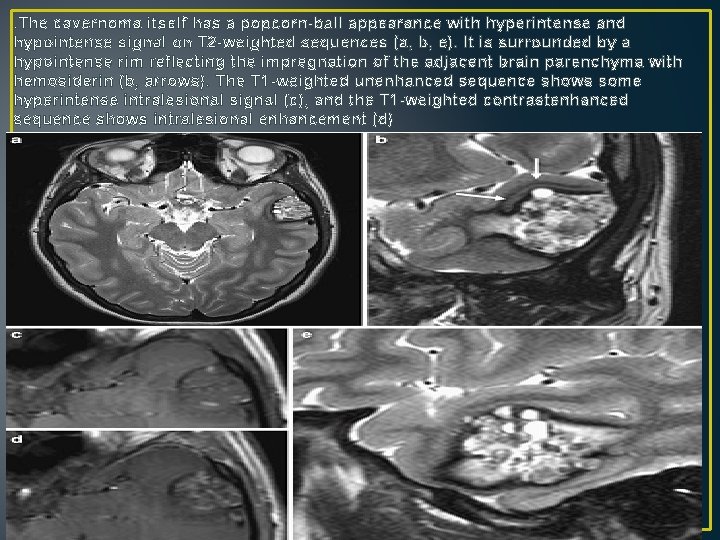
Imaging: MR images typically show a popcornlike pattern with mixed high and low signal intensities in the core and a dark rim of hemosiderin Hyperintense portions on T 1 -weighted images in the core represent subacute (intralesional) hemorrhage or thrombosis within the ‘‘berries’’ of the cavernoma A cavernoma may profoundly bleed into the surrounding brain parenchyma (extralesional hemorrhage)
• In addition to T 2 -weighted fast spin echo sequences, which show the anatomical details best • T 2 -weighted gradient echo or susceptibility-weighted sequences are mandatory • because of susceptibility artefacts at bone–brain interfaces, small cortical/subcortical cavernomas can be easily missed on T 2 -weighted gradient echo sequence • Since up to 20 % of cavernomas are associated with DVAs, T 1 -weighted contrast-enhanced images should be additionally obtained
. The cavernoma itself has a popcorn-ball appearance with hyperintense and hypointense signal on T 2 -weighted sequences (a, b, e). It is surrounded by a hypointense rim reflecting the impregnation of the adjacent brain parenchyma with hemosiderin (b, arrows). The T 1 -weighted unenhanced sequence shows some hyperintense intralesional signal (c), and the T 1 -weighted contrastenhanced sequence shows intralesional enhancement (d)
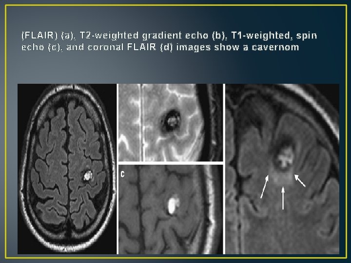
(FLAIR) (a), T 2 -weighted gradient echo (b), T 1 -weighted, spin echo (c), and coronal FLAIR (d) images show a cavernom
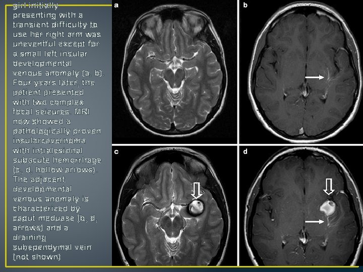
girl initially presenting with a transient difficulty to use her right arm was uneventful except for a small left insular developmental venous anomaly (a, b). Four years later, the patient presented with two complex focal seizures. MRI now showed a pathologically proven insularcavernoma with intralesional subacute hemorrhage (c, d, hollow arrows). The adjacent developmental venous anomaly is characterized by caput meduase (b, d, arrows) and a draining subependymal vein (not shown)
Arteriovenous Malformations • Arteriovenous malformation (AVM) is the most common symptomatic vascular malformation • AVMs are typically solitary lesions. • In multiple lesions, hereditary hemorrhagic telangiectasia (Rendu–Osler–Weber syndrome) , Wyburn–Mason syndrome, and cerebrofacial arteriovenous metameric syndromes (types 1– 3) should be considered • seizures are the most common presentation.
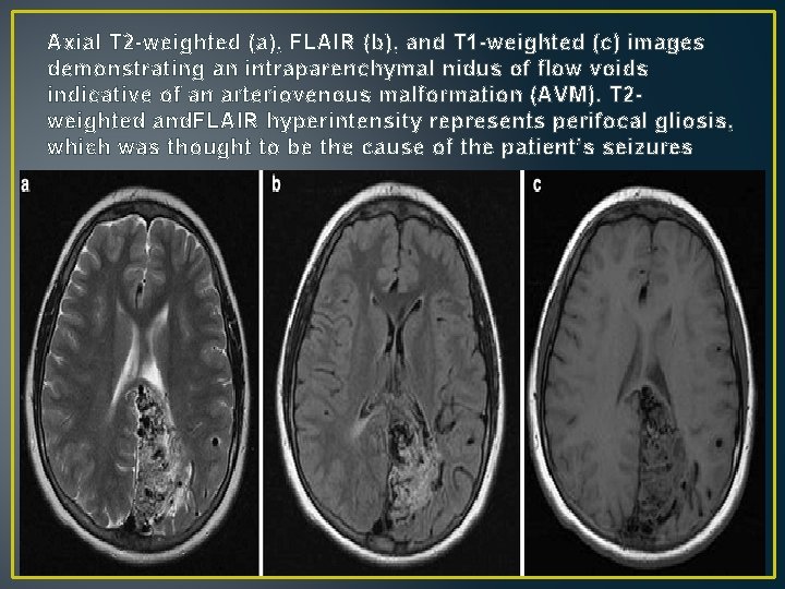
Imaging • On MRI AVMs appear as a ‘‘bag of black worms’’ (flow void structures) with minimal or no mass effect. • It is important to look for intranidal or flow-related aneurysms of the feeding arteries, which may bleed ‘‘instead’’ of the AVM. • Best T 2 fast spin echo
Axial T 2 -weighted (a), FLAIR (b), and T 1 -weighted (c) images demonstrating an intraparenchymal nidus of flow voids indicative of an arteriovenous malformation (AVM). T 2 weighted and. FLAIR hyperintensity represents perifocal gliosis, which was thought to be the cause of the patient’s seizures
Dural Arteriovenous Fistulae • Dural arteriovenous fistulae arteriovenous shunts that are located in the wall of a venous sinus and are typically fed by meningeal arteries • In adult : acquired and may develop following, e. g. , a minor trauma and/or sinus thrombosis • In children: congenital • arteriovenous fistulae are communications between dural branches of the internal carotid artery, the external carotid artery, the vertebral artery, and intracerebral veins and/or sinuses
• The clinical symptoms depend on the size, the location, and the venous drainage pattern • Risk of intracerebral hemorrhage is high in anterior skull base arteriovenous fistulae and risk of intracerebral hemorrhage and epileptic seizures is high in fistulae with retrograde filling of cortical veins
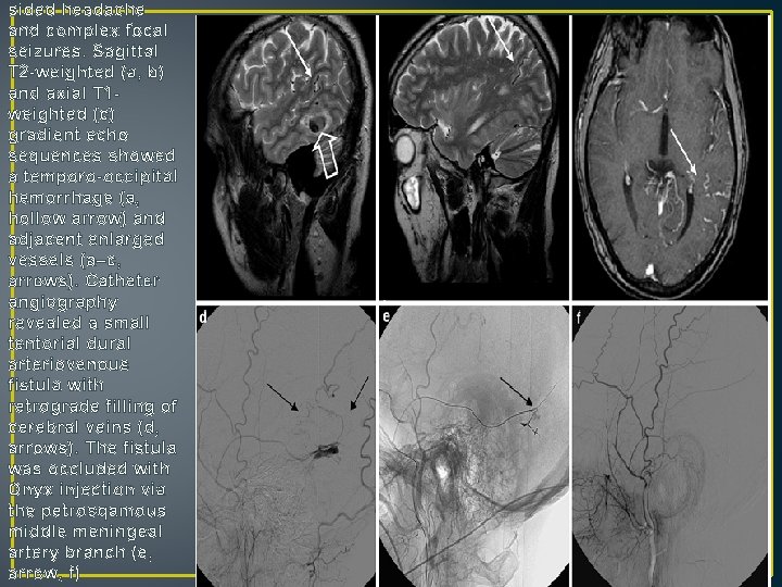
Imaging: Vasogenic edema, hemorrhage, tubular/tortuous flow void (T 2 -weighted) and contrast-enhanced (T 1 -weighted contrast-enhanced sequences) structures are suggestive of a dural arteriovenous fistul Coronal three-dimensional phase contrast angiography is needed to show venous sinus anatomy and may reveal a thrombosed or partially open venous sinus.
sided headache and complex focal seizures. Sagittal T 2 -weighted (a, b) and axial T 1 weighted (c) gradient echo sequences showed a temporo-occipital hemorrhage (a, hollow arrow) and adjacent enlarged vessels (a–c, arrows). Catheter angiography revealed a small tentorial dural arteriovenous fistula with retrograde filling of cerebral veins (d, arrows). The fistula was occluded with Onyx injection via the petrosqamous middle meningeal artery branch (e, arrow, f)
Developmental Venous Anomalies/venous angioma. ’ • DVAs are the most common vascular malformation on contrast-enhanced T 1 -weighted MR images • DVAs are typically incidental findings. If they cause symptoms, they are likely due to associated cavernoma(20%)
• Imaging: DVAs are characterized by dilated medullary white matter • veins (caput medusae) converging on a collector vein which drains into a dural sinus or ependymal vein. Around 5 % of DVAs are atypical, with some shunting • between the arterial and venous system.
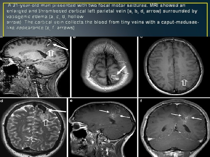
A 21 -year-old man presented with two focal motor seizures. MRI showed an enlarged and thrombosed cortical left parietal vein (a, b, d, arrow) surrounded by vasogenic edema (a, c, d, hollow arrow). The cortical vein collects the blood from tiny veins with a caput-medusaelike appearance (e, f, arrows)
Capillary Telangiectasias Hereditary hemorrhagic telangiectasia (Rendu–Osler– Weber syndrome), ataxia telangiectasia, and past radiation therapy in children in lesions outside the brainstem should be considered.
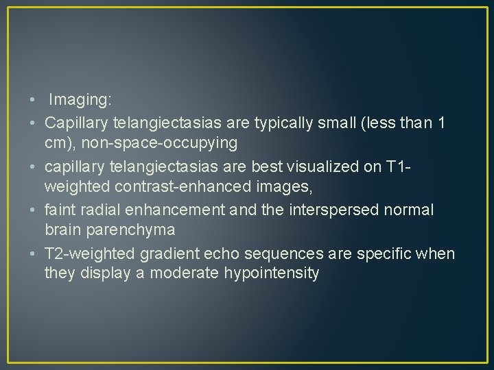
• Imaging: • Capillary telangiectasias are typically small (less than 1 cm), non-space-occupying • capillary telangiectasias are best visualized on T 1 weighted contrast-enhanced images, • faint radial enhancement and the interspersed normal brain parenchyma • T 2 -weighted gradient echo sequences are specific when they display a moderate hypointensity
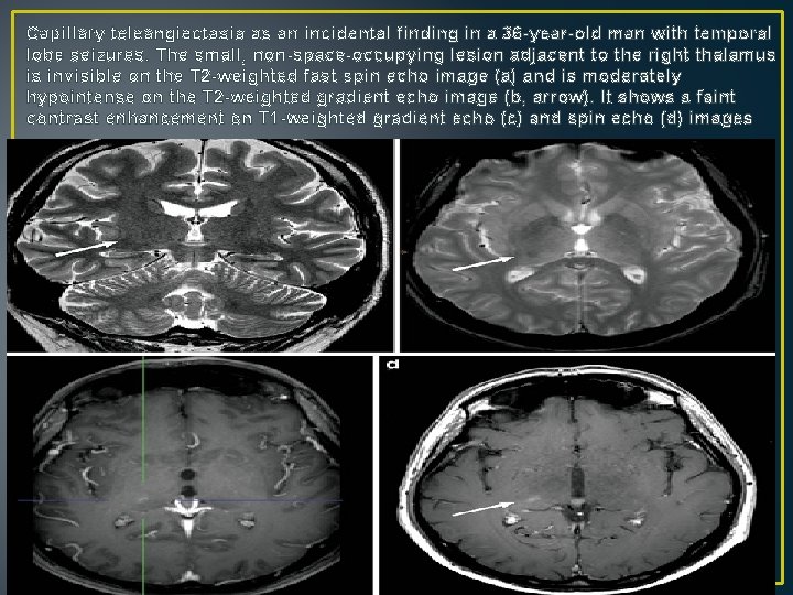
Capillary teleangiectasia as an incidental finding in a 36 -year-old man with temporal lobe seizures. The small, non-space-occupying lesion adjacent to the right thalamus is invisible on the T 2 -weighted fast spin echo image (a) and is moderately hypointense on the T 2 -weighted gradient echo image (b, arrow). It shows a faint contrast enhancement on T 1 -weighted gradient echo (c) and spin echo (d) images
Malformations of Cortical Development
Lissencephaly Type 1, Subcortical Band Heterotopia Lissencephaly means smooth brain , affected brains may be without convolutions (agyria) or with broad and shallow convolutions (pachygyria) o two distinct forms: type 1 (also known as classic) lissencephalies type 2 (also known as cobblestone) lissencephalies
• Type I lissencephaly is a neuronal migration disorder • Moreover, intrauterine infections (particularly CMV) and toxins may cause type I lissencephaly
• Imaging: • On MRI, the brain surface appears smooth with areas of absent (agyria) and abnormally wide gyri (pachygyria). • The cortex is abnormally thick • Common associated malformations include: • Enlarged posterior portions of the lateral ventricles, • rounded hippocampus, • flat anterior portion of the corpus callosum, • very variable hypoplasia of the cerebellum, especially the midline vermis
• In SBH(subcortical band hetrotopia)the brain surface appears superficially normal, except that the sulci between gyri are shallow, and the cortex is normal and not thick • just beneath the cortex, often separated from it by just a few millimeters of white matter, lies a smooth band of neurons that never reached the true cortex. This band has a variable thickness • Most lissencephalies have a genetic cause, however, periventricular and subcortical calcifications suggest an infectious (particularly CMV) cause
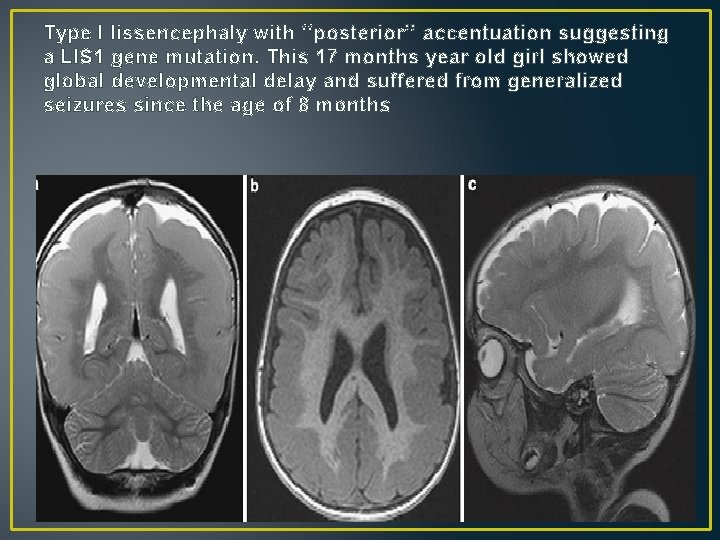
Type I lissencephaly with ‘‘posterior’’ accentuation suggesting a LIS 1 gene mutation. This 17 months year old girl showed global developmental delay and suffered from generalized seizures since the age of 8 months
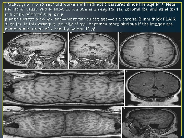
Pachygyria in a 30 year old woman with epileptic seizures since the age of 7. Note the rather broad and shallow convolutions on sagittal (a), coronal (b), and axial (c) 1 mm thick reformations, on a planar surface view (d), and—more difficult to see—on a coronal 3 mm thick FLAIR slice (e). In this example, paucity of gyri becomes more obvious if the images are compared to those of a healthy person (f, g)
Cobblestone Lissencephaly, Congenital Muscular Dystrophies • Congenital muscular dystrophy syndromes or so-called dystroglycanopathies represent a heterogeneous group of congenital diseases affecting the muscles and frequently the brain and eyes, which are characterized by defective protein glycolization. • The clinical spectrum comprises severe [Walker–Warburg syndrome (WWS), Fukuyama congenital muscular dystrophy, muscle–eye–brain disease] and milder forms with or without brain involvement (congenital muscular dystrophy CMD, merosin-deficient congenital muscular dystrophy, merosin-positive congenital muscular dystrophy C 1 C, merosin-positive congenital muscular dystrophy C 1 D, limb girdle muscular dystrophies LGMD 2 I, LGMD 2 K, LGMD 2 L, LGMD 2 M).
• Walker–Warburg Syndrome • the most severe congenital muscular dystrophy syndrom • Symptoms and signs are already present at birth, • children often die within the first 12 months and rarely live longer than 5 year • Clinical Presentation: • profound muscular hypotonia at birth (‘‘floppy newborn’’), epileptic seizures, • anterior (cataracts, shallow anterior chamber, microcornea, microphthalmia, lens defects) • posterior eye anomalies (retinal detachment or dysplasia, hypoplasia or atrophy of the optic nerve and macula and coloboma). • Glaucoma or buphthalmos may be present, • small genitalia in boys, • occasionally cleft lip and palate.
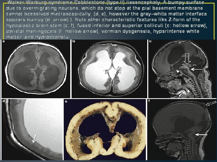
• Imaging: • Distinct hydrocephalus • The brain surface is smooth(either agyric or contains only some broad and shallow gyrus) • The brain parenchyma consists of a thin band of bumpy gray and hypomyelinated and thus hyperintense white matter • The bumpy surface is explained with the fact that migrating neurons do not stop at the pial basement membrane but migrate too far (cobblestone or type II lissencephaly). • Polymicrogyria • corpus callosum is thin • inferior and superior colliculi are fused • the brainstem is hypoplastic with a kinking between the pons and mesencephalon (Z-form) • posterior fossa is small • vermis dygenesis resembling a Dandy–Walker variant • in some cases there is a posterior meningo- or encephalocele
Walker-Warburg syndrome, Cobblestone (type II) lissencephaly, A bumpy surface due to overmigrating neurons, which do not stop at the pial basement membrane cannot beesolved macroscopically, (d, e), however the gray–white matter interface appears bumpy (d: arrow) ). Note other characteristic features like Z-form of the hypoplastic brain stem (c, f), fused inferior and superior colliculi (c: hollow arrow), parietal meningocele (f: hollow arrow), vermian dysgenesis, hyperintense white matter, and hydrocephalu
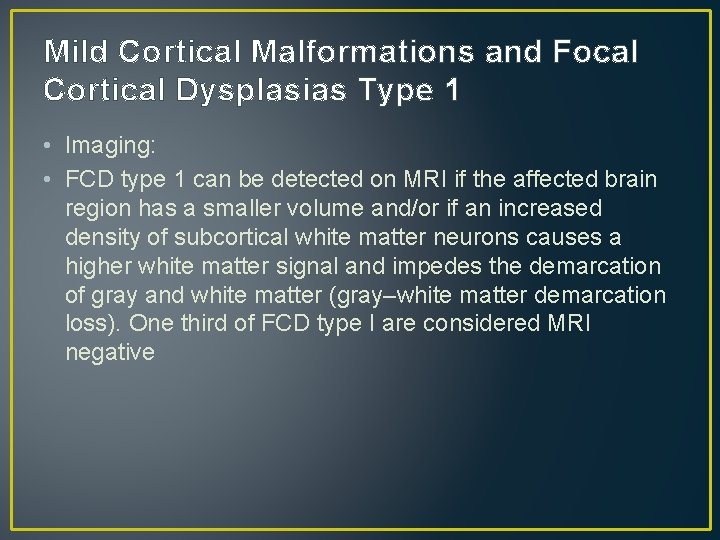
Focal Cortical Dysplasias • FCDs type 1 are so called cytoarchitectural abnormalities without (type 1 A according to Palmini) or with giant or immature neurons (type 1 B according to Palmini), but without dysmorphic neurons or balloon cells. • FCDs type 2 contain dysmorphic neurons (type 2 A) or dysmorphic neurons and balloon cells (type 2 B).
Mild Cortical Malformations and Focal Cortical Dysplasias Type 1 • Imaging: • FCD type 1 can be detected on MRI if the affected brain region has a smaller volume and/or if an increased density of subcortical white matter neurons causes a higher white matter signal and impedes the demarcation of gray and white matter (gray–white matter demarcation loss). One third of FCD type I are considered MRI negative
Focal Cortical Dysplasia Type 2 A • type 2 A are distinctly less frequent than FCD type 2 B • Drug-resistant epilepsy with focal (complex focal [simple focal) without or with secondarily generalized seizures.
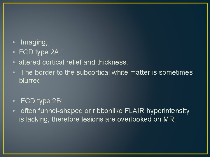
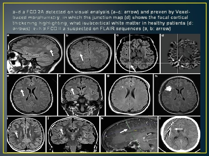
• • Imaging; FCD type 2 A : altered cortical relief and thickness. The border to the subcortical white matter is sometimes blurred • FCD type 2 B: • often funnel-shaped or ribbonlike FLAIR hyperintensity is lacking, therefore lesions are overlooked on MRI
a–d a FCD 2 A detected on visual analysis (a–c: arrow) and proven by Voxelbased morphometry, in which the junction map (d) shows the focal cortical thickening highlighting, what isubcortical white matter in healthy patients (d: arrows). e–h a FCD II a suspected on FLAIR sequences (a, b: arrow)
Focal Cortical Dysplasia Type FCD with balloon cells, 2 B/ Taylor type dysplasia/ transmantle dysplasia. • Drug-resistant epilepsy with focal (complex focal [simple focal) without or with secondarily generalized seizures. • Following surgery, [80 % of patients are seizure-free.
Imaging: MRI hallmark is a distinct funnel-shaped FLAIR hyperintensity tapering towards the lateral ventricle, which has been called the transmantle sign and which is found in 90 % of cases In some instances, the FLAIR hyperintensity can be followed towards the lateral ventricle; in other instances it appears as a penlike line along the inner cortical surface. The dysplastic cortex is isointense on T 1 -weighted FCD 2 B are therefore only visible on T 1 -weighted images if the cortex is markedly thickened or if they are distinct adjacent to white matter changes
• The dysplastic cortex is isointense (1/3) or slightly hyperintense (2/3) on T 2 -weighted, and slightly hyperintense on FLAIR fast spin echo sequences. • FCDs 2 B single neocortical lesions • the most common location is—likely due to the lobe size is frontal • If there is more than one dysplasia, one should consider tuberous sclerosis and carefully look for subependymal giant cell astrocytoma and subependymal nodules.
The bottom of sulcus dysplasia can be easily overlooked, especially on axial FLAIR images. Due to their spatial orientation, coronal and sagittal FLAIR sequences, isotropic 3 D FLAIR sequences may be needed Calcification within the subcortical lesion parts may occur in larger lesions; contrast enhancement is typically absent.
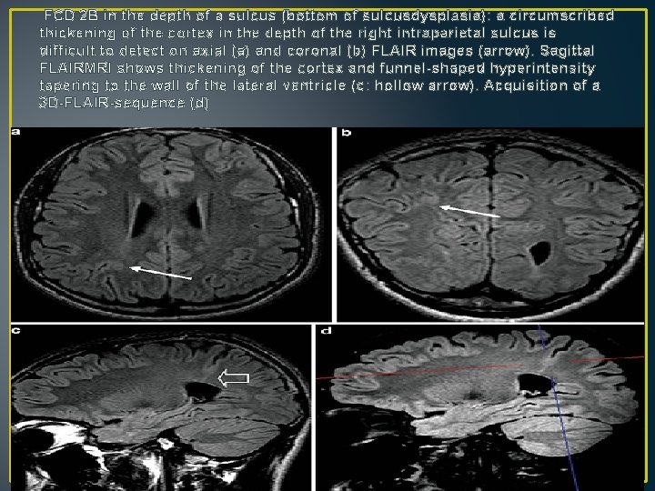
FCD 2 B in the depth of a sulcus (bottom of sulcusdysplasia): a circumscribed thickening of the cortex in the depth of the right intraparietal sulcus is difficult to detect on axial (a) and coronal (b) FLAIR images (arrow). Sagittal FLAIRMRI shows thickening of the cortex and funnel-shaped hyperintensity tapering to the wall of the lateral ventricle (c: hollow arrow). Acquisition of a 3 D-FLAIR-sequence (d)
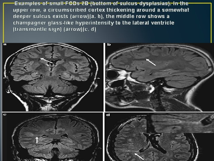
Examples of small FCDs 2 B (bottom of sulcus dysplasias). In the upper row, a circumscribed cortex thickening around a somewhat deeper sulcus exists (arrow)(a, b), the middle row shows a champagner glass-like hyperintensity to the lateral ventricle (transmantle sign) (arrow)(c, d)
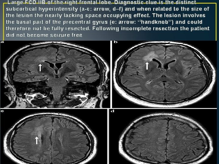
Large FCD IIB of the right frontal lobe. Diagnostic clue is the distinct subcortical hyperintensity (a-c: arrow, d–f) and when related to the size of the lesion the nearly lacking space occupying effect. The lesion involves the basal part of the precentral gyrus (e: arrow: ‘‘handknob’’) and could therefore not be fully resected. Following incomplete resection the patient did not become seizure free
Hemimegalencephaly • 1. Isolated form without systemic involvement. • 2. Syndromic form which may occur as hemihypertrophy • of part or all of the ipsilateral body. It has been described in patients with organoid (formerly: epidermal) nevus syndrome, Proteus syndrome (? Neurocutaneous Diseases, neurofibromatosis type 1 , hypomelanosis of Ito , Klippel–Trenaunay–Weber syndrome, and tuberous sclerosis • 3. Total hemimegalencephaly with enlargement of the ipsilateral half of the brainstem and cerebellum
Imaging: The abnormal hemisphere is larger than the contralateral one, and the midline is pushed to the contralateral side cortex appears thickened and enlarged, the gyri are usually broad and flat and the sulci shallow Sylvian fissure is short and thickened and the posterior end is open. Gray and white matter are difficult to delineate from each other White matter volume is increased and the white matter signal on T 2 -weighted images is clearly abnormal In neonates, the white matter signal on T 2 -weighted images is low as opposed to normal neonates, in which the white matter signal is higher than that of gray matter
• In older children with hemimegalencephalies, the white matter signal becomes higher than normal reflecting lack of myelination • If only parts of the hemisphere show enlargement and dysplastic features, the disease may be called focal megalencephaly or large FCD 2 B
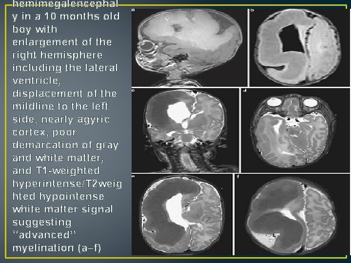
hemimegalencephal y in a 10 months old boy with enlargement of the right hemisphere including the lateral ventricle, displacement of the mildline to the left side, nearly agyric cortex, poor demarcation of gray and white matter, and T 1 -weighted hyperintense/T 2 weig hted hypointense white matter signal suggesting ‘‘advanced’’ myelination (a–f)
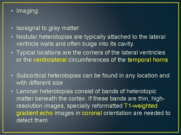
Heterotopia • Heterotopias are conglomerate masses of gray matter in an abnormal location • . They can be uni- or bilateral, • nodular, ribbonlike, chainlike, ball-like, or curvilinear configuration • located attached to the ventricle wall, within the white matter, or attached to the cortex
• Imaging: • Isosignal to gray matter • Nodular heterotopias are typically attached to the lateral ventricle walls and often bulge into its cavity. • Typical locations are the corners of the lateral ventricles or the ventrolateral circumferences of the temporal horns. • Subcortical heterotopias can be found in any location and with different size • Laminar heterotopias consist of bands of heterotopic matter beneath the cortex. If these bands are thin, highresolution images, specially reformatted T 1 -weighted gradient echo images in coronal orientation are needed to detect them
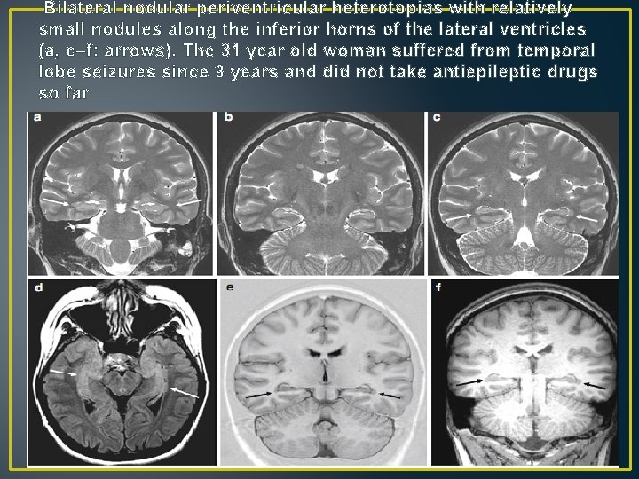
Bilateral nodular periventricular heterotopias with relatively small nodules along the inferior horns of the lateral ventricles (a, c–f: arrows). The 31 year old woman suffered from temporal lobe seizures since 3 years and did not take antiepileptic drugs so far
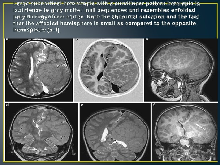
Large subcortical heterotopia with a curvilinear pattern. heteropia is isointense to gray matter inall sequences and resembles enfolded polymicrogyriform cortex. Note the abnormal sulcation and the fact that the affected hemisphere is small as compared to the opposite hemisphere (a–f)
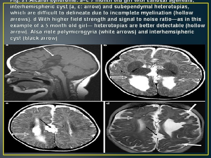
Aicardi Syndrome • Callosal agenesis • interhemispheric cysts are usually detected by ultrasound in utero. • Newborns are typically blind and suffer from infantile spasms. Asignificant number of girls, however, seem to develop normally until around the age of 3 months, when they begin to have infantile spasms. • Ocular examination reveals zones of depigmentation of the pigmented epithelium characterized as chorioretinal lacunae. • Only 40 % of girls get older than 15 years of age.
• Imaging: • In a blind female newborn with epileptic seizures look for callosal agenesis, interhemispheric cyst(s), and cortical dysplasias (heterotopias, polymicrogyria)
Fig. 21 Aicardi syndrome: a–c 7 month old girl with callosal agenesis, interhemispheric cyst (a, c: arrow) and subependymal heterotopias, which are difficult to delineate due to incomplete myelination (hollow arrows). d With higher field strength and signal to noise ratio—as in this example of a 5 month old girl— heterotopias are better detectable (hollow arrow). Also note polymicrogyria (white arrows) and interhemsipheric cyst (black arrow)
Kallmann Syndrome • Hyposmia or anosmia, hypogonadism due to hypothalamic insufficiency, involuntary movements of a body segment in reply to voluntary movements of the similar contralateral segment (mirror movements) and renal abnormalities (e. g. , unilateral agenesis) in X-linked Kallmann syndrome. • Kallmann syndrome appears as olfactory bulb, tract, and sulcus a-/hypoplasia, and small adenohypophysis due to insufficient hypothalamic stimulation, posterior pituitary lobe is normal
• Thank you