MRI Ashwaq Albalawi What is MRI Magnetic resonance

MRI Ashwaq Albalawi
 is an imaging technique used primarily in")
What is MRI Magnetic resonance imaging (MRI) is an imaging technique used primarily in medical settings to produce high quality images of the soft tissues of the human body. It is based on the principles of nuclear magnetic resonance (NMR)

History of MRI *Nikola Tesla discovered the Rotating Magnetic Field in 1882 in Budapest, Hungary. This was a fundamental discovery in physics. All MRI machines are calibrated in "Tesla Units". The strength of a magnetic field is measured in Tesla or Gauss Units. The stronger the magnetic field, the stronger the amount of radio signals which can be elicited from the body's atoms and therefore the higher the quality of MRI images. 1 Tesla = 10, 000 Gauss Low-Field MRI= Under. 2 Tesla (2, 000 Gauss) Mid-Field MRI=. 2 to 0. 6 Tesla (2, 000 Gauss to 6, 000 Gauss) High-Field MRI= 1. 0 to 1. 5 Tesla (10, 000 Gauss to 15, 000 Gauss)

History of MRI In 1937, Columbia University Professor Isidor I. Rabi working in the Pupin Physic Laboratory in Columbia University, New York City, observed the quantum phenomenon dubbed nuclear magnetic resonance (NMR). He recognized that the atomic nuclei show their presence by absorbing or emitting radio waves when exposed to a sufficiently strong magnetic field. The method is based on measuring the spin of the protons in the atom’s core, a phenomenon known as nuclear magnetic moments the. Professor Isidor I. Rabi received the Nobel Prize for his work. He is one of 28 Nobel Laureates from the Pupin Physics Laboratory in New York City. Raymond Damadian, a physician and experimenter working at Brooklyn's Downstate Medical Center discovered that hydrogen signal in cancerous tissue is different from that of healthy tissue because tumors contain more water. More water means more hydrogen atoms. When the MRI machine was switched off, the bath of radio waves from cancerous tissue will linger longer then those from the healthy tissue. Less than two

Application An MRI scan be used as an extremely accurate method of disease detection throughout the body and is most often used after the other testing fails to provide sufficient information to confirm a patient's diagnosis. In the head, trauma to the brain can be seen as bleeding or swelling. Other abnormalities often found include brain aneurysms, stroke, tumors of the brain, as well as tumors or inflammation of the spine. Neurosurgeons use an MRI scan not only in defining brain anatomy but in evaluating the integrity of the spinal cord after trauma. It is also used when considering problems associated with the vertebrae or intervertebral discs of the spine. An MRI scan evaluate the structure of the heart and aorta, where it can detect aneurysms. It provides valuable information on glands and organs within the abdomen, and accurate information about the structure of the joints, soft tissues, and bones of the body. Often, surgery can be deferred or more accurately directed after knowing the results of an MRI scan.

The difference between MRI and CT-Scan MRI Principle used for imaging CT-Scan it does not use radiation. It Uses large Uses X-rays for imaging and uses radiation. This radiation is harmful if external field, RF pulse and 3 there is repeated exposure. different gradient fields. AT Scan Suited for bone injuries, Application MRI Suited for Soft tissue evaluation, e. g. , ligament and tendon injury, Lung and Chest imaging, cancer spinal cord injury, brain tumors, etc. detection. MRI costs a lot of money , which is cost usually more expensive than CT CAT Scan costs (about half the price of MRI). scans. MRIDepending on what the MRI is looking Time CAT Scan CTUsually completed within 5 for, and where it is needing to look, the scan minutes. Actual scan time usually less may be quick (finished in 10 -15 minutes) or than 30 seconds , so. CT scans are widely may take a long time (2 hours). used in emergency room.

MRI CT-scan

• An MRI consists of: – a primary magnet: creates the magnetic field by coiling electrical wire and running a current through the wire – gradient magnets: allow for the magnetic field to be altered precisely and allow image slices of the body to be created. – a coil: emits the radio frequency pulse allowing for the alignment of the protons.

How does MRI Work Background Information • Our bodies are made up of roughly 63% water • MRI machines use hydrogen atoms • The hydrogen atoms act like little magnets, which have a north and south pole • In our body, normally the direction that these tiny magnet point is randomly distributed

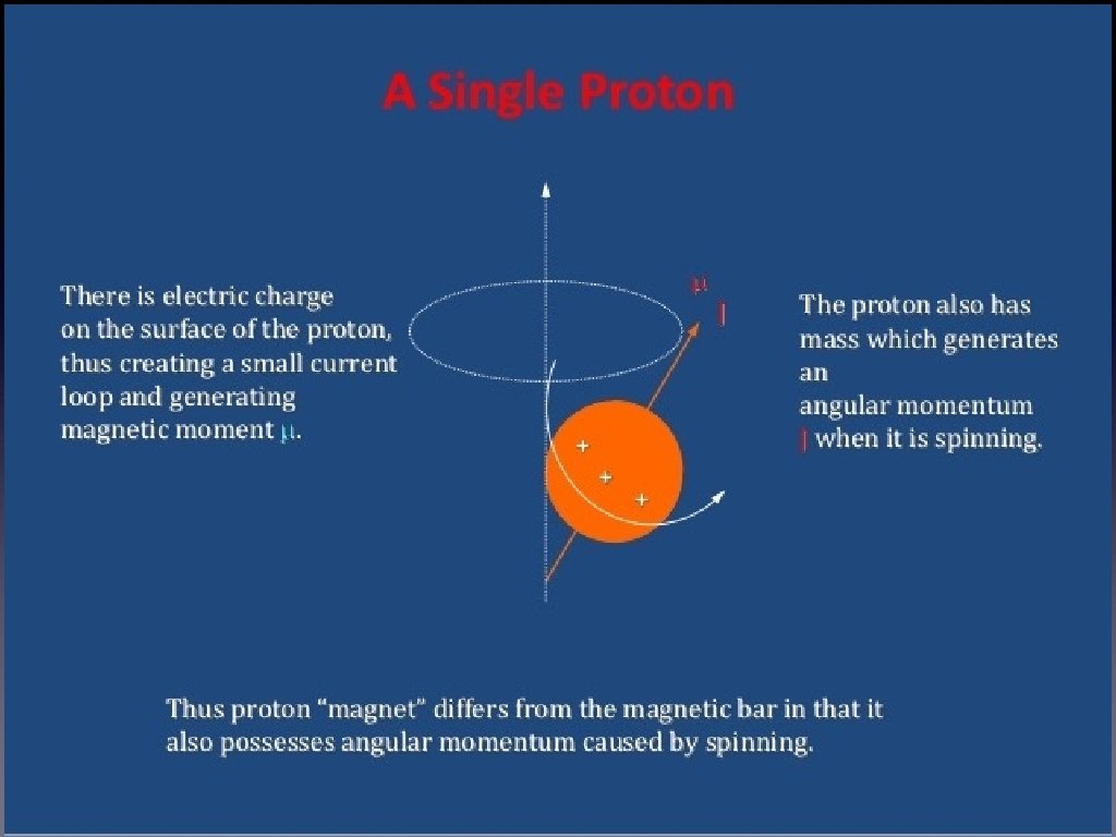
Why Are Protons Important to MRI? Positively charged. Spin about a central axis A moving (spinning) charge creates a magnetic field. The straight arrow (vector) indicates the direction of the magnetic field.

Magnetic Field When we apply external magnitic Field Some of the protons align with the field and some actually align against the field canceling each other out. A slight excess will align with the field so that the net result is an alignment with the external field The frequency of the precession is directly proportional to the strength of the magnetic field and is defined by the Larmor Equation: . At room temperature, the ratio of anti parallel versus parallel proton is roughly 100, 000 to 100, 006 / tesla of B 0

Resonance The RF coil produces a radio frequency simultaneously to the magnetic field • This radio frequency vibrates at the perfect frequency (resonance frequency) which helps align the atoms in the same direction • the radio frequency coil sent out a signal that resonates with the protons. The radio waves are then shut off. The protons continue to vibrate sending signals back to the radio frequency coils that receive these signals.

• The signals are then ran through a computer and go through a Fourier equation to produce an image. • Tissues can be distinguished from each other based on their densities.

Imaging – When the RF pulse is turned off the hydrogen protons slowly return to their natural alignment within the magnetic field and release their excess stored energy. This is known as relaxation. T 1 AND T 2 RELAXATION • When RF pulse is stopped higher energy gained by proton is retransmitted and hydrogen nuclei relax by two mechanisms • T 1 or spin lattice relaxation- by which original magnetization (Mz) begins to recover. • T 2 relaxation or spin relaxation - by which magnetization in X-Y plane decays towards zero in an exponential fashion. It is due to incoherence of H nuclei. • T 2 values of CNS tissues are shorter than T 1 values.

T 2 Relaxation • When the tipped spine are precessing , they “dephase” as they do not spin at precisely the same speed. As they get out of phase, the magnetization is no longer coherent and signal decays.

T 1 RELAXATION After protons are Excited with RF pulse They move out of Alignment with B 0 But once the RF Pulse is stopped they Realign after some Time And this is called t 1 relaxation. T 1 is defined as the time it takes for the hydrogen nucleus to recover 63% of its longitudinal magnetic.

T 2* Decay Spin coherence is also sensitive to the fact that the magnetic field is not completely uniform. • Inhomogeneities in the field cause some protons to spin at slightly different frequencies so they lose coherence faster • Factors that change local magnetic field (susceptibility) can change T 2* decay. •
 : time interval in which signals are")
TR AND TE • TE (echo time) : time interval in which signals are measured after RF excitation. • TR (repetition time) : the time between two excitations is called repetition time.

Image Contrast Different tissues have different relaxation times. These relaxation time differences can be used to generate image contrast. • T 1 - Gray/White matter • T 2 - Tissue/CSF • T 2* - Susceptibility (functional MRI)

KINDS o. F MRI • Brain MRI • An MRI of the brain produces very detailed pictures of the brain. It is commonly used to study patients with headaches, seizures, weakness, blurry vision, etc. It also can further evaluate an abnormality seen on a CT scan. During the brain MRI, a special device called a head coil is placed around the patient's head. It does not touch the patient, and the patient can see through large gaps in the coil. This device is what helps to produce the very detailed pictures of the brain. • Cardiac MRI can evaluate the size and thickness of the chambers of the heart, the extent of damage caused by a heart attack or progressive heart disease, and build-up of plaque and blockages in the blood vessels. It is an invaluable tool for detecting and evaluating coronary artery disease and the function of the heart muscles, valves and vessels. • Spine MRI This test is most commonly used to look for a herniated disc or narrowing of the spinal canal (spinal stenosis) in patients with neck, arm, back and/or leg pain. It is also the best to look for a recurrent disc herniation in a patient who has had prior back surgery.

KINDS OF MRI • Bone and Joint MRI can evaluate virtually all of the bones and joints, as well as the soft tissues. Tendon, ligament, muscle, cartilage and bone injuries can be diagnosed using MRI scans. It can also be used to look for infections and masses. • Abdomen MRI of the abdomen is most frequently used to further evaluate an abnormality seen on another test, such as an ultrasound or CT scan. Thus, the exam is usually tailored to look at specific organs or tissues, such as the liver, adrenal glands or pancreas. • Pelvic MRI For women, pelvic MRI is used to evaluate the ovaries and uterus as follow-up to an ultrasound exam which showed an abnormality. It is also used to evaluate endometrial cancer. For men, pelvic MRI is sometimes used to evaluate prostate cancer. #MRA An MRA evaluates blood vessels. The blood vessels in the neck (carotid and vertebral arteries)

RISKS • The magnet may cause pacemakers, artificial limbs, and other implanted medical devices that contain metal to malfunction or heat up during the exam. • Any loose metal object may cause damage or injury if it gets pulled toward the magnet. • Dyes from tattoos or tattooed eyeliner can cause skin or eye irritation. • Medication patches can cause a skin burn. • The wire leads used to monitor an electrocardiogram (ECG) trace or respiration during a scan must be placed carefully to avoid causing a skin burn. • Prolonged exposure to radio waves during the scan could lead to slight warming of the body.

MRI Today MRI 7 Tesla The scanner will produce high-resolution images of microscopic structures within the human body and brain, allowing researchers to measure subtle changes in the size, function, and metabolism of specific brain structures associated with disease. For example, the scanner will let researchers measure tiny fluctuations in blood flow and metabolic processes that signal differences in brain activity. UI researchers will use the 7 T scanner to investigate how the brain processes sound information; look at subtle abnormalities in white matter caused by brain disease and trauma; and detect age-related brain changes that affect decision-making.
 offers new")
MRI Today • prostate cancer. The development of modern multiparametric-high-field-magnetic-imaging (m. MRI) offers new possibilities and approaches in detection, localization and staging of prostate cancer due to its high resolution and soft-tissue contrast. m. MRI can provide information about the morphological, metabolic and cellular changes and characterize tissue- and tumour - vascularity and correlate it with tumour aggressiveness. This helps to locate and stage a possible tumour and to guide targeted-biopsies towards disease-suspicious areas. Internationally published data support the rapidly growing use of multiparametric MRI, as being the most sensitive and specific imaging tool for prostate cancer patients.

Resource • http: //www. simplyphysics. com/page 2_4. html • http: //www. fda. gov/Radiation. Emitting. Productsand. Procedures/Med ical. Imaging/ucm 200086. htm#rb • http: //www. diffen. com/difference/CT_Scan_vs_MRI • http: //www. medicinenet. com/mri_scan/article. htm#what_is_an_mri_scan • http: //www. miriamhospital. org/centers-and-services/diagnosticimaging/magnetic-resonance-imaging-mri/types-of-mri-exams. html

Outline • Definition of MRI • History • Application • The deference between MRI & CT-Scan • How does MRI work • Kind of MRI • RISKS • MRI Today
- Slides: 27