MOVEMENT OF MATERIAL ACROSS CELL MEMBRANE Cell Transport


MOVEMENT OF MATERIAL ACROSS CELL MEMBRANE Cell Transport System

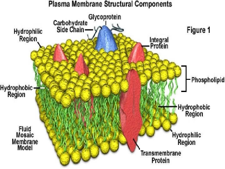
�Structure of cell membrane: !. Act as permeability barrier between interior and exterior environment. (7 -10 nm) thick !. Permit selective exchange of substances between intra and extracellular spaces. !. It has lipid bilayer , the polar ends face outward and inwards (hydrophobic) and non-polar (hydrophilic) face each other. AMPHIPATHIC
 ii. Glycolipids (gangliosides-cerebrocides). iii. Cholesterol. As result of this")
�Lipids are: i. Phospholipids (lecithin-cephalins) ii. Glycolipids (gangliosides-cerebrocides). iii. Cholesterol. As result of this structure, cell membrane 1. Allow fat soluble substances to pass 2. Not allow water soluble substances across it and are obstructed.

�Proper cell functioning require v Water, AA, Proteins, salts, vitamins, glucose , FA pass into cells v Metabolic waste products pass out of cell to avoid chemical damage.

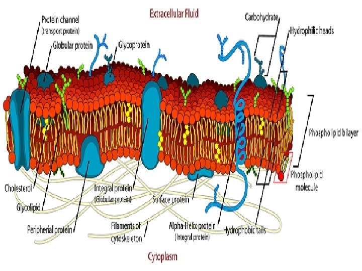
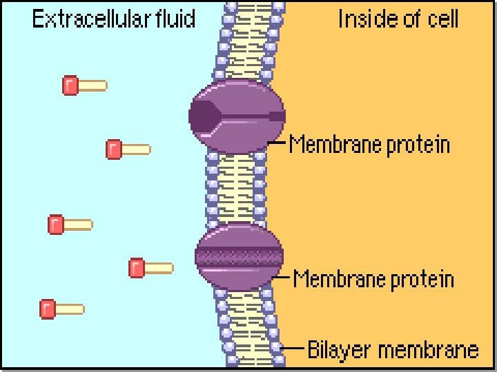
�Proteins in cell membrane a. Integral or intrinsic proteins: are Globular proteins: embedded in lipid bilayer at irregular intervals held by covalent linkages. These act as transporter of molecule, receptors and G. proteins Irregular distribution of proteins give mosaic appearance to membrane , which is not constant but is fluid i. e changeable from moment to moment. Fluidity s due to weak non-covalent interactions.

b. Peripheral proteins or extrinsic proteins are weakly bound and protrude out of membrane. Other substances: Carbohydrates are either linked to lipids (glycolipids) or to proteins(glycoproteins).

Functions of proteins i. Serve as transporter ii. Energy dependant pumps iii. Pores iv. Gates v. Receptors vi. Enzymes vii. Energy transducer

�Substances are transported in two stages 1. Enters the Lipid bi-layer 2. Enter cytoplasm which is aqueous medium. Water soluble substance: has to cross the obstruction by expenditure of energy. After entering membrane, then it easily passes into cell. The energy required is provided by the hydrolysis of ATP into ADP i. e metabolic energy.

Lipid soluble: can pass the membrane but entrance into cytosol require energy as the bonds exist between various components of the membrane Energy provided is not metabolic energy but is that due to Brownian movements of particles


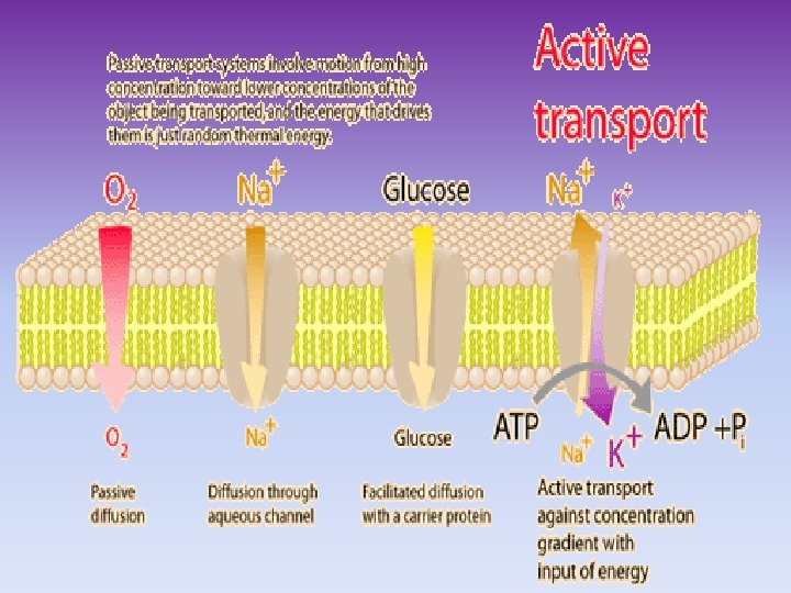
�Transport of solutes/substances occurs A. Passive transport or diffusion show movement of solutes along concentration gradient(chemical or electrical) i. Simple diffusion ii. Facilitated diffusion

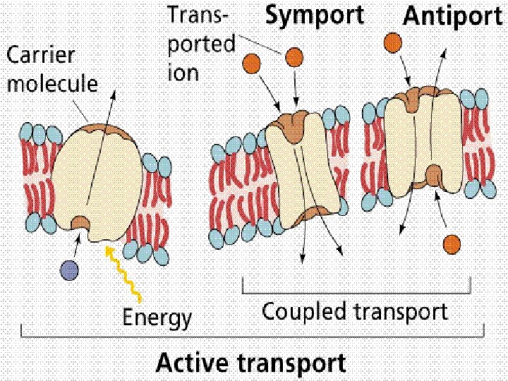
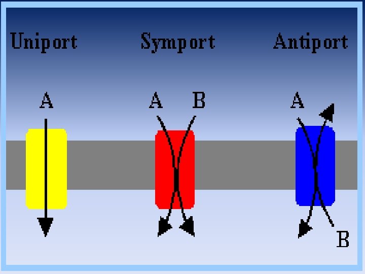
B. Active Transport: Transport from low to higher gradients Pumps are utilized that need energy derived from the hydrolysis of ATP(high energy Phosphate bonds) Small ion or molecules movement occurs by three types of : 1. Uniport 2. Symport 3. Anti-port

�Uniport: Substance move across plasma membrane singly and independently. If a transporter is involved it is called UNIPORTER: e. g. i. actively moves Ca++ from cytosol to ECF. ii. Facilitated diffusion HCO 3 - transporter
 e. g")
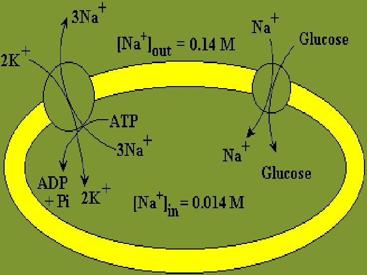
�Symport : Two substances move across membrane together in same direction (symporter) e. g Absorption of glucose along with Na+ from Proximal Convoluted tubules in kidney. �Antiport: counter transport (antiporter) Two substances are moved in different directions e. g : Na+K+ATPase { efflux of 3. Na+ to cell exterior and influx of 2. K+ ions.
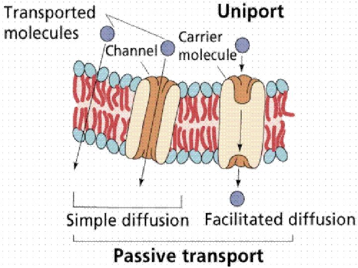

PASSIVE OR SIMPLE DIFFUSION


PASSIVE DIFFUSION �Simplest transport across gradients i. Either leak channels ii. Channel proteins- specialized proteins Ø rate depend upon * solubility of solute * diffusion is ∞ to concentration Ø no metabolic energy-Brownian movements of molecules Ø Rise in temperature increases while fall decreases diffusion

ØHydrostatic pressure also controls it. > pressure > diffusion. ØElectrical gradient: +vely charged move towards – vely charged. Membrane having same charge as of solute will not allow the diffusion. ØSmaller sized molecules (Cl-) diffuse more rapidly than larger sized (Na+)

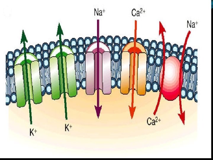
Movements of ions through ion channels: Ions Na+ , K+, Cl-, Ca++ have vital functions �Of excitable tissues-neurons, muscle cells Their concentration differ in ICF /ECF. They are transported through channels/ leaks. These ion channels so called gates, are flexible energy barrier and prevent or allow ion passage by opening or closing.

�Channels are gateways for solutes: v. Na+ Channels: influx of Na+ produces depolarization of excitable tissues Quinindine block this channel, is used in the treatment of arrhythmias. v K+ Channels: efflux of K+ causes repolarization of tissues(neuons and muscle cells) v Ca++ channels: used for normal tone of cardiac muscle and most of skeletal muscle. Ca++ blockers are used in hypertension
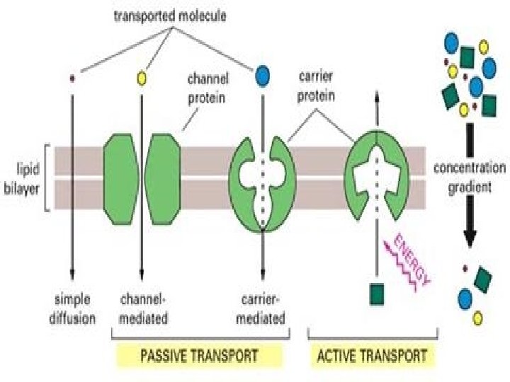

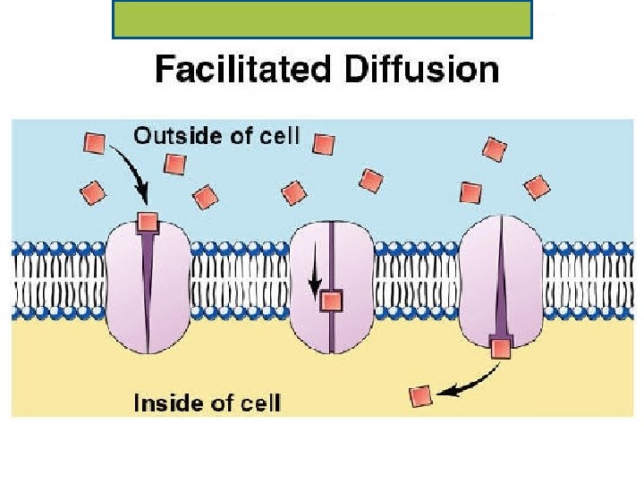
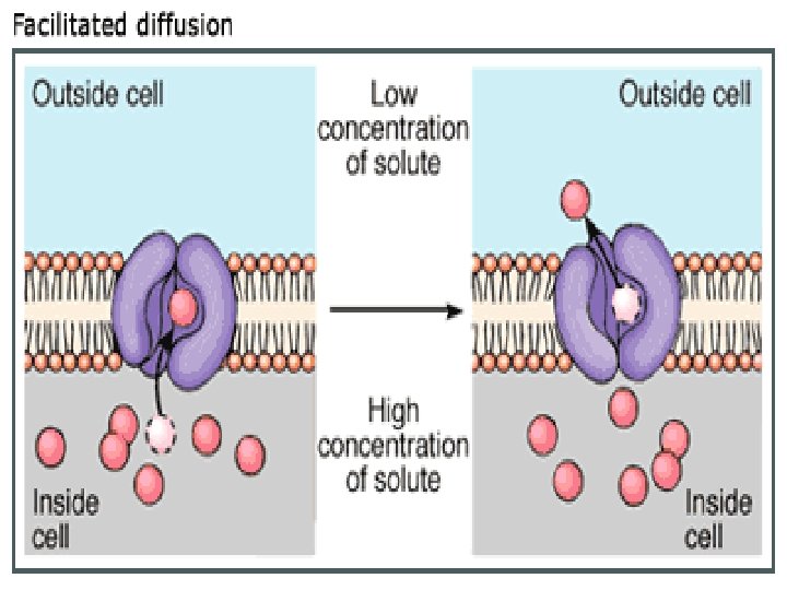
FACILITATED DIFFUSION �Some solute do not diffuse at faster rate �It is also called carrier mediated �Integral membrane protein serve to provide specific “aqueous” route. �Solute molecule gets bound to protein on higher concentration site AND then trans located to side with lower concentration. NO SOURCE OF METABOLIC ENERGY NEEDED

�First there is rapid flow but due to raised concentration the diffusion stops. �Facilitated diffusion inhibited by the competitive inhibitors as in enzymes. �Proteins undergo conformation changes while loading or unloading. �Diffusion can be increased by increasing the carrier proteins { up-regulation of receptors in insulin action} �Glucose uptake by brain, RBCs, Liver Kidneys, cardiac muscles

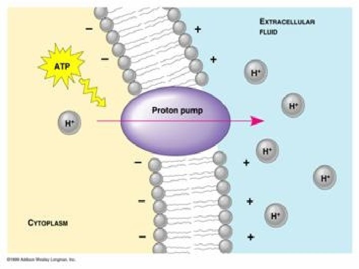
ACTIVE TRANSPORT �Process by which solute particles move against the concentration gradient. �From lower to higher gradient (up-hill transport) �It depends upon the metabolic energy and is seen in only metabolic active cells. �Restriction of metabolism i. deprivation of O 2, OR inhibitors (cyanides) stops active transport

�Presence of transport specific protein is essential for this. � Na+ K+ ATPase of cell membrane: ECF has high Na+ while K+ is more intra-cellulary. This pump is example of anitporter. This pump is an enzyme It require energy by hydrolysis of ATP to ADP and Pi.

�H+ K+ ATPase Are called proton pump as they exchange one H+ ion for one K+ ions. Pumps are present in endosomes, lysosomes, mitochondria and some epith-elial cells. �Plasma membrane Ca++ pump �Endoplasmic reticulum Ca++ Pump �Sarcoplasmic reticulum Ca++ pump.

ENDOCYTOSIS and EXOCYTOSIS

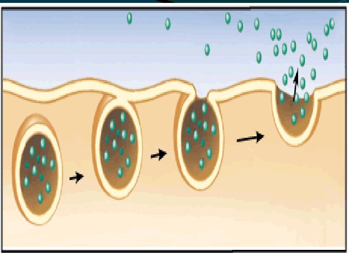
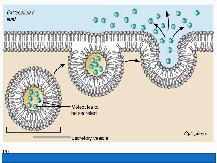
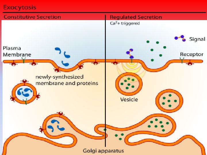
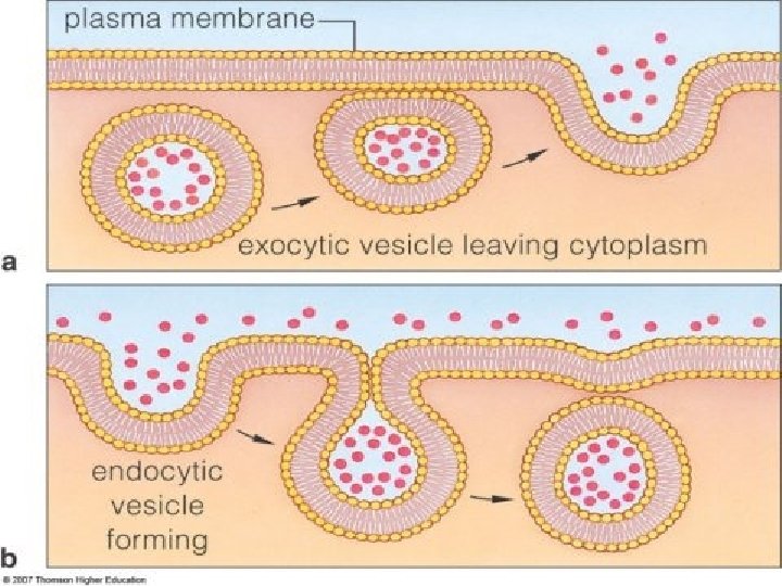
movement of large molecules and particulate matter across cell membrane. Exocytosis is extrusion of material from cell while endocytosis is entry of material into cell

� Exocytosis: �Responsible for secretion of cytoplasmic proteins stored in granules/vesicles. �Vesicles are bound membrane as that of cell. �The membrane fuses with cell membrane , followed by breakdown area of fusion. �The contents are thus poured out of cells. �Exocytosis need energy, Ca++ and certain proteins.

�Fate of exocytosed material: i. some are bound to cell membranes forming peripheral proteins acting as receptors for hormones ii. some become part of extracellular matrix— collagen iii. Molecules like hormones/insulin, PTH enter circulation to act on target. iv. Neurotransmitter , acetylcholine from presynaptic neurons , bind with post- ganglionic neurons produce action

1. Mitochondrion 2. Synaptic vesicle with neurotransmitters 3. Autoreceptor 4. Synapse with neurotransmitter released (serotonin) 5. Postsynaptic receptors activated by neurotransmitter 6. Calcium channel 7. Exocytosis of a vesicle 8. Recaptured neurotransmitter

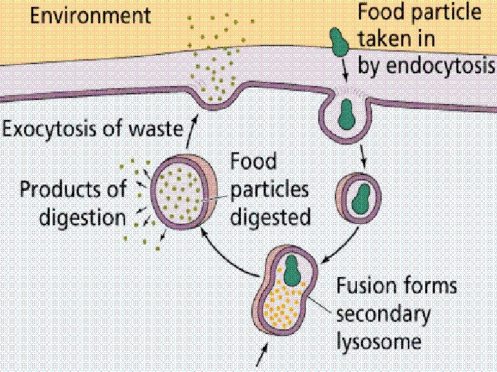
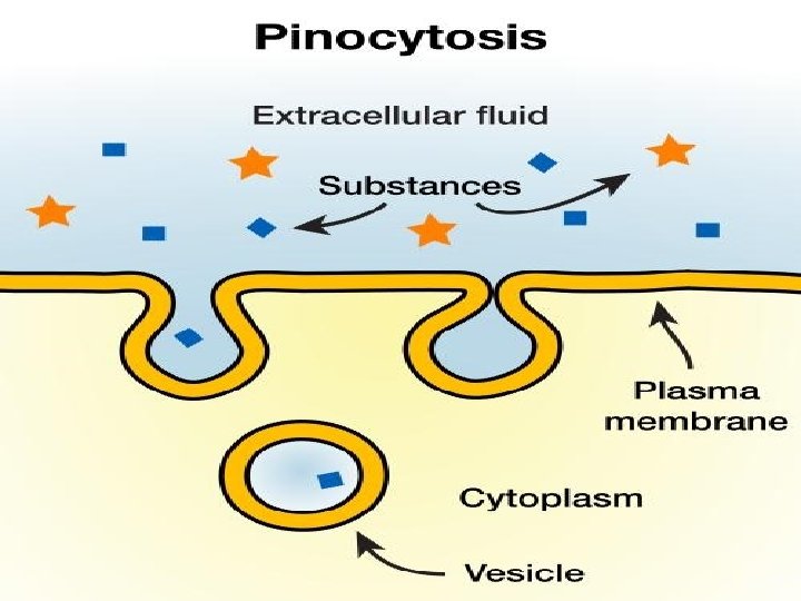
�Endocytosis : �the taking in of matter by a living cell by invagination of its membrane to form a vacuole. �Endocytosis is an energy-using process by which cells absorb molecules (such as proteins) by engulfing them. It is used by all cells of the body because most substances important to them are large polar molecules that cannot pass through the hydrophobic plasma or cell membrane.

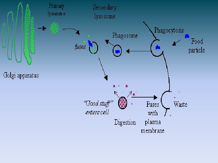
A. Fluid phase or non-selective endocytosis:

�It is non-selective. Uptake of solute depends upon its concentration in ECF. � Membrane invigilates internally to form vesicle, followed by uptake of ECF along with its contents like proteins/ polysaccharides and polynucleotides. �The vesicle and its contents is internalized by its separation from origin. �Portion of cell membrane that gave rise to vesicle regenerate to maintain integrity.

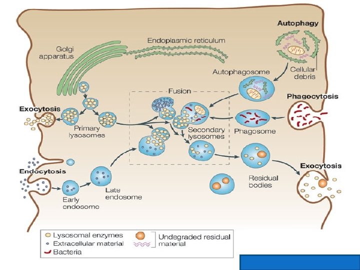
�The vesicle become attached to primary lysosome which are now called secondary lysosomes. �The hydrolytic enzymes in lysosomes cause breakdown of the macromolecules, the products (AA, sugars, nucleotides etc) released to cytoplasm for use. �Some energy/Ca++ are needed for this endocytosis.

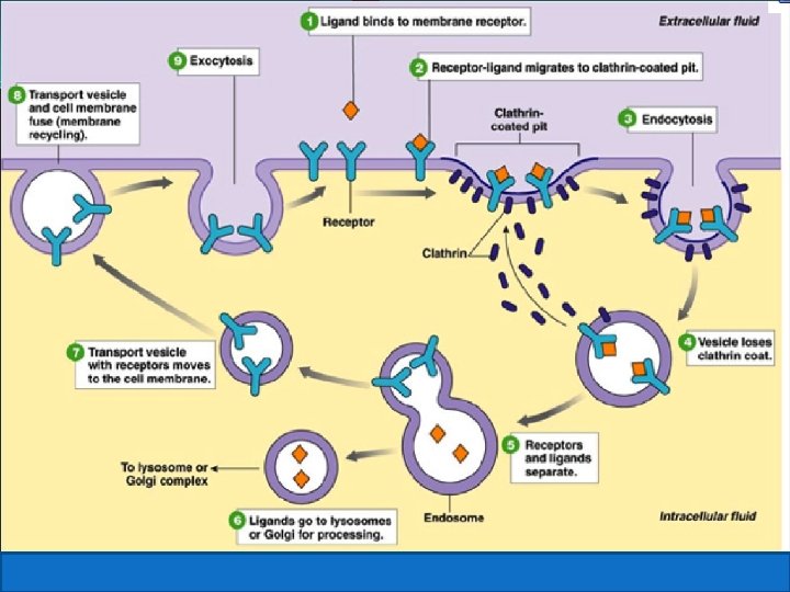
B. Selective or receptor mediated endocytosis: or Absorptive endocytosis: Selective as process starts with binding of substance to be ingested with its specific receptors.

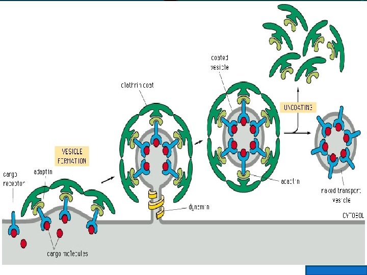
These receptors are present in coated pits on exterior of cell membrane. Pits are lined with protein “ Clathrin” and “adaptin”. Pit after taking the material form small coated endocytic vesicle , which is pinched off from cell surface and internalized.

�Factors needed for vesicle formation and internalization are i. adopter proteins ii phosphatidyl-inositol iii protein DYNAMIN that binds and hydrolyses GTP for releasing energy.
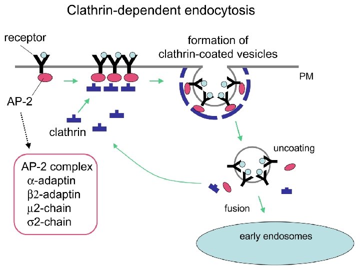

�Later vesicle lose clathrin coat and fuse with early endosomes. �Receptor molecule release bound ligand. �Early endosome become late endosomes after passing through stage of multi-vesicular bodies. �Endosomes interact with lysosomes, p. H is acidic that activate acid hydrolases in lysosomes. �Breakdown products are either passed out or retained in endosomes

Endocytosis is divided into two types depending upon size of material 1. Phagocytosis: shown by neutrophil cells, macrophages= ingestion of large particles as bacteria, viruses, cell debris. 2. Pinocytosis: property of all cells, uptake of ECF and its contents by cells.

Types of endocytosis:

- Slides: 67