Motor Control Lecture 2 Michael S Beauchamp Ph

Motor Control: Lecture 2 Michael S. Beauchamp, Ph. D. Assistant Professor Department of Neurobiology and Anatomy University of Texas Health Science Center at Houston, TX Michael. S. Beauchamp@uth. tmc. edu

Hierarchical Organization and Functional Segregation of Central Motor Structures Level 4: Association Cortex Level 3: Motor Cortex Side Loop 1: Basal Ganglia (Caudate Nucleus, Putamen, Globus Pallidus, Substantia Nigra, Subthalamic Nucleus) Thalamus Level 2: Brain Stem (Red Nucleus, Reticular Formation, Vestibular Nuclei, Tectum, Pontine Nuclei, Inferior Olive) Level 1: Spinal Cord (VA, VL, CM) Side Loop 2: Cerebellum
![Spinal Reflexes Myotatic reflex [myo- + G. tasis, a stretching] a. k. a. stretch](http://slidetodoc.com/presentation_image_h2/2f5a342a242dda674b8c3bc522390ebd/image-3.jpg "Spinal Reflexes Myotatic reflex [myo- + G. tasis, a stretching] a. k. a. stretch")
Spinal Reflexes Myotatic reflex [myo- + G. tasis, a stretching] a. k. a. stretch reflex Muscle spindles (Ia) + alpha motor neurons

Myotatic reflex

Myotatic Reflex Important for Posture Maintenance Continuous feedback from muscle spindles allows automatic postural maintenance and adjustments
 The Human Brain, 3 rd Edition")
“Knee-jerk” Response From J. Nolte (1993) The Human Brain, 3 rd Edition

Reciprocal reflexes Reciprocal inhibition in the stretch reflex

Flexor and extensor in opposition

Reciprocal inhibition in the stretch reflex + – +
 + alpha motor neurons Autogenic inhibition Golgi")
Spinal Reflexes Myotatic reflex Muscle spindles (Ia) + alpha motor neurons Autogenic inhibition Golgi tendon organs (Ib) - alpha motor neurons

Flexor and extensor: co-contraction

Autogenic Inhibition + –

Reciprocal reflexes Reciprocal inhibition in the stretch reflex Reciprocal excitation in the autogenic inhibition reflex

Reciprocal excitation in the autogenic inhibition reflex + + + –

Flexor and crossed extension reflexes Flexor reflex Cutaneous and nociceptive receptors (II, III, and IV) + alpha motor neurons

Flexor Reflex

Flexor and crossed extension reflexes Flexor reflex Cutaneous and nociceptive receptors (II, III, and IV) + alpha motor neurons Reciprocal inhibition in the flexor reflex Crossed extension reflex

Crossed extension reflex

Recurrent inhibition of motor neurons Renshaw Cell

Renshaw cell + –

Alpha motor neuron Clinically, often called Lower Motor Neurons
 Rostral midbrain Caudal pons Upper")
Descending Spinal Pathways Precentral gyrus (from Nolte, p. 438) Rostral midbrain Caudal pons Upper Motor Neurons Rostral medulla Cervical spinal cord

Anatomy and Physiology of Descending Spinal Pathways Brain structures influence spinal motor neurons and spinal circuits through descending pathways Flexor-extensor rule Proximal-distal rule

Organization of spinal tracts

Flexor-Extensor Rule and Proximal-Distal Rule

Two Groups of Descending Pathways Lateral pathways control proximal and distal muscles Lateral corticospinal Rubrospinal Medial pathways control axial muscles Vestibulospinal Reticulospinal Tectospinal Anterior corticospinal

Corticospinal Tracts

Corticospinal tracts Control of distal musculature, esp. fine control of extremities Control of axial muscles Corticospinal and corticobulbar tracts constitute the major voluntary drive to the brain stem and spinal motor systems Lesions produce initial paralysis, with eventual recovery of function (except fine control of distal musculature)

Rubrospinal Tract

Rubrospinal tract Alternate route to corticospinal tract Receives input from cerebellum and cerebral cortex Encodes movement velocity Excitation of flexors and inhibition of extensors Relatively small in humans

Vestibulospinal Tracts

Vestibulospinal tracts Mediate postural adjustments and head movements Control balance Lateral vestibulospinal tract Excites antigravity muscles Controls postural changes to compensate for tilts and movements of body Medial vestibulospinal tract Innervates neck muscles to stabilize head position Coordinates head and eye movements

Reticulospinal Tracts
 to spinal motor neurons Regulate")
Reticulospinal tracts Major alternate route (to the corticospinal tract) to spinal motor neurons Regulate sensitivity to flexor reflexes, such that only noxious stimuli elicit them Reticular formation contains circuitry for complex movements, including complex postures, orienting, and stretching Integration of sensory input to guide motor output

Tectospinal Tract

Tectospinal tract Originates in Superior Colliculus Innervates neck and proximal muscles Presumably involved in reflex orienting to visual stimuli Relatively minor in humans

Influence of descending pathways on spinal mechanisms • voluntary movement • reflex modulation
, in Principles of Neural Science, 4 th")
From K. Pearson & J. Gordon (2000), in Principles of Neural Science, 4 th Edition (Kandel, Schwartz, & Jessel, Eds. )
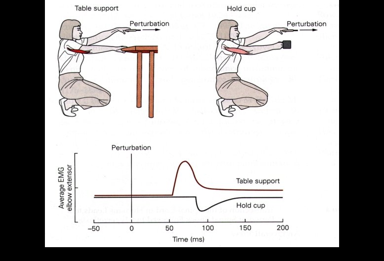

Influence of descending pathways on spinal mechanisms • voluntary movement • reflex modulation • gamma bias alpha-gamma coactivation

Hierarchical Organization and Functional Segregation of Central Motor Structures Level 4: Association Cortex Level 3: Motor Cortex Side Loop 1: Basal Ganglia (Caudate Nucleus, Putamen, Globus Pallidus, Substantia Nigra, Subthalamic Nucleus) Thalamus Level 2: Brain Stem (Red Nucleus, Reticular Formation, Vestibular Nuclei, Tectum, Pontine Nuclei, Inferior Olive) Level 1: Spinal Cord (VA, VL, CM) Side Loop 2: Cerebellum
- Slides: 41