Motifs Domains 1 Protein domains Pairwise sequence comparison


. Motif")


 method is similar to DALI in that it")


 which are")


 method uses double dynamic programming to produce")
![TOPOFIT In the TOPOFIT method [1], similarity of protein structures is analyzed using three-dimensional](https://slidetodoc.com/presentation_image_h2/082dc94cda5a02d36515a77063e8427c/image-71.jpg "TOPOFIT In the TOPOFIT method [1], similarity of protein structures is analyzed using three-dimensional")


 Ø Flex. Prot – Flexible alignment Ø c_alpha_match")


 Ø Multi. Prot Ø MASS 92")

- Slides: 97
Motifs & Domains 1
Protein domains Pairwise sequence comparison of proteins led to strange results • A domain is an independent folding unit • A domain is the next step up in complexity from a motif • There appear to be a limited number of folds (domains) that can be made from the 20 natural aa’s • Domain unit of evolution • Mixing and matching can create new function and regulation • Most proteins involved in cell signalling consist exclusively of small domains interspersed by linker regions. The linkers may be unstructured as described in the following section.
How proteins are made from domains. Some proteins consist only of domains that have no enzymatic activity. It is thought that they function as scaffolds for specific complex formation. SH 3 GRB 2 BRCT domains are a good example of divergent evolution. An ancient domain found in pro- and eukaryotes, it is characterised by a conserved fold despite significant sequence divergence. BRCTs are known to bind DNA and other proteins. Protein-protein interactions included self binding, binding BRCTs on other proteins, binding non. BRCT domains and binding to phoserine peptides.
Determining Domain Structure by Limited Proteolysis
Protein regulation by coordinated action of domains Having multiple domains in one protein can serve a variety of functions, one of which is illustrated here. The kinases, Src, Lck and Hck, all of which can cause aberrent growth signalling, are regulated by an internal Y phophorylation. When Y 527 is phosphorylated, SH 2 and SH 3 are “locked”, forcing lobes of kinase down and blocking access to the active site. Young et al. , 2001, Cell, v. 105, p. 115
A continuum of protein structures
Motifs & Domains Family is a set of sequences that are related (functionally/structurally). Motif is a simple combination of a few secondary structures, that appear in several different proteins in nature. 7
Motifs & Domains A collection of motifs forms a domain. Domain is a more complex combination of secondary structures, that is common in a family. It has a very specific function. A protein may contain more than one domain. Beta-Alpha-Beta motif For further reading: http: //www. expasy. org/swissmod/course/text/chapter 4. htm http: //www. ii. uib. no/~inge/talks/sverige 00/sld 003. htm 8
Motifs in Protein Analysis http: //www. ii. uib. no/~inge/talks/ebi-nov-99/sld 009. htm 9
Grouping of Secondary Structures Elements Super-secondary Structures or Motifs: bab b-hairpin aa Certain arrangements of two or three consecutive secondary structural elements. -barrels 10
Structural Alignment 11
Why structural alignment? Ø In evolutionary related proteins structure is much better preserved than sequence Ø Structural motifs may predict similar biological function Ø Getting insight into protein folding 12
Applications: Ø Classification of protein databases by structure Ø Search of partial and disconnected structural patterns in large databases Ø Comparison and detection of drug receptor active sites (structurally similar receptor cavities could bind similar drugs) Ø Similar substructures in drugs acting on a given receptor 13
Ø Ø Ø The superimposition pattern is unknown – pattern discovery The matching recovered can be inexact We are not necessarily looking largest superimposition, since matching may have biological for the other meaning 14
Human Hemoglobin alpha-chain pdb: 1 jeb. A Human Myoglobin pdb: 2 mm 1 Sequence id: 27% Structural id: 90% 15
What is the best transformation that superimposes the unicorn on the lion? 16
Solution: Regard the shapes as sets of points and try to “match” these sets using a transformation 17
This is not a good result…. 18
Good result: 19
Kinds of transformations: Ø Rotation Ø Translation Ø Scaling and more…. 20
Translation: Y X 21
Rotation: Y X 22
Scale: Y X 23
We represent a protein as a geometric object in the plane. The object consists of points represented by coordinates (x, y, z). Lys Met Gly Thr Glu Ala 24
The aim: Given two proteins Find the transformation that produces the best Superimposition of one protein onto the other 25
Correspondence is Unknown Given two configurations of points in the three dimensional space: + 26
find those rotations and translations of one of the point sets which produce “large” superimpositions of corresponding 3 -D points ? 27
The best transformation: T 28
Simple case – two closely related proteins with the same number of amino acids. + Question: how do we asses the quality of the transformation? 29
Scoring the Alignment Two point sets: A={ai} i=1…n B={bj} j=1…m • Pairwise Correspondence: (ak 1, bt 1) (ak 2, bt 2)… (ak. N, bt. N) (1) Bottleneck max ||aki – bti|| (2) RMSD (Root Mean Square Distance) Sqrt( Σ||aki – bti||2/N) 30
RMSD – Root Mean Square Deviation Given two sets of 3 -D points : P={pi}, Q={qi} , i=1, …, n; rmsd(P, Q) = √ S i|pi - qi |2 /n Find a 3 -D transformation T* such that: rmsd( T*(P), Q ) = min. T √ S i|T(pi) - qi |2 /n Find the highest number of atoms aligned with the lowest RMSD 31
Pitfalls of RMSD Ø all atoms are treated equally (residues on the surface have a higher degree of freedom than those in the core) Ø best alignment does not always mean minimal RMSD Ø does not take into account the attributes of the amino acids 32
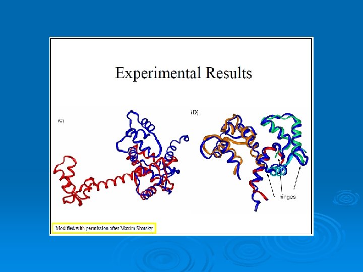
Flexible alignment vs. Rigid alignment Flexible alignment 33
Structure alignment approaches: Geometric-intermolecular Algorithms may do this by minimizing the RMSD in superimposed alpha-carbon positions. Ø Geometric-intramolecular Algorithms minimize the difference between aligned contact maps or distance matrices. Ø Non-Geometric Algorithms align structural properties, such as %buried, or secondary structure type. Ø 34
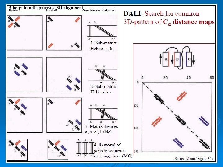
DALI: an intramolecular geometric structural alignment algorithm http: //www. ebi. ac. uk/dali/ 47
DALI Ø Generate a distance matrix for each protein. Ø The distance matrix contains all pairwise distances. (symmetrical) Ø Dij = distance between Calpha i and Calpha j. 48
49
FSSP - fold classification and structural alignments The Dali database is based on exhaustive all-against-all 3 D structure comparison of protein structures currently in the Protein Data Bank http: //ekhidna. biocenter. helsinki. fi/dali/start 52
53
Structural neighbors 54
The superimposition in PDB format. RMSD 55
Methods of Structural alignment DALI A common and popular structural alignment method is the DALI, or distance alignment matrix method, which breaks the input structures into hexapeptide fragments and calculates a distance matrix by evaluating the contact patterns between successive fragments. Secondary structure features that involve residues that are contiguous in sequence appear on the matrix's main diagonal; other diagonals in the matrix reflect spatial contacts between residues that are not near each other in the sequence. When these diagonals are parallel to the main diagonal, the features they represent are parallel; when they are perpendicular, their features are antiparallel. This representation is memory-intensive because the features in the square matrix are symmetrical (and thus redundant) about the main diagonal.
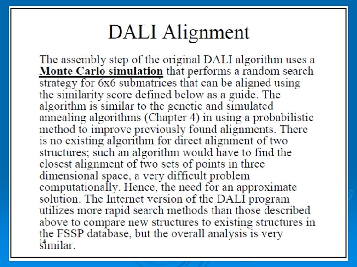
When two proteins' distance matrices share the same or similar features in approximately the same positions, they can be said to have similar folds with similar-length loops connecting their secondary structure elements. DALI's actual alignment process requires a similarity search after the two proteins' distance matrices are built; this is normally conducted via a series of overlapping submatrices of size 6 x 6. Submatrix matches are then reassembled into a final alignment via a standard score-maximization algorithm - the original version of DALI used a Monte Carlo simulation to maximize a structural similarity score that is a function of the distances between putative corresponding atoms. In particular, more distant atoms within corresponding features are exponentially downweighted to reduce the effects of noise introduced by loop mobility, helix torsions, and other minor structural variations. [13] Because DALI relies on an all-to-all distance matrix, it can account for the possibility that structurally aligned features might appear in different orders within the two sequences being compared. The DALI method has also been used to construct a database known as FSSP (Fold classification based on Structure-Structure alignment of Proteins, or Families of Structurally Similar Proteins) in which all known protein structures are aligned with each other to determine their structural neighbors and fold classification. There is an searchable databased on DALI as well as a downloadable program and web search based on a standalone version known as Dali. Lite. 57
Combinatorial extension The combinatorial extension (CE) method is similar to DALI in that it too breaks each structure in the query set into a series of fragments that it then attempts to reassemble into a complete alignment. A series of pairwise combinations of fragments called aligned fragment pairs, or AFPs, are used to define a similarity matrix through which an optimal path is generated to identify the final alignment. Only AFPs that meet given criteria for local similarity are included in the matrix as a means of reducing the necessary search space and thereby increasing efficiency. [15] A number of similarity metrics are possible; the original definition of the CE method included only structural superpositions and inter-residue distances but has since been expanded to include local environmental properties such as secondary structure, solvent exposure, hydrogen-bonding patterns, and dihedral angles. [15] An alignment path is calculated as the optimal path through the similarity matrix by linearly progressing through the sequences and extending the alignment with the next possible high-scoring AFP pair. The initial AFP pair that nucleates the alignment can occur at any point in the sequence matrix. Extensions then proceed with the next AFP that meets given distance criteria restricting the alignment to low gap sizes. The size of each AFP and the maximum gap size are required input parameters but are usually set to empirically determined values of 8 and 30 respectively
GANGSTA+ is a combinatorial algorithm for non-sequential structural alignment of proteins and similarity search in databases (http: //gangsta. chemie. fu-berlin. de). It uses a combinatorial approach on the secondary structure level to evaluate similarities between two protein structures based on contact maps. Different SSE assignment modes can be used. The assignment of SSEs can be performed respecting the sequential order of the SSEs in the polypeptide chains of the considered protein pair (sequential alignment) or by ignoring this order (non-sequential alignment). Furthermore, SSE pairs can optionally be aligned in reverse orientation. The highest ranking SSE assignments are transfered to the residue level by a pointmatching approach[16]. To obtain an initial common set of atomic coordinates for both proteins, pairwise attractive interactions of the C-alpha atom pairs are defined by inverse Lorentzians and energy minimized. For more algorithmic details see [17].
60
61
62
MAMMOTH MAtching Molecular Models Obtained from THeory. As its name suggests, MAMMOTH was originally developed for comparing models coming from structure prediction (THeory) since it is tolerant of large unalignable regions, but it has proven to work well with experimental models, especially when looking for remote homology. Benchmarks on targets of blind structure prediction (the CASP experiment) and automated GO annotation have shown it is tightly rank correlated with human curated annotation. A highly complete database of mammoth-based structure annotation for the predicted structures of unknown proteins Covering 150 genomes facilitates genomic scale normalization.
MAMMOTH-based structure alignment methods decomposes the protein structure into short peptides (heptapeptides) which are compared with the heptapeptides of another protein. Similarity score between two heptapeptides is calculated using a unit-vector RMS (URMS) method. These scores are stored in a similarity matrix, and with a hybrid (local-global) dynamic programming the optimal residue alignment is calculated. Protein similarity scores calculated with MAMMOTH is derived from the likelihood of obtaining a given structural alignment by chance. MAMMOTH-mult is an extension of the MAMMOTH algorithm to be used to align related family of protein structures. This algorithm is very fast and produces consistent and high quality structural alignments. Multiple structural alignments calculated with MAMMOTH-mult produces structurally-implied sequence alignments that can be further used for multiple-template homology modeling, HMM-based protein structure prediction, and profile-type PSI-BLAST searches. 64
65
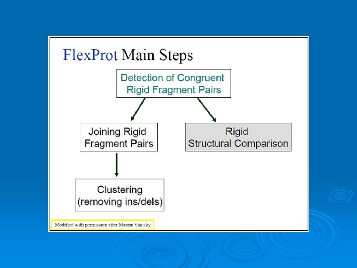
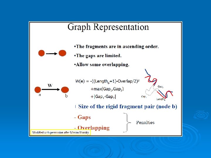
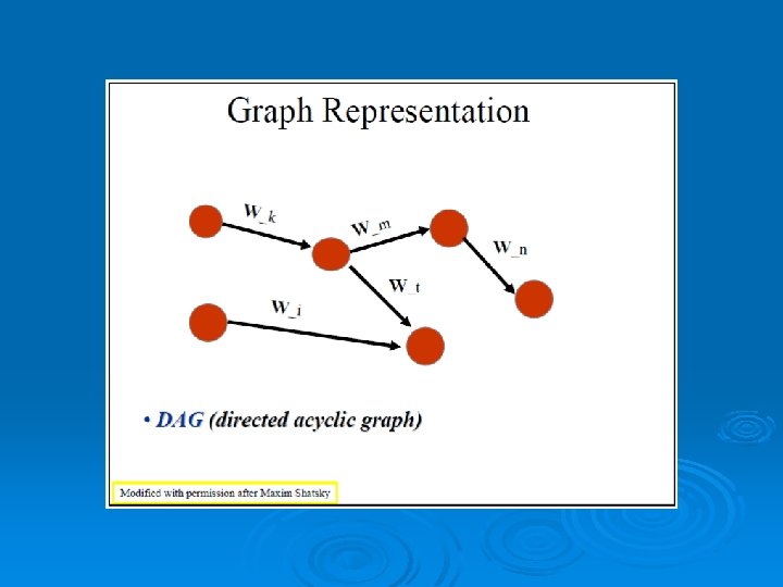
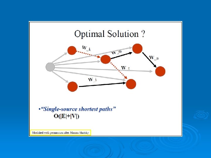
RAPIDO Rapid Alignment of Proteins In terms of DOmains. RAPIDO is a web server for the 3 D alignment of crystal structures of different protein molecules, in the presence of conformational changes. Similar to what CE does as a first step, RAPIDO identifies fragments that are structurally similar in the two proteins using an approach based on difference distance matrices. The Matching Fragment Pairs (MFPs) are then represented as nodes in a graph which are chained together to form an alignment by means of an algorithm for the identification of the longest path on a DAG (Directed Acyclic Graph). The final step of refinement is performed to improve the quality of the alignment. After aligning the two structures the server applies a genetic algorithm for the identification of conformationally invariant regions These regions correspond to groups of atoms whose interatomic distances are constant (within a defined tolerance). In doing so RAPIDO takes into account the variation in the reliability of atomic coordinates by employing weighting-functions based on the refined B-values. The regions identified as conformationally invariant by RAPIDO represent reliable sets of atoms for the superposition of the two structures that can be used for a detailed analysis of changes in the conformation. In addition to the functionalities provided by existing tools, RAPIDO can identify structurally equivalent regions even when these consist of fragments that are distant in terms of sequence and separated by other movable domains.
67
SABERTOOTH uses structural profiles to perform structural alignments. The underlying structural profiles expresses the global connectivity of each residue. Despite the very condensed vectorial representation, the tool recognizes structural similarities with accuracy comparable to established alignment tools based on coordinates and performs comparably in quality. Furthermore, the algorithm has favourable scaling of computation time with chain length. Since the algorithm is independent of the details of the structural representation, the framework can be generalized to sequence-to-sequence and sequence-to-structure comparison within the same setup, and it is therefore more general than other tools. SABERTOOTH can be used online at http: //www. fkp. tu-darmstadt. de/sabertooth/
69
SSAP The SSAP (Sequential Structure Alignment Program) method uses double dynamic programming to produce a structural alignment based on atom-to-atom vectors in structure space. Instead of the alpha carbons typically used in structural alignment, SSAP constructs its vectors from the beta carbons for all residues except glycine, a method which thus takes into account the rotameric state of each residue as well as its location along the backbone. SSAP works by first constructing a series of inter-residue distance vectors between each residue and its nearest noncontiguous neighbors on each protein. A series of matrices are then constructed containing the vector differences between neighbors for each pair of residues for which vectors were constructed. Dynamic programming applied to each resulting matrix determines a series of optimal local alignments which are then summed into a "summary" matrix to which dynamic programming is applied again to determine the overall structural alignment. SSAP originally produced only pairwise alignments but has since been extended to multiple alignments as well. It has been applied in an all-to-all fashion to produce a hierarchical fold classification scheme known as CATH (Class, Architecture, Topology, Homology), which has been used to construct the CATH Protein Structure Classification database. 70
TOPOFIT In the TOPOFIT method [1], similarity of protein structures is analyzed using three-dimensional Delaunay triangulation patterns derived from backbone representation. It has been found that structurally related proteins have a common spatial invariant part, a set of tetrahedrons, mathematically described as a common spatial sub-graph volume of the three-dimensional contact graph derived from Delaunay tessellation (DT). Based on this property of protein structures we present a novel common volume superimposition (TOPOFIT) method to produce structural alignments of proteins. The superimposition of the DT patterns allows one to objectively identify a common number of equivalent residues in the structural alignment, in other words, TOPOFIT identifies a feature point on the RMSD/Ne curve, a topomax point, until which two structures correspond to each other including backbone and inter-residue contacts, while the growing number of mismatches between the DT patterns occurs at larger RMSD (Ne) after topomax point. The topomax point is present in all alignments from different protein structural classes; therefore, the TOPOFIT method identifies common, invariant structural parts between proteins. The TOPOFIT method adds new opportunities for the comparative analysis of protein structures and for more detailed studies on understanding the molecular principles of tertiary structure organization and functionality. .
72
73
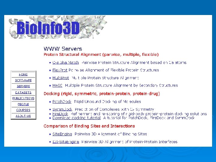
Structural alignment tools Ø Bioinfo 3 D tools – l l l The Structural Bioinformatics group at Tau of Nussinov-Wolfson pairwise & multiple alignments Geometric-intermolecular Ø DALI – l l European Bioinformatics Institute Geometric-intramolecular 74
Bioinfo 3 D Tools (pairwise alignment) Ø Flex. Prot – Flexible alignment Ø c_alpha_match – The protein's backbones are structurally compared using their calpha atoms coordinates. Ø Site. Engine - Recognizes regions on the surface of one protein that resemble a specific binding site of another. 75
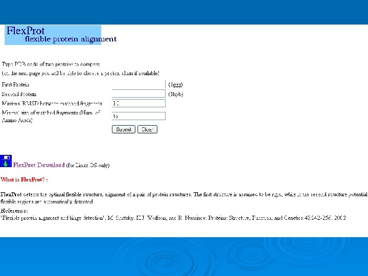
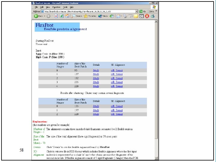
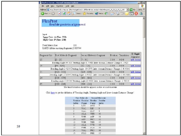
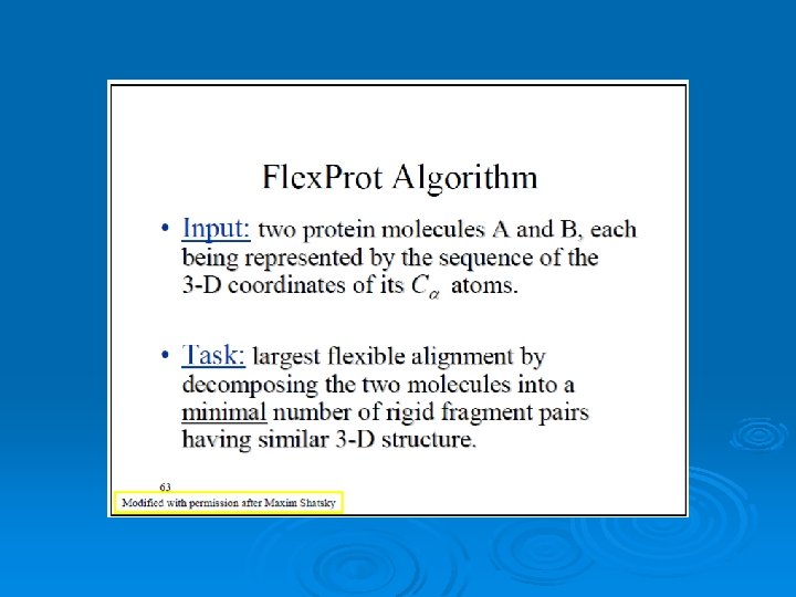
Flex. Prot - Flexible protein alignment: Ø The first structure is assumed to be rigid, while in the second structure potential flexible regions are automatically detected. Ø Input: two pdb ids (specific chain) Ø Output: list of alignments according the number of hinges. 76
77
Result: 78
Result without hinge: 79
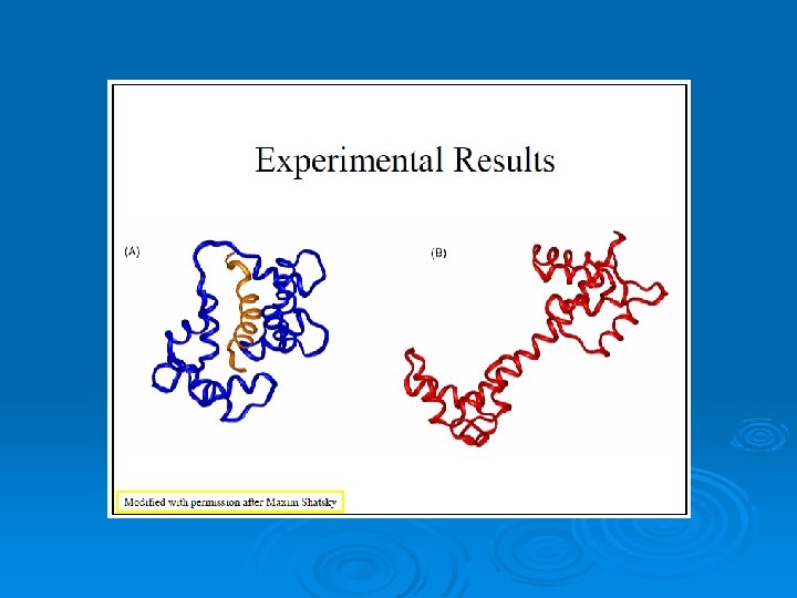
Result with one hinge: 80
Visualization of the 3 D alignment Ø The results can be viewed with PDB viewers: Protein explorer or Rasmol. The aligned proteins are marked as different chains. 81
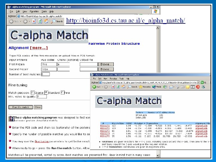
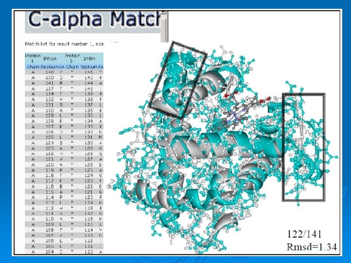
C_alpha_match Ø Rigid pairwise alignement Ø Based on c-alpha atoms Ø Input: l l Two pdb files or ids These protein's backbones are structurally compared using their c-alpha atoms coordinates. Ø Output: l l A set of high scoring conformations May be viewed in a PDB viewer. 82
83
Result: 84
Result: 85
Site. Engine Ø Ø Ø Based in surfaces Recognizes regions on the surface of one protein that resemble a specific binding site of another. This may suggest the similarity of their binding partners and biological functions. Takes into account the physico-chemical properties of both the backbone and the side-chains. Therefore it can recognize similar binding patterns shared by proteins that have no sequence or fold similarity. Highly efficient and suitable for large scale database searches of the entire PDB. 86
searches the structure of the complete molecule for regions similar to the binding site of interest 87
88
Results: Ø 10 top ranking solutions are presented Ø Each solution includes the surface of the complete molecule that are recognized to be most similar to the binding site of interest Ø Pdb file of the superimposition 89
Result: 90
Property: Ø The physico-chemical property Ø Representation of each amino acid of a protein as a set of features that are important for its interaction with other molecules. Ø The abbreviations of these features are: l l l DON - Hydrogen bond donor ACC - Hydrogen bond acceptor DAC - Hydrogen bond donor and acceptor (e. g in histidine) ALI - Aliphatic Hydrophobic property PII - Aromatic property (pi contacts) 91
Bioinfo 3 D Tools (multiple alignment) Ø Multi. Prot Ø MASS 92