Morphology and histology of the urinary passages pelvis


")


")


- Slides: 40
Morphology and histology of the urinary passages, pelvis, ureter, bladder and urethra Dr. Gallatz Katalin
Metanephros Primordia: ureteric bud and metanephric tissue
Mesonephric duct ureteric bud → ureters, renal pelvis, calyces, collecting tubules
Formation of Bladder and Urethra Primordium: urogenital sinus upper part: urinary bladder → continuous with allantois lower part: urethra
Renal sinus: calyces + pelvis + renal artery + renal vein + adipose tissue
Renal calyces and pelvis
Retrograde pyelogram
Ureter Ø A 25 – 30 cm long muscular tube transporting urine from kidney to urinary bladder. Ø Begins as a continuation of renal pelvis, descends into the pelvis and drains into the bladder Ø CROSSINGS § It crosses: § the gonadal vessels, (post. ) § the bifurcation of common iliac a. (ant. ), § vas deferens in male § uterine artery in female
Ø COURSE IN PELVIS & TERMINATION: § Runs downward in front to internal iliac artery, reaches ischial spine § Turns forward and medially , enters the upper lateral angle of urinary bladder § Near its termination, is crossed by the vas deferens in male § Passes obliquely through the wall of bladder before opening into the bladder cavity. Bladder muscles contraction mechanically close the orifice of the ureter preventing the backflow of urine toward the kidney
Ureteric Constrictions Ø The ureter has constrictions at three points (sites of obstruction and stone 1 impaction) 1. At the ureteropelvic junction 2. At the crossing of external/common iliac artery 3. 2 At site of entrance to bladder 3
Arterial Supply Ø Ureter is supplied by multiple arteries throughout its course Ø 1 These are: : 1. Renal artery 2. Gonadal artery 3. Internal iliac artery 2 3 4
Urinary Bladder Ø Located immediately behind the pubic symphysis Ø Shape and relations vary according to the amount of urine. l In adults, an empty bladder is entirely a pelvic organ; as it is filled with urine, rises up. l In young children, it projects above the pelvic inlet
Urinary Bladder Ø An empty bladder is pyramidal in shape. Ø Parts l apex l base (posterior surface) l superior surface l infrolateral surfaces l neck
Urinary Bladder
Apex Ø Directed forward Ø Lies behind the upper margin of the symphysis pubis § Is connected to umbilicus by the median umbilical ligament (remnant of urachus)
Base or Posterior surface Ø Triangular in shape Ø Upper part covered by peritoneum Ø Lower part related to: l in males: vas deferent and seminal vesicles l in females: vagina
Superior surface Ø Completely covered by peritoneum. Ø Related to the coils of ileum or sigmoid colon in males and to uterus in females Male pelvis Female pelvis
Female pelvis Male pelvis
Infrolateral surfaces: Ø Related in front to the retropubic pad of fat and the pubic bones Ø Posteriorly it is in contact with the obturator internus above and levator ani below
Neck: Ø Lies inferiorly, and is the most fixed part of the bladder Ø In male, rests on the upper surface of prostate. Ø Here, the smooth muscle fibers of the bladder are continuous with those of the prostate Ø The circular muscle fibers thickened to form the sphincter vesicae
Inner surface of the urinary bladder Ø Mucous membrane is folded except, between the openings of the two ureters and the urethra. This region is called the vesical trigone. Here the mucous membrane is smooth. Ø Uvula vesicae, a small elevation located just at the orifice of the urethra.
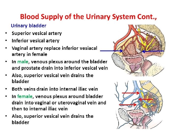
Blood supply Ø Arteries: from internal iliac artery • superior vesical artery • inferior vesical artery Ø Venous drainage: into internal iliac vein Ø Lymphatics: into internal iliac lymph nodes
INNERVATION Ø The nerves form the vesical nerve plexus that contains: l Sympathetic fibers derived mainly from L 1, 2 l Parasympathetic fibers derived from pelvic splanchnic nerves S 2, 3, 4 l Sensory fibers from the bladder are transmit pain sensation resulting by overdistention
Ø The normal capacity of bladder is about 300 -500 ml. Ø As bladder fills, the superior surface bulges upward into abdominal cavity.
Male urethra Ø About 20 cm long Ø Parts: l intramural l prostatic l membranous l spongious (penile)
Prostatic part Ø length=3 cm Ø widest part Ø inside prostate gland Ø structures opening into prostatic urethra: § ejaculatory ducts (seminal colliculus) § ducts of prostatic glands Membranous part Ø Length=1 cm Ø Passes trough the urogenital diaphragm Ø Surrounded by external urethral sphincter
Ø Spongious part urethra Ø length=16 cm Ø narrowest part of the urethra § extends inside penis, in the glans it dilates – navicular fossa § It opens through external urethral orifice
Female urethra § It is short and straight, § starts from neck of urinary bladder and ends at the external urethral orifice (anteriorly to the vaginal opening)
FEMALE URETHRA
HISTOLOGY OF THE URINARY PASSAGES
MINOR CALYX Transitinal epithelium (urothelium)
MINOR CALYX PAPILLA
URETER Layers: 1. Tunica mucosa transitional epithelium lamina propria 2. Tunica muscularis inner and outer longitudinal middle circular 3. Tunica adventitia
URETER
Transitinal epithelium (urothelium)
URINARY BLADDER Layers: 1. Tunica mucosa 2. Tunica muscularis 3. Tunica adventitia
URINARY BLADDER
URINARY BLADDER
Thank you for your attention! TUBULUS PROXIMALIS EM KÉPE