Morphology and histology of small intestine and pancreas

")

 and ileum (B) from a cadaver where the superior")
")


 Goblet cells Intestinal villus Crypt of Lieberkühn propria Lamina muscularis mucosae")
 2.")


- Slides: 16
Morphology and histology of small intestine and pancreas Dr. Ágota Ádám
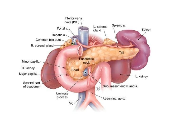
Duodenum and pancreas (drawing!)
jejunum ileum - thicker wall - wider mesentery - 2 arteries enter the bowel wall Capsule endoscopy
Specimens of the jejunum (A) and ileum (B) from a cadaver where the superior mesenteric artery was injected with red-coloured gelatin before fixation. The largest vessels present are the jejunal and ileal branches of the superior mesenteric artery and these are succeeded by anastomotic arterial arcades, which are relatively few in number (1– 3) in the jejunum, becoming more numerous (2– 6) in the ileum. From the arcades, straight arteries (arteriae recta) pass towards the gut wall; frequently, successive straight arteries are distributed to opposite sides of the gut. Note the denser vascularity of the jejunal wall. A B
Meckel’s diverticulum – remnant of the vitteline duct (2 -3% of the individuals)
Abdomen, axialis CT 1. 2. 3. 4. 5. 6. 7. 8. 9. 10. 11. Rectus hüvely/M. rectus abdominis Costa Lobus sin. hepatis, segmentum med. IV. Lobus sin. hepatis, segmenta latt. II-III. Duodenum Gaster (antrum) Colon Art. hepatica V. portae Pancreas (collum) Jejunum 12. 13. 14. 15. 16. 17. 18. 19. 20. 21. 22. V. lienalis Cauda pancreatis Gl. suprarenalis sin. V. cava inf. Crus diaphragmatis A. mesenterica sup. Aorta Vertebra thorac. XII. M. erector spinae Ren sinister Lien
Histology of the small intestine – basic features INTESTINAL VILLI – with culticle Simple columnar epithelium + goblet cells Lacteal (lymph vessel) CRYPTS of LIEBERKÜHN Tunica mucosa (epitelium + propria + lamina muscularis mucosae) Submucosa Tunica muscularis Tunica serosa (adventitia)
Cuticule (brush border) Goblet cells Intestinal villus Crypt of Lieberkühn propria Lamina muscularis mucosae
Epithelial cell types in the small intestine 1. Enterocytes (simple columnar, with cuticule) 2. Goblet cells 3. Enteroendocrine cells (→ bioactive peptides: gastrointestinal hormones, eg: cholecystokinin, secretin, gastric inhibitory polipeptide, motilin, etc. ) 4. Paneth cells – at the bottom of the crypts; large eosinophilic granules with anti-microbial agents (lysozyme) – Protection and regulation of the normal bacterial flora 5. Stem cells (multipotent – they can create all cell types in the intestinal epithelium)
Which part of the small intestine? - Check the subucosa! Duodenum Jejunum Ileum Kerckring valves (folds) Brunner’s glands Peyer’s patch Brunner’s glands in the submucosa ! – mucus-rich alkaline secretion to protect the duodenal mucosa (neutralizing the stomach acid) ‘nothing special’ Peyer’s patches in the submucosa! - aggregated lyphoid nodules; members of MALT; B-dependent zone Only in the anti-mesenterial side!
Histology of the pancreas Exocrine component Endocrine component Serous gland – pancreatic acini (basophilic cytoplasm & zymogen granules – contain digestive enzymes ) Islets of Langerhans Hormone secretion, regulates blood sugar level (glucagon: ⇑ ; Centroacinar cells= intercalated duct cells located in insulin: ⇓ ) the acinus Secretion ⇓ PP: self-regulate pancreatic secretion activities
Histology of the pancreas
Serous acini Islet of Langerhans Paccinian corpuscles Islet of Langerhans
Why am I studying this…? I will be a dentist! There are several diseases of the GI system that couse signs and symptoms in the oral cavity. Here a few examples: - Gastric reflux – chronic reflux couses characteristic enamel erosion - Gardner syndrome (genetic disease characterized by intestinal polyposis with a very high risk of malignant transformation into adenocarcinoma): - multiple enostoses of the jaws - supernumary and/or unerupted teeth - increased risk of odontomas Enamel erosion in a bulimic patient Oral lesion in ulcerative colitis
Thank you for your attention!