Morphology and histology of kidney Dr Zita Puskr

")

 fascia prerenal Posterior (Zuckerkandl’s)")


 corpuscle Glomerular")
 Vascular pole Urinary pole")


and")


 2. Lamina propria: loose")


")


 Urethra feminina Structure: Tunica mucosa 1. Epithelium mucosae: urothelium (near the bladder) stratified")

- Slides: 34
Morphology and histology of kidney Dr. Zita Puskár
Kidney (Ren)
Syntopy of kidney Anterior Aspects 12 cm length, 6 cm width, 3 cm thickness Descending Colon Ascending Colon
Renal fascia Kidneys are retroperitoneal organs Lateroconal fascia Anterior (Gerota’s) fascia prerenal Posterior (Zuckerkandl’s) fascia retrorenal
Capsules of the kidney Fibrous capsule Adipose capsule
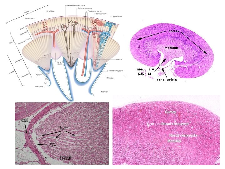
Coronal section of the kidney
Blood circulation of the kidney
Blood circulation of the kidney Interlobular artery Interlobular vein
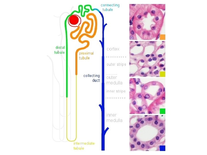
Functional unit of the kidney: Nephron Cortex Cortical nephron 1. Renal (Malpighian) corpuscle Glomerular (Bowman’s) capsule Glomerulus Juxta medullary nephron 2. Proximal convoluted tubule 3. Loop of Henle Proximal straight tubule (descending thick limb) Medulla Descending (thin) limb of loop of Henle Ascending (thin) limb of loop of Henle Distal straight tubule (ascending thick limb) 4. Distal convoluted tubule Proximal and distal tubule: convoluted part - pars convoluta, straight part - pars recta
Renal corpuscle (Malpighi’s body) Vascular pole Urinary pole
Podocytes
Filtration membrane
Mesangial cells and their matrix fill intersticies between capillaries that lack podocytes Functions: 1. Structural support and contraction 2. Phagocytosis (proteins, antibody-antigen complexes) 3. Secretion (cytokines, prostaglandins)
Tubular system
Proximal convoluted tubule
Histology of proximal convoluted tubule PAS
Loop of Henle 1. collecting tubule 2. thick ascending limb 3. thin loop 4. vasa recta 1. thick descending limb 2. collecting tubule 3. thick ascending limb
Distal convoluted tubule
Juxtaglomerular apparatus Distal Convoluted Tubule Lacis cells Functions: autoregulation of glomerular filtration rate (GFR)and controlling blood pressure Macula densa: coumnar cells of distal convoluted ducts (monitoring ion concentrations, secretion of ATP, adenosine and vasoactive molecules → contraction of afferent arteriole → decrease GFR Juxtaglomerular granular cells (JG): secretory phenotype of smooth muscle cell in the tunica media of the AA (baroreceptor function, renin secretion that cleaves angiotensinogen to angiotensin I and angiotensib II by angiotensin converting enzyme (ACE) → increased systemic blood presure (Extraglomerular mesangial) Lacis cells: supportive function and signal transmission, Erythropoetin secretion
Collecting tubules and ducts
Cell types of the collecting tubules and ducts Principal cells Intercalated cells
Medullary ray
Minor Calyx, Major Calix and Pelvis Renalis Umbrella cell Pear shaped cell Structure: Tunica mucosa 1. Epithelium mucosae: urothelium (transitional epithelium) 2. Lamina propria: loose connective tissue with blood vessels No lamina muscularis mucosae and tela submucosa!!! Tunica muscularis (peristalsis) Inner longitudinal Outer circular Tunica adventitia Loose connective tissue with blood and lymph vessels and nerves connected to the adipose tissue of the renal sinus (interlobar arteries, veins)
Ureter Structure: Tunica mucosa 1. Epithelium mucosae: urothelium (transitional epithelium) 2. Lamina propria: loose connective tissue (blood vessels, nerves, small tubular mucous glands near the pelvis ) No lamina muscularis mucosae and tela submucosa!!! Tunica muscularis (peristalsis) Inner longitudinal Middle circular Outer longitudinal Tunica adventitia Loose connective tissue with adipocytes, blood and lymph vessels and nerves
Ureter
Quiz 1 2 3 2 1 3 1 2 5 1 4
Quiz 7 6 8 9
Urinary bladder (vesica urinaria)
Urinary bladder Structure: Tunica mucosa 1. Epithelium mucosae: urothelium 2. Lamina propria: loose connective tissue (blood and lymp vessels, nerves) 3. lamina muscularis mucosae (It is missing in the trigonum vesicae) Tela submucosa – thick, loose connective tissue with elastic fibers, lymph and blood vessels, nerves and lymph follicles Tunica muscularis (elastic fibers, lymph and blood vessels, vegatative nerves, ganglions) Inner longitudinal Middle circular (at the origine of urethra: sphincter vesicae) Outer longitudinal Subserosa-serosa /Tunica adventitia
Urethra férfi Male női Female
(Female) Urethra feminina Structure: Tunica mucosa 1. Epithelium mucosae: urothelium (near the bladder) stratified cuboidal or columnar epithelium (middle part) stratified squamous non-keratinized epithelium with endoepithelial glands 2. Lamina propria: loose connective tissue (blood and lymph vessels, nerves, sinuses with intima cushions - cavernous tissue, mucous (Skene) glands - paraurethral ducts) Tunica muscularis Inner circular (together with the inner circular layer of the bladder: m. sphincter vesicae) (Where the urethra passes throug the urogenital diaphragm a striated muscle ring appears: external urethral sphincter) Outer longitudinal Tunica adventitia
Reference: Röhlich Pál: Szövettan, Budapest, 2006 Anthony L. Mescher: Junqueira’s Basic Histology, New York, 2010 Michael Ross and Lynn J. Romrell: Histology, Baltimore, 1989 Geoffrey M. Cooper and Robert E. Hausman: The Cell, A molecular Approach, (ASM, Sinauer), Washington, Sunderland, 2009