
MorbidityMortality meeting Presenter Supervisor Moderator At Triage 1019
Morbidity&Mortality meeting Presenter 翁一銘 Supervisor 吳孟書 Moderator 邱德發
At Triage (10/19 am 00: 23) n n n Age: 1 -month-5 -day-old Sex: Female Triage grade: 3級 Vital sign: BT: 37. 6, HR: 158, RR: 28, BPC. C: 病患來診為頸部腫脹
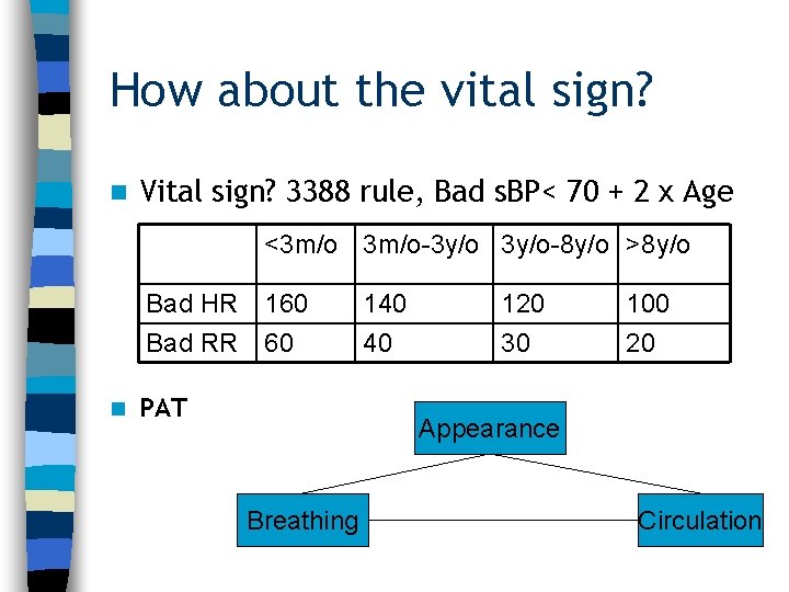
How about the vital sign? n Vital sign? 3388 rule, Bad s. BP< 70 + 2 x Age <3 m/o-3 y/o-8 y/o >8 y/o Bad HR Bad RR n 160 60 PAT 140 40 120 30 100 20 Appearance Breathing Circulation
Present illness Progressive swelling over right pre-auricle area in 2 days; Rapid expand to chin and neck tonight n Decreased appetite: 90 ml --> 60 ml q 4 h today n Brought to LMD 5 days ago due to irritable crying and mild fever (37. 8 C) was told with medicine prescribed n Some black, tarry? stool since 1 week ago n
Past history n n n n Allergy: NKA No previous admission or OP Birth History: 25 y/o-G 1 P 1 mother via C/S GA: 39 weeks Birth Weight: 2780 gm Newborn screening: negative BW: 4 kg (3 rd-10 th percentile) Vaccination history: On schedule FHx: No hereditary disease No similar symptoms of family members
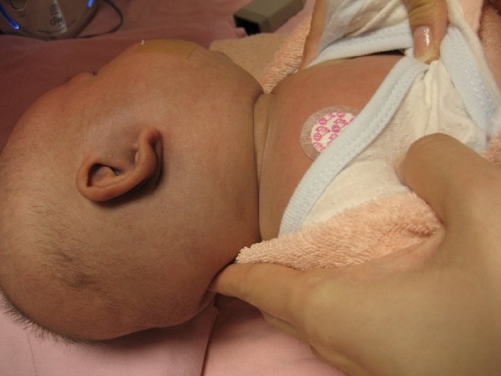
Physical Examination n n n Activity: fair, Cons: Alert HEENT: no icteric, no conjunctivitis, no throat injection, no tonsil enlarge, dry lip&mucosa Palpable mass over per-auricle area, Swelling over R’t cheek & chin c ecchymosis Heart : RHB , no murmur Chest : clear breath sound , no wheeze , no retraction , no crackle , no nasal flaring Abdomen : soft&flat, normoactive bowel sound, no tender or rebound pain, no hepatosplenomegaly Extremity : freely movable, no edema
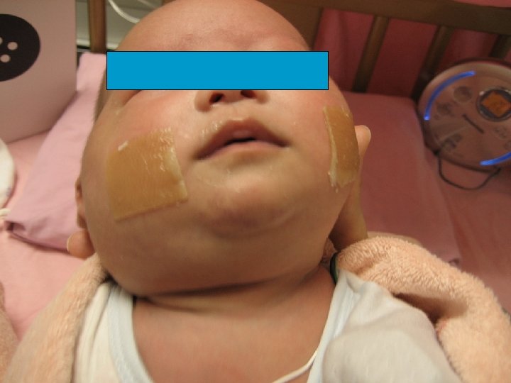
Pics
Pediatric neck mass n How to approach this patient? n Something (Hx, PE) more you want to know?
How to approach this patient? Neck Mass? Character, Size, Multiple, Asymmetry, Onset, Duration, Progression, Accompany symptoms Content: Tissue fluid, Blood, Pus, LNs, Tumor Etiology: 1. Congenital cyst 2. Infection 3. Trauma 4. Tumor n Uptodate 2008 V. 16. 2



Impression n IMP: 1. Suspect Vascular lesion c Hematoma (trauma? ) 2. R/O Infection c Pus Origin: LAPs Tonsil/ Pharynx/ Glottic Salivary gland Odontogenic 3. R/O Malignancy c Rupture
Impression&Order (10/19 am 01: 00) n IMP: Facial cellulitis n CBC/DC, Na/K/Cl, (am 02: 00) n IV: D 5 S 1/4 run 10 cc/hr, n CXR n BUN/Cr, B/Cx 1, (am 02: 25) n AST/ALT, Bil(T/D), CRP, (am 03: 20)
n Do you agree?
Do you agree? n Airway/Breathing: C-spine protection? n Circulation: IVF: D 5 S 1/4 run 10 cc/hr? (BW: 4 kg) n Prepare blood n NPO n C-spine, Skull&long bone X-ray survey n Bedside Echo? CT?
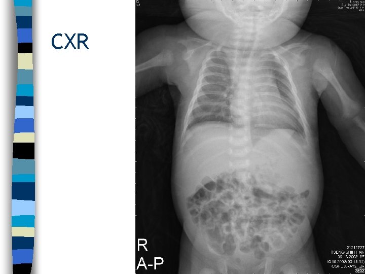
CXR
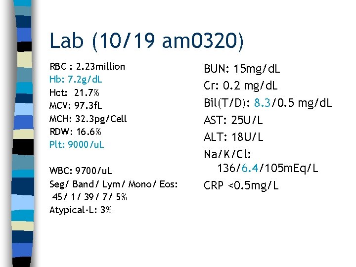
Lab (10/19 am 0320) RBC : 2. 23 million Hb: 7. 2 g/d. L Hct: 21. 7% MCV: 97. 3 f. L MCH: 32. 3 pg/Cell RDW: 16. 6% Plt: 9000/u. L WBC: 9700/u. L Seg/ Band/ Lym/ Mono/ Eos: 45/ 1/ 39/ 7/ 5% Atypical-L: 3% BUN: 15 mg/d. L Cr: 0. 2 mg/d. L Bil(T/D): 8. 3/0. 5 mg/d. L AST: 25 U/L ALT: 18 U/L Na/K/Cl: 136/6. 4/105 m. Eq/L CRP <0. 5 mg/L
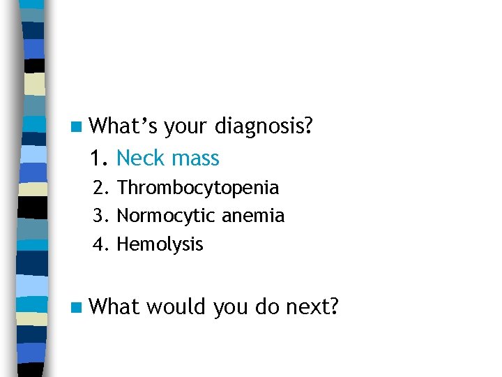
n What’s your diagnosis? 1. Neck mass 2. Thrombocytopenia 3. Normocytic anemia 4. Hemolysis n What would you do next?
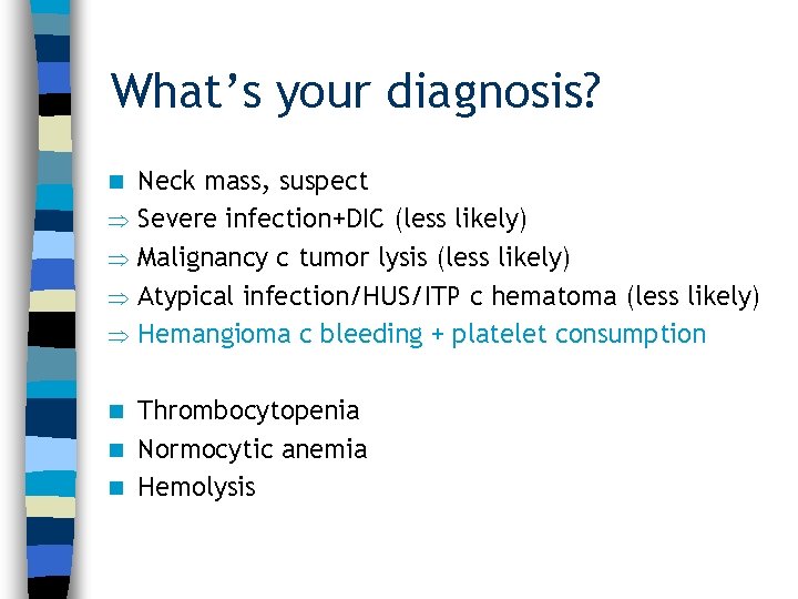
What’s your diagnosis? n Neck mass, suspect Severe infection+DIC (less likely) Malignancy c tumor lysis (less likely) Atypical infection/HUS/ITP c hematoma (less likely) Hemangioma c bleeding + platelet consumption Thrombocytopenia n Normocytic anemia n Hemolysis n
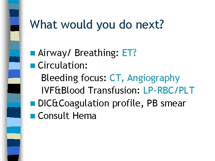
What would you do next? n Airway/ Breathing: ET? n Circulation: Bleeding focus: CT, Angiography IVF&Blood Transfusion: LP-RBC/PLT n DIC&Coagulation profile, PB smear n Consult Hema
Disposition (10/19 am 03: 45) n Diagnosis: 1. Thrombocytopenia 2. Facial cellulitis n Admission to NICU
NICU Course (10/19 am 04: 12) n n n n On critical DIC&Coagulation profile: INR: 1. 76, Ddimer>10000, Fibrinogen<50, FDP>80 PB smear: Suspect fragmentation of RBCs Blood Transfusion c LP-RBC/PLT/FFP Skull & Long bone survey X-ray: Negative ABX: Fortum + Oxacilline On ET+MV (10/20 pm) Head&Neck CT (10/21 am)
CT
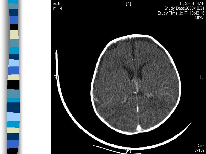
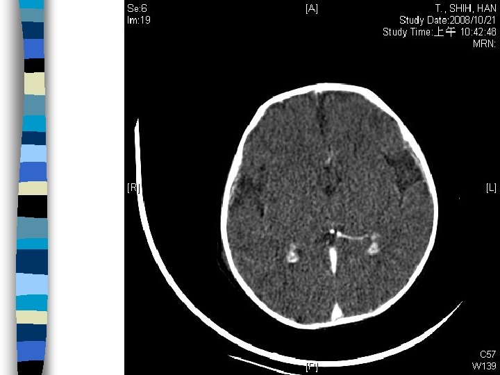
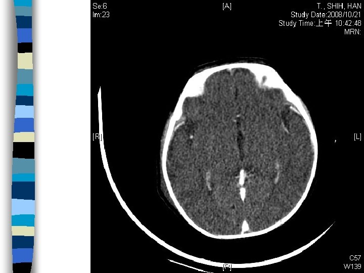
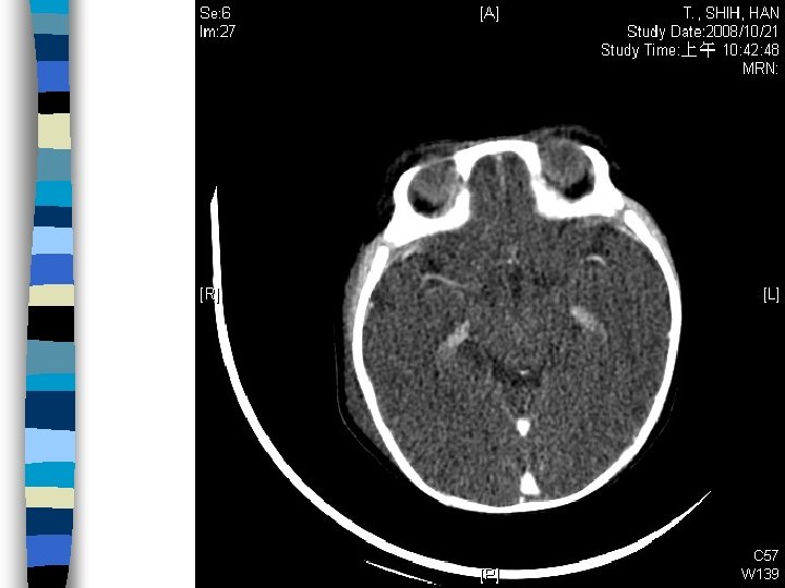
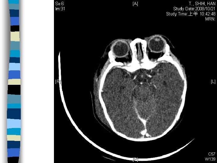
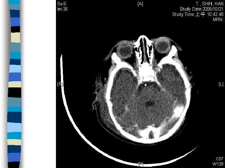
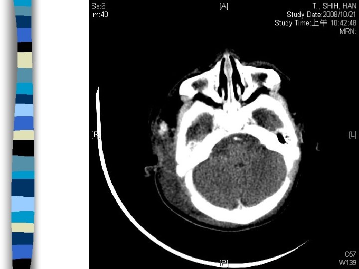
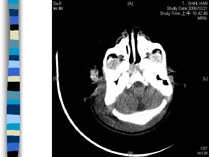
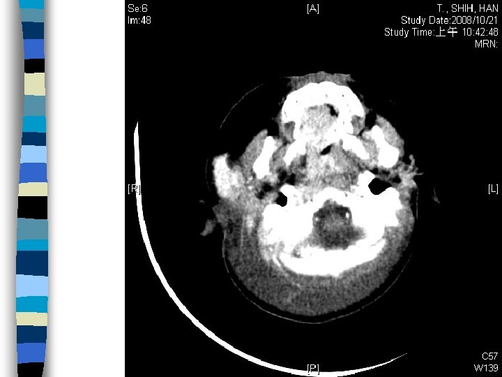
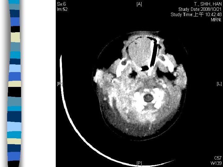
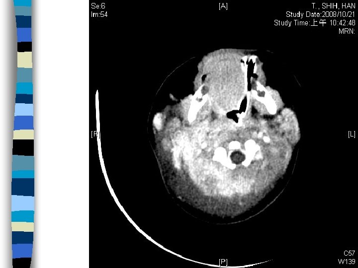
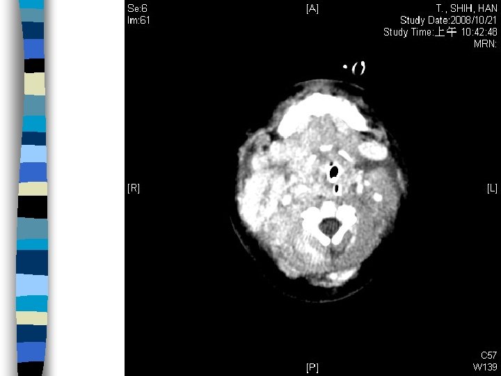
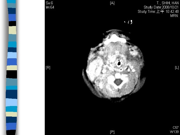
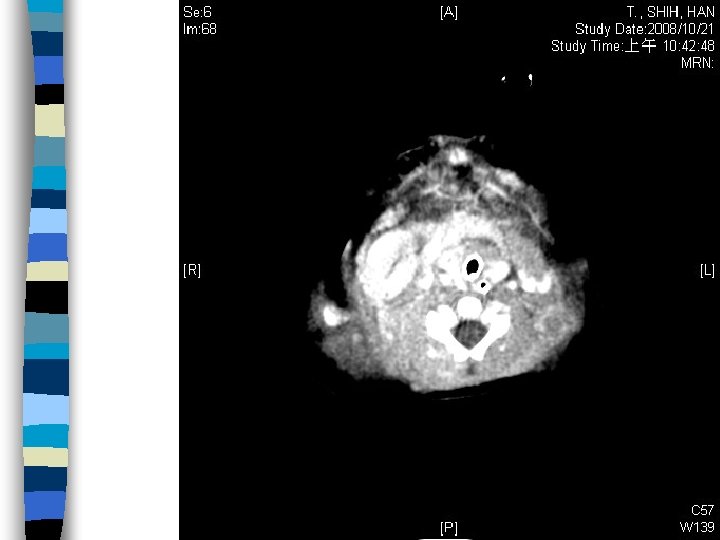
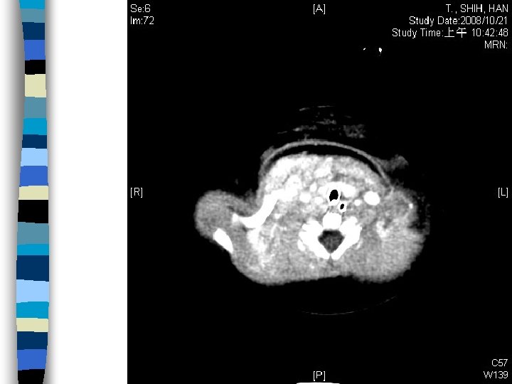
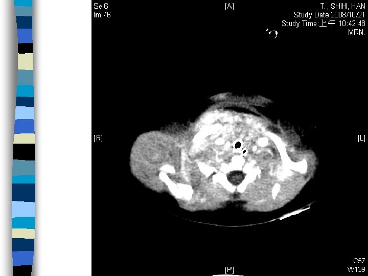
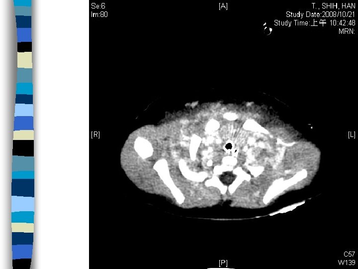
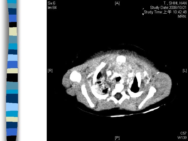
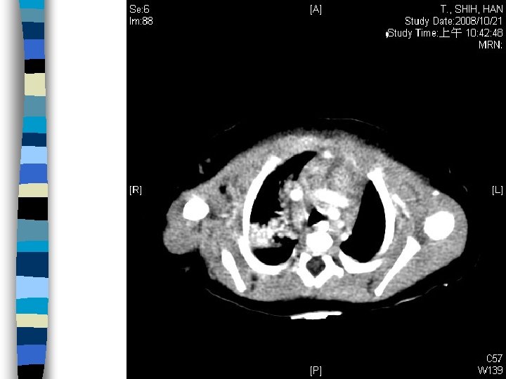
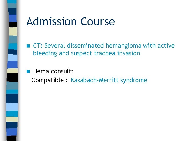
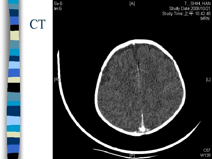
Admission Course n CT: Several disseminated hemangioma with active bleeding and suspect trachea invasion n Hema consult: Compatible c Kasabach-Merritt syndrome
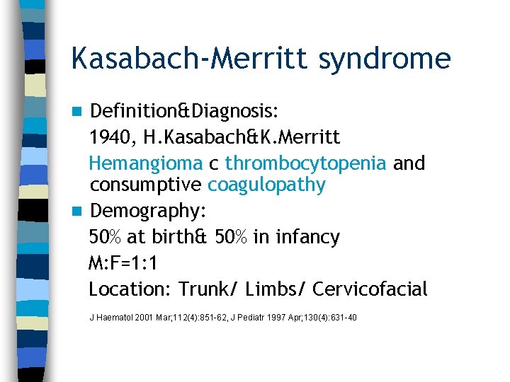
Kasabach-Merritt syndrome Definition&Diagnosis: 1940, H. Kasabach&K. Merritt Hemangioma c thrombocytopenia and consumptive coagulopathy n Demography: 50% at birth& 50% in infancy M: F=1: 1 Location: Trunk/ Limbs/ Cervicofacial n J Haematol 2001 Mar; 112(4): 851 -62, J Pediatr 1997 Apr; 130(4): 631 -40
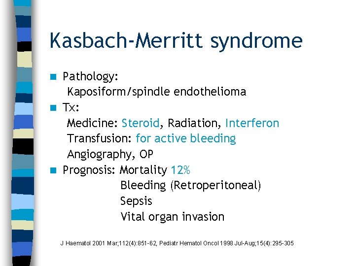
Kasbach-Merritt syndrome Pathology: Kaposiform/spindle endothelioma n Tx: Medicine: Steroid, Radiation, Interferon Transfusion: for active bleeding Angiography, OP n Prognosis: Mortality 12% Bleeding (Retroperitoneal) Sepsis Vital organ invasion n J Haematol 2001 Mar; 112(4): 851 -62, Pediatr Hematol Oncol 1998 Jul-Aug; 15(4): 295 -305
Admission Course n Solumedrol + BT c LP-RBC/PLT/FFP n Interferon-alpha added n Extubation 1 week latter n Transfer to ward on 11/1
Take home message 1. 針對嬰兒及新生兒病患, 頸部腫塊大部分為良性, 但 可有致命併發症, 如同本例. 2. 仔細問診及理學檢查幫助判斷頸部腫塊的內容及成 因. 3. 小心迅速擴大腫塊, 注意呼吸道及伴隨外傷或兒虐 的可能. “Killing” neck mass in an infant ~Most lesions are benign. However, life-threaten complications could occur. In our case, Kasbach-Merritt syndrome was considered. Detail history taking and physical examination help us defined the content and the etiologies of the lesion. Watch out rapid expanding mass, risk of airway compromise and evidence of trauma&child abuse n
At Triage (11/1 pm 18: 43) n n n Age: 4 -year-4 -month-old Sex: Male Triage grade: 1級 Vital sign: BT: 36. 5, HR: 156, RR: 22, BP-, E 4 V 5 M 6 C. C: 病患來診為發燒, 年齡>3歲, 體溫 >38 ℃且 無病容(表現活動力佳)
Present Illness Fever up to 38 degrees since 10/30 n Mild cough and rhinorrhea. Mild nasal stuffiness. n Neck pain with swelling and erythematous change today n No significant change in activity and appetite. n
Past History 09/24 -10/02 1. Mis-swallowing of alkali(p. H > 10) 2. Corrosive esophagitis, grade 2 3. Pharyngitis&Stomatitis n 10/27 -10/30 1. Esophageal stricture with stenosis S/P ballon dilation, 12 cm from incisors. n Allergy: NKA n Travel history: no n Current medication: no n
Birth History Birth Hx: G 1 P 1, fullterm, birth BW: 3100 gm n Newborn screening: Severe Pul. stenosis s/p balloon dilatation PDA s/p obliteration, ASD, Heart failure Congestive hepatitis n Growth: BW: 14 kg (<3 rd percentile) BH: 103 cm (25 -50 th percentile) n Vaccination: as schedule n Development as milestone n
Physical Examination General: acute ill looking, dehydration n Activity: fair, Appetite: poor n HEENT: Conj: not injected, Throat: injected, no oral ulcers or vesicles, Tonsils: not injected, Ear drums: intact n Neck: swelling over anterior part, erythematous, no pus formation, no wound, supple, no LAPs n
Physical Examination Chest: symmetric expansion, no retraction, regular heart beat, coarse breathing sound, no crackles, no wheezing n Abdomen: soft and not distended, normoactive bowel sounds no tenderness, no muscle guarding n Extremities: freely movable, no pitting edema, no rash, no petechia n
n How to approach this patient? n Something (Hx, PE) more you want to know?
Alkali injuries n n As lesion severity increases, stricture formation incidence increases. Following a grade 2 b burn, stricture incidence may be as high as 71%, and after a grade 3 burn, as high as 100%. Emerg Md Clin North Am 207; Vol. 2(2): 459 -476
How to approach this patient? Fever, focus? Neck Mass? Character, Size, Multiple, Asymmetry, Onset, Duration, Progression, Accompany symptoms =>Content: Tissue fluid, Pus, LAPs, Tumor, Blood Etiology: Congenital cyst, Infection, Tumor, Trauma n Severity: Involving level, abscess formation (Dermis, Subcutaneous, Fascia)
Cellulitis/ Abscess/ Deep neck inf. n Deep neck inf. ~PI: Fever, Neck mass, Stiffness, Sorethroat, Poor intake, Dysphagia, Drooling, LAPs ~Risk: Pharyngitis, Tonsilitis, Odontogenic, Immunocompromised, Poor hygiene Otolaryngol Clin N AM 2008(41)459 -483
Imagine for deep neck infection ~Imagine: CECT vs X-ray (vs Echo vs MRI) CECT: Accurancy: CECT+clinical examination: 89% CECT alone: 63% - 95% (differentiating a drainable abscess from cellulitis) CXR: CECT is superior for evaluating cellulitis or abscess within the mediastinum Neck Lat. : Sensitivity: Neck lat: 83% vs CECT: 100% (for retropharyngeal and parapharyngeal abscesses) Otolaryngol Clin N AM 2008(41)459 -483
Impression & Order (pm 19: 00) n Impression: Neck cellulitis n Neck AP-Lat CBC/DC, AST/ALT, Na/K/Cl, CRP Cr/BUN B/Cx 1 On lock? NPO CT? n n n
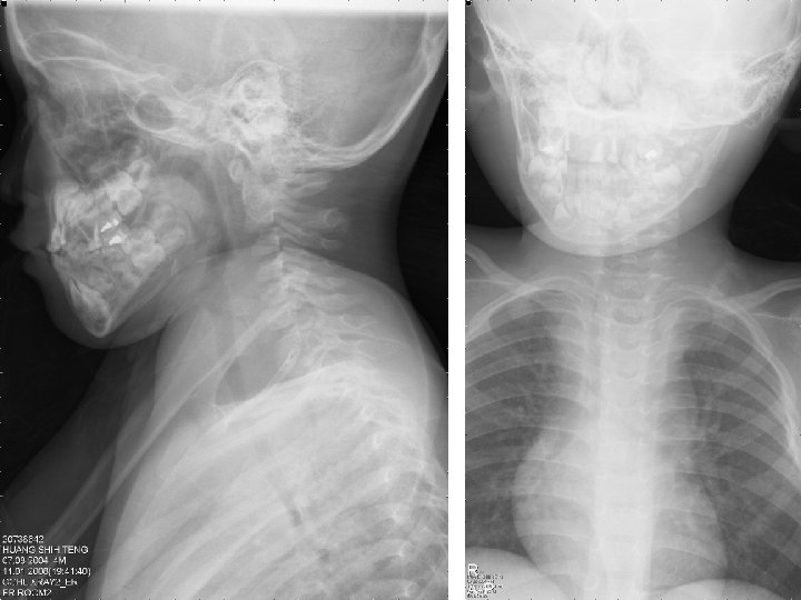
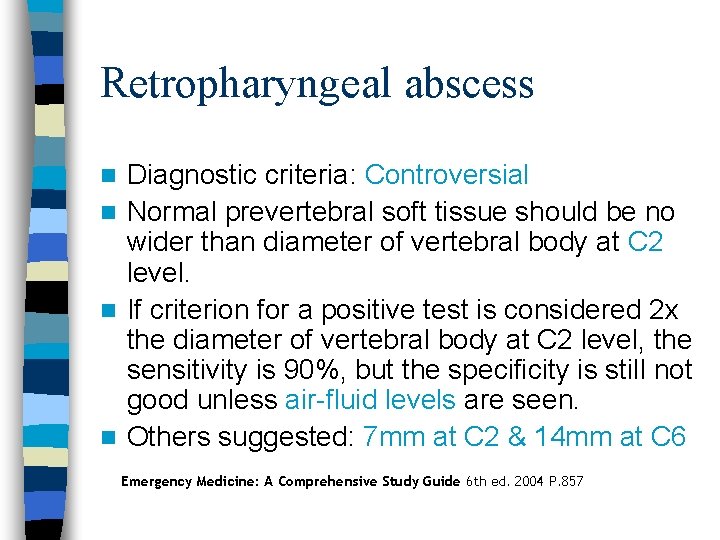
Retropharyngeal abscess Diagnostic criteria: Controversial n Normal prevertebral soft tissue should be no wider than diameter of vertebral body at C 2 level. n If criterion for a positive test is considered 2 x the diameter of vertebral body at C 2 level, the sensitivity is 90%, but the specificity is still not good unless air-fluid levels are seen. n Others suggested: 7 mm at C 2 & 14 mm at C 6 n Emergency Medicine: A Comprehensive Study Guide 6 th ed. 2004 P. 857
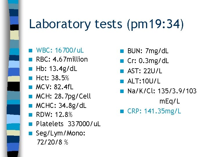
Laboratory tests (pm 19: 34) n n n n n WBC: 16700/u. L RBC: 4. 67 million Hb: 13. 4 g/d. L Hct: 38. 5% MCV: 82. 4 f. L MCH: 28. 7 pg/Cell MCHC: 34. 8 g/d. L RDW: 12. 8% Platelets 337000/u. L Seg/Lym/Mono: 72/20/8 % n n n BUN: 7 mg/d. L Cr: 0. 3 mg/d. L AST: 22 U/L ALT: 10 U/L Na/K/Cl: 135/3. 9/103 m. Eq/L CRP: 141. 35 mg/L
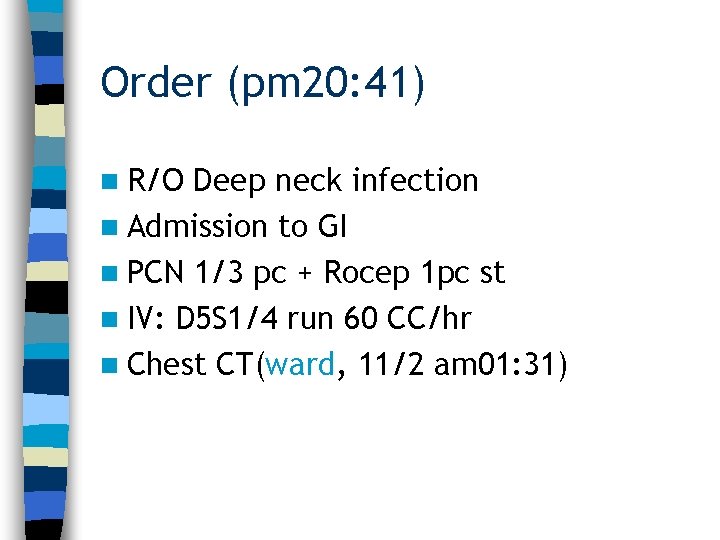
Order (pm 20: 41) n R/O Deep neck infection n Admission to GI n PCN 1/3 pc + Rocep 1 pc st n IV: D 5 S 1/4 run 60 CC/hr n Chest CT(ward, 11/2 am 01: 31)
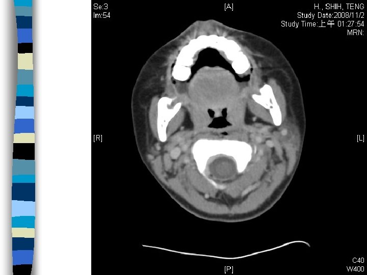
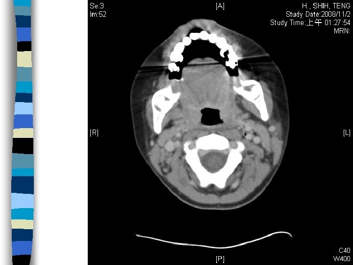
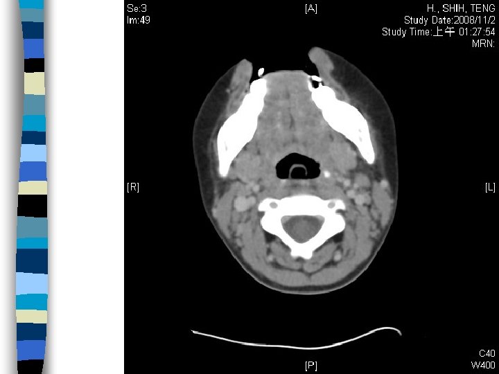
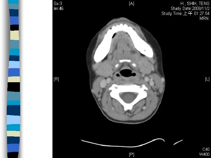
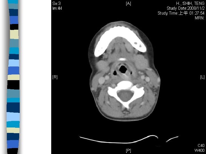
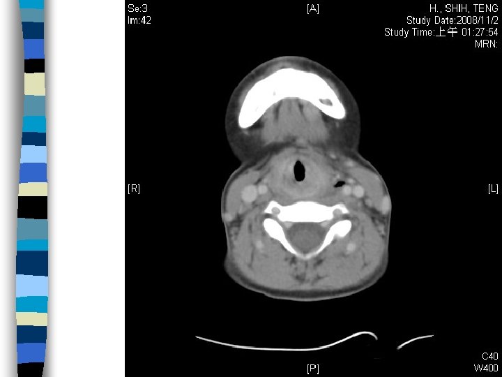
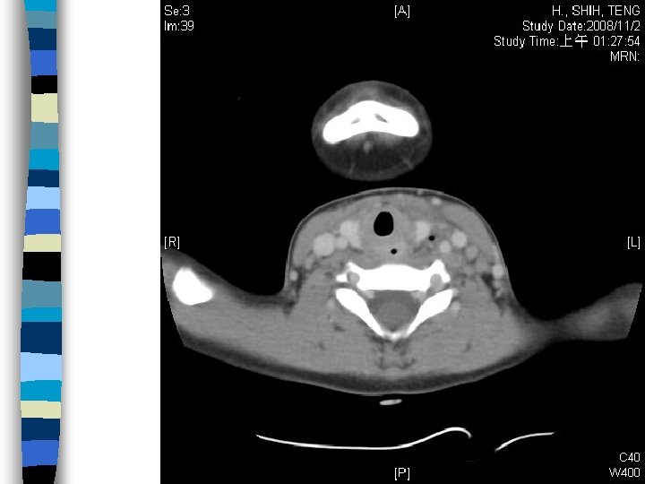
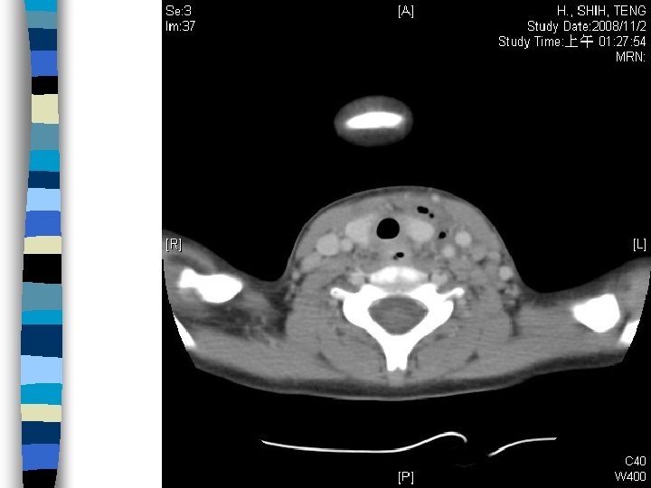
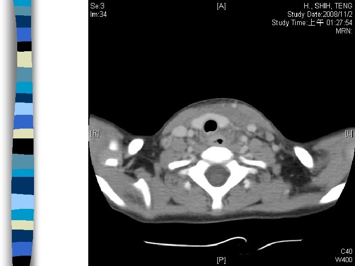
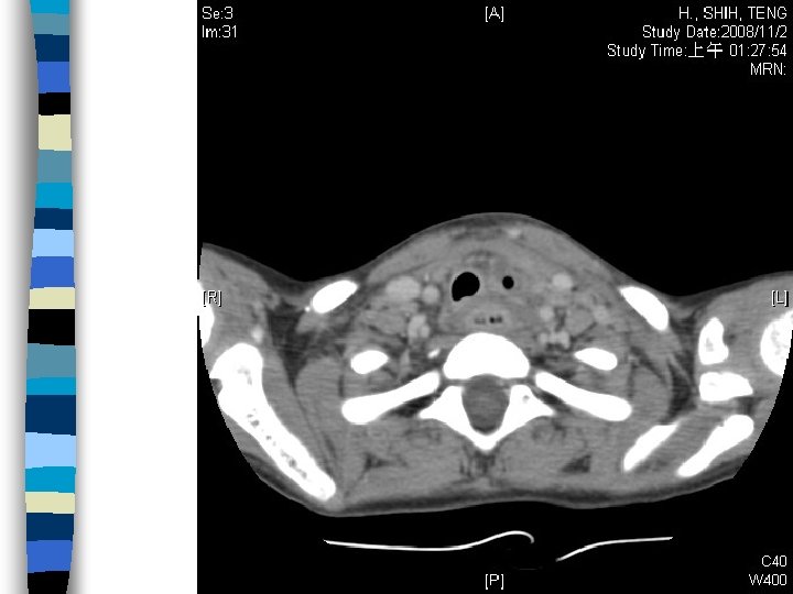
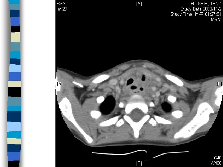
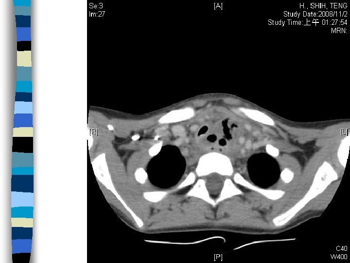
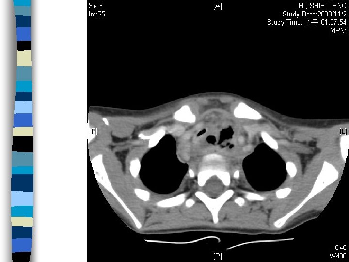
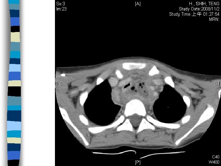
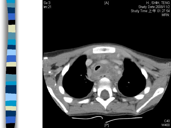
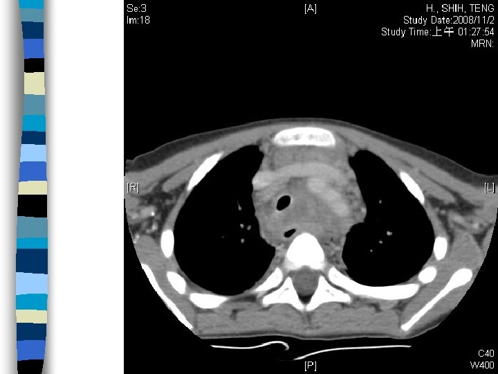
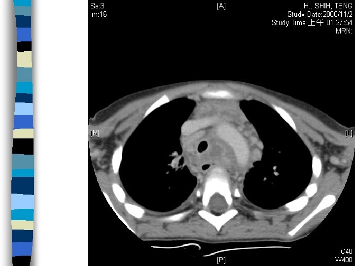
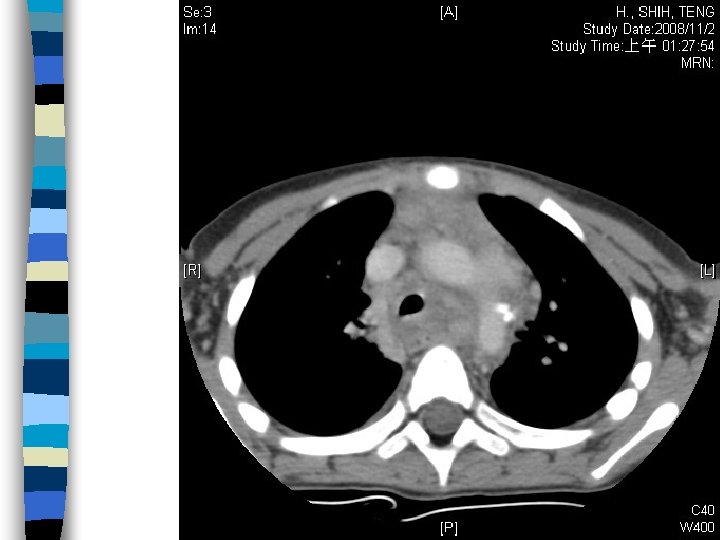
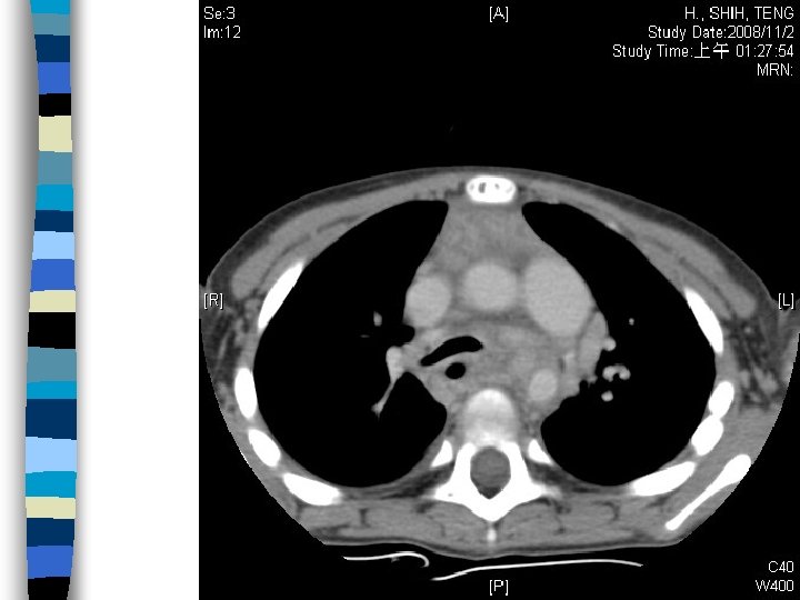
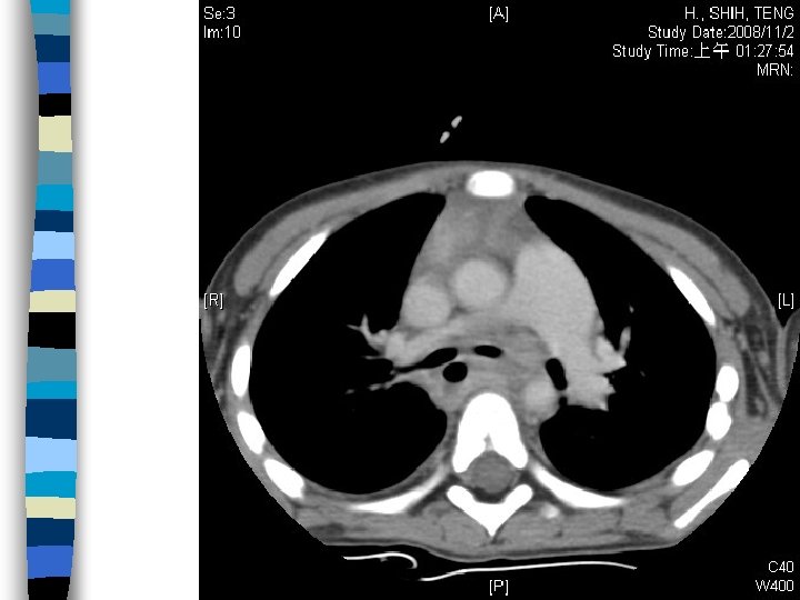
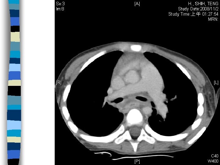
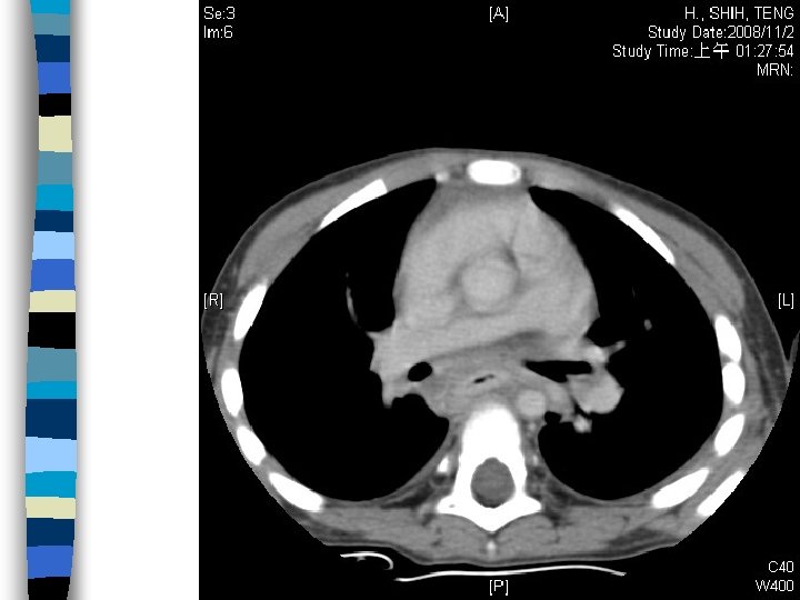
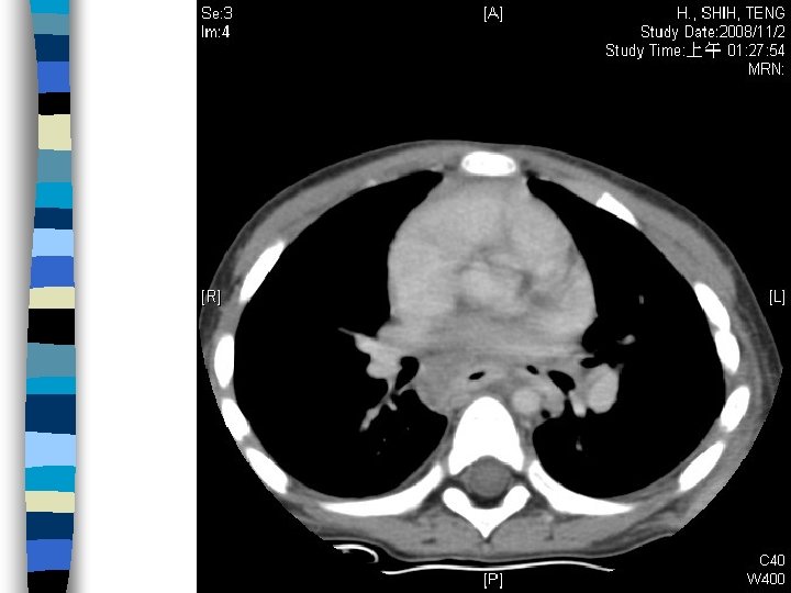
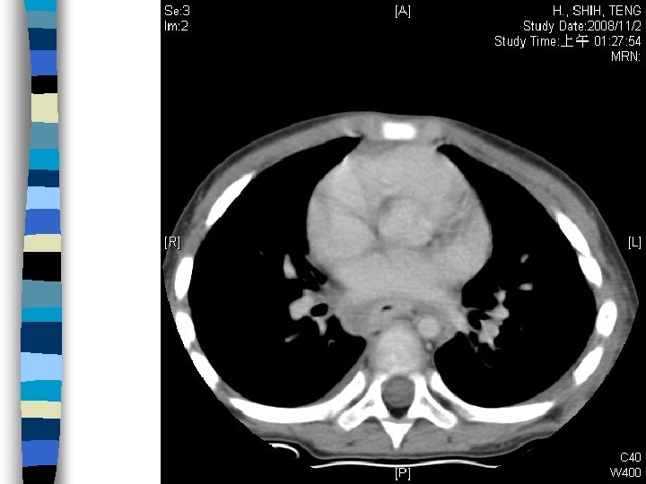
CT report (11/4) Findings: Free air at Lt lower neck. Encapsulated fluid extending from thoracic inlet to subcarinal level, in favor of mediastinitis and abscess formation. Swelling of middle to lower third esophagus, compatible with corrosive injury. n Conclusion: Corrosive esophagitis with esophageal perforation, mediastinitis and abscess formation. n
OP Note (11/2) Operative findings: a much pus out beneath the esophagus and just under the sterum, penrose drain was insetrted, esophagus was fibrotic condition from outside looking and some infected and granumatous tissue found below the trachea, laparoscopic gastrostomy was done. n Operative method: 1. Drainage over the esophageal space. 2. laparoscopic gastrostomy n
Admission Course Extubation on 11/5 n Transferred to ward & Try gastrostomy feeding on 11/6, n Advancing & remove penrose n Abx: Ampicillin + Ceftriaxone (11/1~11/10) Augmentin (11/10~15) n MBD on 11/15 c oral form Augmentin n
Take home message 1. 針對兒科病患, 仔細的問診, 理學檢查幫助判斷 深頸感染的可能. 2. 對食道腐蝕性傷害, 及食道氣球擴張術後者, 應 將食道破裂列入可能鑑別診斷. n Esophageal rupture should be considered in patients post corrosive injury or balloon dilation. Infection with deep neck space involvement could be easily miss or underestimated in child. Detail history taking, and examination help us to find another cause.
- Slides: 95