Monoclonal Antibodies Monoclonal antibodies m Ab or mo

Monoclonal Antibodies
 are monospecific antibodies [specificity to")
• Monoclonal antibodies (m. Ab or mo. Ab) are monospecific antibodies [specificity to antigens is singular (mono- +specific) in any of several ways: antibodies that all have affinity for the same antigen; antibodies that are specific to one antigen or one epitope; or antibodies specific to one type of cell or tissue. Regarding antibodies, monospecific and monovalent overlap in meaning; both can indicate specificity to one antigen, one epitope, or one cell type (including one microorganism species)] that are made by identical immune cells that are all clones of a unique parent cell, in contrast to polyclonal antibodies which are made from several different immune cells.

• Monoclonal antibodies have monovalent affinity, in that they bind to the same epitope. • Given almost any substance, it is possible to produce monoclonal antibodies that specifically bind to that substance; they can then serve to detect or purify that substance. This has become an important tool in biochemistry, molecular biology and medicine. When used as medications, the non-proprietary [not registered] drug name ends in -mab.

Discovery • The idea of a magic bullet was first proposed by Paul Ehrlich, who, at the beginning of the 20 th century, postulated that, if a compound could be made that selectively targeted a disease-causing organism, then a toxin for that organism could be delivered along with the agent of selectivity. He and Elie Metchnikoff received the 1908 Nobel Prize for Physiology and Medicine for this work, which led to an effective syphilis [bacterial skin infection] treatment by 1910. • In the 1970 s, the B-cell cancer, multiple myeloma [a cancer of plasma cells, a type of white blood cells normally responsible for producing antibodies] was known, and it was understood that these cancerous B-cells all produce a single type of antibody

• This was used to study the structure of antibodies, but it was not yet possible to produce identical antibodies specific to a given antigen. Building on the work of many others, in 1975, Georges Kohler and Cesar Milstein succeeded in making fusions of myeloma cell lines with B cells to produce hybrodomas that made antibodies to known antigens and that were immortalized. They shared the Nobel Prize for Physiology and Medicine in 1984 for the discovery. In 1988, Greg Winter and his team pioneered the techniques to humanize monoclonal antibodies, removing the reactions that many monoclonal antibodies caused in some patients.
 is a selection medium for")
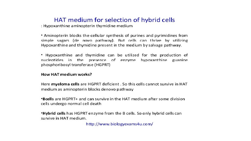
Hybridoma cell production • HAT Medium (Hypoxanthine Aminopterin Thymidine) is a selection medium for mammalian cell culture which relies on the combination of: • Aminopterin, a drug that acts as a powerful folate metabolism inhibitor by inhibiting dihydrofolate reductase • Hypoxanthine (a purine derivative) • Thymidine (a deoxynucleoside) which are intermediates in DNA synthesis.

• The trick is that aminopterin blocks DNA de novo synthesis, which is absolutely required for cell division to proceed, but hypoxanthine and thymidine provide cells with the raw material to evade the blockage (the salvage pathway), if they have the right enzymes, which means having functioning copies of the genes that encode them. • The enzyme dihydrofolate reductase, which produces Tetra Hydro Folate (THF) by the reduction of dihydrofolate, is specifically blocked by aminopterin.

• One of the important targets for cellular reproduction is Thymidylate Synthase (Tetra Hydro Folate is a cofactor), which creates Thymidine Monophosphate (TMP) from Deoxy Uridine Monophosphate (d. UMP). By additional phosphorylation reactions (by Thymidine Kinase), TMP can be used to make Thymidine Tri Phosphate (TTP), one of the four nucleotide precursors that are used by DNA polymerase to create DNA. Without the THF required to convert d. UMP, there can be no TTP, and DNA synthesis cannot proceed, unless TMP can be produced from another source. The alternative source is the Thymidine present in the HAT medium that can be absorbed by the cells and phosphorylated by Thymidine Kinase (TK) into TMP.

• The synthesis of IMP, (precursor to GMP and GTP, and to AMP and ATP) also requires THF, and also can be bypassed. In this case Hypoxanthine Guanine Phosphoribosyl Transferase (HGPRT) reacts hypoxanthine absorbed from the medium with PRPP (Phosphoribosyl pyrophosphate), liberating pyrophosphate, to produce IMP by a salvage pathway. • Therefore, the use of HAT medium for cell culture is a form of artificial selection for cells containing working TK and HGPRT

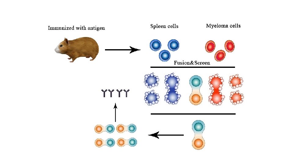
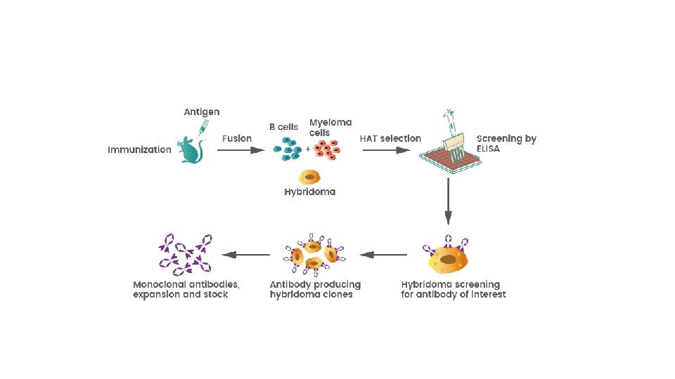
Monoclonal antibodies are typically made by cell culture that involves fusing myeloma cells with the spleen cells from a mouse that has been immunized with the desired antigen. However, recent advances have allowed the use of rabbit B-cells to form a rabbit hybrodoma. Polyethylene glycol is used to fuse adjacent plasma membranes, but the success rate is low so a selective medium in which only fused cells can grow is used.

This is possible because myeloma cells have lost the ability to synthesize hypoxanthine-guanine-phosphoribosyl transferase (HGPRT), an enzyme necessary for the salvage synthesis of nucleic acids. The absence of HGPRT is not a problem for these cells unless the de novo purine synthesis pathway is also disrupted. By exposing cells to aminopterin (a folic acid analogue, which inhibits dihydrofolate reductase), they are unable to use the de novo pathway and become fully auxotrophic for nucleic acids, thus requiring supplementation to survive.

The selective culture medium is called HAT medium because it contains hypoxanthine, aminopterin, and thymidine. This medium is selective for fused hybridoma cells. Unfused myeloma cells cannot grow because they lack HGPRT, and thus cannot replicate their DNA. Unfused spleen cells cannot grow indefinitely because of their limited life span. Only fused hybrid cells, referred to as hybridomas, can grow indefinitely in the media because the spleen cell partner supplies HGPRT and the myeloma partner has traits that make it immortal (like a cancer cell).

This mixture of cells is then diluted and clones are grown from single parent cells on microtitre wells. The antibodies secreted by the different clones are then assayed for their ability to bind to the antigen (with a test such as ELISA or Antigen Microarray Assay) or immuno-dot blot. The most productive and stable clone is then selected for future use. The hybridomas can be grown indefinitely in a suitable cell culture medium. They can also be injected into mice (in the peritoneal cavity, surrounding the gut). There, they produce tumors secreting an antibodyrich fluid called ascites fluid.

Purification of monoclonal antibodies The sample is first conditioned, or prepared for purification. Cells, cell debris, lipids, and clotted material are first removed, typically by centrifugation followed by filtration with a 0. 45 µm filter. These large particles can cause a phenomenon called membrane fouling in later purification steps. In addition, the concentration of product in the sample may not be sufficient, especially in cases where the desired antibody is one produced by a low-secreting cell line. The sample is therefore condensed by ultrafiltration and dialysis.

Most of the charged impurities are usually anions such as nucleic acids and endotoxins and are often separated by ion exchange chromatography. A much quicker, single-step method of separation is Protein A/G affinity chromatography (Protein A/G is a recombinant fusion protein that combines Ig. G binding domains of both Protein A and Protein G). The antibody selectively binds to protein A/G, so a high level of purity (generally >80%) is obtained. However, this method may be problematic for antibodies that are easily damaged, as harsh conditions are generally used.

Types of Monoclonal Antibodies Different types of monoclonal antibodies are used in cancer treatments. • Naked monoclonal antibodies Naked m. Abs are antibodies that work by themselves. There is no drug or radioactive material attached to them. These are the most common type of m. Abs used to treat cancer. Most naked m. Abs attach to antigens on cancer cells, but some work by binding to antigens on other, noncancerous cells, or to even free-floating proteins.

• Naked m. Abs can work in different ways. • Some boost a person’s immune response against cancer cells by attaching to them and acting as a marker for the body’s immune system to destroy them. An example is Alemtuzumab (Campath®) (Intravenous infusion), which is used to treat some patients with Chronic Lymphocytic Leukemia (CLL). Alemtuzumab binds to the CD 52 antigen, which is found on cells called lymphocytes (which include the leukemia cells). Once attached, the antibody attracts immune cells to destroy these cells.

Other naked m. Abs work mainly by attaching to and blocking antigens that are important signals for cancer cells (or other cells that help cancer cells grow or spread). Trastuzumab (Herceptin®) (Intravenous) is an antibody against the HER 2 (Human Epidermal Growth Factor Receptor 2) protein. In some types of cancer, such as breast and stomach cancer, the cells sometimes have large amounts of this protein on their surface. When HER 2 is activated, it helps these cells grow. Trastuzumab binds to these proteins and stops them from becoming active.

Conjugated monoclonal antibodies Monoclonal antibodies joined to a chemotherapy drug, another kind of toxin or a radioactive particle are called conjugated monoclonal antibodies. The m. Ab is used as a homing device to take one of these substances directly to the cancer cells. The m. Ab circulates in the body until it can find and hook onto the target antigen. It then delivers the toxic substance where it is needed most. This lessens the damage to normal cells in other parts of the body. Conjugated m. Abs are also sometimes referred to as tagged, labeled, or loaded antibodies

They can be divided into groups depending on what they are linked to. Radiolabeled antibodies: Radiolabeled antibodies have small radioactive particles attached to them. Ibritumomab tiuxetan (Zevalin®) (Intravenous) (either yttrium-90 or indium 111) is an example of a radiolabeled m. Ab. The antibody binds to the CD 20 antigen found on the surface of normal and malignant B cells (but not B cell precursors), allowing radiation from the attached isotope (mostly beta emission) to kill it and some nearby cells.

• This eliminate B cells from the body, allowing a new population of healthy B cells to develop from lymphoid stem cells. It can be used to treat some types of non-Hodgkin lymphoma. Treatment with this type of antibody as radioimmunotherapy (RIT). is sometimes known
 drugs attached to them.")
Chemolabeled antibodies These m. Abs have powerful chemotherapy (or other) drugs attached to them. They are also known as antibody-drug conjugates (ADCs). (The drug is often too powerful to be used on its own – it would cause too many side effects if not attached to an antibody. ) Chemolabeled antibodies approved by the FDA to treat cancer now include:
, an antibody that targets the CD 30 antigen (found on lymphocytes),")
Brentuximab Vedotin (Adcetris®), an antibody that targets the CD 30 antigen (found on lymphocytes), attached to a chemo drug called MMAE. This drug is used to treat Hodgkin lymphoma and anaplastic large cell lymphoma that is no longer responding to other treatments. Brentuximab vedotin consists of the Chimeric monoclonal antibody brentuximab (c. AC 10, which targets the cellmembrane protein CD 30) linked to cathepsin cleavable linker (valinecitrulline), para-aminobenzylcarbamate spacer three to five units of the antimitotic agent, monomethyl auristatin E (MMAE, reflected by the 'vedotin' in the drug's name).

• The peptide-based linker bonds the antibody to the cytotoxic compound in a stable manner so the drug is not easily released from the antibody under physiologic conditions to help prevent toxicity to healthy cells and ensure dosage efficiency. • The peptide antibody-drug bond facilitates rapid and efficient drug cleavage inside target tumor cell. The antibody c. AC 10 part of the drug binds to CD 30 which often occurs on diseased cells but rarely on normal tissues.

The antibody portion of the drug attaches to CD 30 on the surface of malignant cells, delivering MMAE which is responsible for the antitumour activity. Once bound brentuximab vedotin is internalised by endocytosis and thus selectively taken up by targeted cells. The vesicle containing the drug is fused with lysosomes and lysosomal cysteine proteases, particularly cathepsin B start to break down valine-citrulline linker and MMAE is no longer bound to the antibody and is released directly into the tumor environment.

Biospecific monoclonal antibodies These drugs are made up of parts of 2 different m. Abs, meaning they can attach to 2 different proteins at the same time. An example is blinatumomab (Blincyto), which is used to treat some types of Acute Lymphocytic Leukemia (ALL). One part of blinatumomab attaches to the CD 19 protein, which is found on some leukemia and lymphoma cells. Another part attaches to CD 3, a protein found on immune cells called T cells. By binding to both proteins, this drug brings the cancer cells and immune cells together, which is thought to cause the immune system to attack the cancer cells.

Possible side effects of monoclonal antibodies Monoclonal antibodies are given intravenously. The antibodies themselves are proteins, so giving them can sometimes cause something like an allergic reaction. Possible side effects can include: Fever Chills Weakness Headache Nausea Vomiting Diarrhea Rashes Low blood pressure Compared with chemotherapy drugs, naked m. Abs tend to have fewer serious side effects. But they can still cause problems in some people.
- Slides: 30